
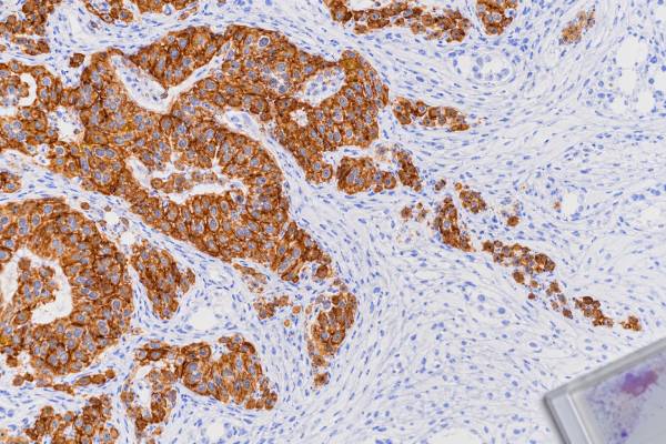
Pancytokeratin immunohistochemistry (IHC) is a broad tissue stain used to look for keratin proteins that are typical of epithelial cells. In a poorly differentiated tumor, positive pancytokeratin staining can strongly support epithelial differentiation and therefore a carcinoma, but it does not identify the organ where the cancer began. “Pancytokeratin” is not one single antibody; laboratories use antibody cocktails such as AE1/AE3 or other broad-spectrum keratin reagents to cover multiple low- and high-molecular-weight cytokeratins. Staining is usually cytoplasmic and can have a filamentous or membranous accent. Most common carcinomas are positive, yet important exceptions exist: some carcinomas can be weak or negative, while several noncarcinomas, including mesothelioma and selected sarcomas or germ cell tumors, may also express keratins. The result is therefore a starting point in tumor lineage classification. Pathologists combine it with morphology and markers for lymphoid, melanocytic, mesenchymal, and organ-specific differentiation before making a final diagnosis.
- What pancytokeratin detects: a broad group of keratin proteins normally found in epithelial cells and many epithelial cancers.
- What positive staining usually supports: epithelial differentiation and carcinoma when the tumor appearance and other markers agree.
- What it cannot determine: pancytokeratin alone cannot identify the primary organ, cancer stage, or treatment response.
- Why negative staining needs caution: poorly differentiated, sarcomatoid, treated, or technically compromised carcinomas can show focal or absent keratin staining.
- Why a panel is needed: some noncarcinomas also express keratins, so CD45, S100/SOX10, mesenchymal markers, and site-specific stains may be required.
Table of Contents
- What the Pancytokeratin IHC Test Detects
- What Positive Pancytokeratin Staining Means
- How Pancytokeratin Helps Identify Carcinoma
- Negative, Focal, and Unusual Staining Patterns
- How Pancytokeratin Fits Into Tumor Marker Panels
- Limitations and Diagnostic Pitfalls
- What Happens After a Pancytokeratin Result
What the Pancytokeratin IHC Test Detects
Keratins are structural proteins that form part of the cytoskeleton of epithelial cells. Different epithelial tissues express different keratin combinations. Pathologists can take advantage of this pattern by using antibodies that recognize individual keratins, such as CK7 or CK20, or broad cocktails that recognize many keratins at once.
The term pancytokeratin refers to these broad-spectrum cocktails. Common examples include AE1/AE3 and other proprietary or laboratory-validated mixtures. Because different cocktails recognize somewhat different keratin sets, “pancytokeratin positive” should always be interpreted in the context of the actual reagent used by the laboratory.
The test is performed on a thin tissue section from a biopsy, resection, cell block, or other pathology specimen. After the antibody reaction is developed, keratin staining typically appears in the cytoplasm of epithelial cells. Depending on the tumor and reagent, the staining may look diffuse, filamentous, dot-like, or partly membranous. The pathologist confirms that the stained cells are the suspicious tumor cells rather than benign epithelium trapped within the specimen.
Broad keratin staining is valuable because many malignant tumors become so poorly differentiated that their tissue of origin is hard to recognize on routine hematoxylin-and-eosin sections. A positive result can move the diagnostic workup toward carcinoma and away from lymphoma, melanoma, or most sarcomas. The tumor IHC panel approach uses this kind of lineage marker before more specific stains are selected.
Pancytokeratin IHC does not test keratin in the blood. It also does not measure a mutation, tumor burden, or a standardized “keratin score.” Its role is visual: it shows which cells in the tissue express the proteins recognized by the antibody cocktail.
What Positive Pancytokeratin Staining Means
A positive pancytokeratin stain means that the tested cells contain keratin proteins recognized by the reagent. In a malignant tumor with epithelial morphology, diffuse cytoplasmic staining strongly supports a carcinoma. Large tissue-microarray studies show that most common epithelial cancers are positive with broad keratin reagents.
Positive staining is especially helpful when a tumor is undifferentiated. For example, a metastatic mass may consist of sheets of large atypical cells without obvious gland formation or squamous maturation. If those malignant cells are strongly pancytokeratin positive and CD45 is negative, carcinoma moves high on the differential diagnosis. The pathologist can then use additional markers to determine a likely subtype and organ of origin.
The pattern and distribution matter. Diffuse staining in most tumor cells is usually more persuasive than rare isolated positive cells. Some cancers show only focal or patchy keratin expression, particularly sarcomatoid or highly dedifferentiated carcinomas. A dot-like pattern can occur in selected neuroendocrine carcinomas. These appearances do not have a universal score; they are interpreted morphologically.
A positive result does not automatically mean “common carcinoma.” Several tumors outside the conventional carcinoma category can express keratins. Mesothelioma is an epithelial or biphasic malignancy that is typically keratin positive. Synovial sarcoma and epithelioid sarcoma can express cytokeratins. Some germ cell tumors, chordomas, and certain other neoplasms may also stain. This is why a broad keratin stain is a lineage clue rather than an absolute classification test.
Normal epithelial structures are also positive. In a lymph node containing a few keratin-positive cells, the pathologist must distinguish metastatic carcinoma from benign epithelial inclusions or contaminating tissue. Morphology and the clinical setting remain essential.
“Positive” also says nothing by itself about stage, grade, or prognosis. Pancytokeratin is primarily a diagnostic marker. A more extensive staining pattern does not necessarily mean a more advanced cancer.
How Pancytokeratin Helps Identify Carcinoma
Carcinomas are malignant tumors with epithelial differentiation. Because keratins are fundamental epithelial proteins, pancytokeratin is one of the most practical first-line stains for identifying this lineage in a poorly differentiated neoplasm.
A large 2022 tissue-microarray study of more than 15,000 tumors found very high pan-keratin positivity across many epithelial tumor types. This supports the stain’s value as a sensitive broad screen. Still, sensitivity varies by tumor type and by antibody cocktail, and no broad keratin reagent covers every epithelial cancer perfectly.
Once epithelial differentiation is established, the question changes from “Is this likely a carcinoma?” to “What kind of carcinoma, and where did it probably start?” Pancytokeratin cannot answer the second question alone. A lung adenocarcinoma, colorectal adenocarcinoma, pancreatic carcinoma, urothelial carcinoma, breast carcinoma, and many other cancers can all be pancytokeratin positive.
Pathologists therefore add narrower markers based on the tumor’s appearance and clinical location. CK7 and CK20 patterns can help organize metastatic adenocarcinomas, although they are not site-specific. TTF-1 and Napsin A may support lung adenocarcinoma. PAX8 can support renal, Müllerian, or thyroid lineage. GATA3 can support breast or urothelial differentiation. CDX2 or SATB2 may support intestinal differentiation in the right setting.
For squamous tumors, p40 IHC can show squamous differentiation more specifically than a broad keratin cocktail. A pancytokeratin-positive, p40-positive lung tumor has a different implication from a pancytokeratin-positive, TTF-1-positive tumor.
This stepwise approach is important in small biopsies. Using every possible stain at once can exhaust the tissue and leave too little material for molecular tests. Experienced pathologists therefore choose the few stains most likely to separate the leading diagnoses.
Negative, Focal, and Unusual Staining Patterns
A negative pancytokeratin result lowers support for epithelial differentiation, but it does not completely exclude carcinoma. Some poorly differentiated and sarcomatoid carcinomas lose keratin expression or express only keratins not well recognized by the selected cocktail. Small biopsies may also miss the positive portion of a heterogeneous tumor.
Technical factors can create false-negative results. Prolonged fixation, poor fixation, decalcification, necrosis, cautery artifact, scant viable tissue, or suboptimal antigen retrieval can weaken staining. Internal controls—such as benign epithelium on the same slide—help show whether the assay worked. If controls fail, the negative tumor result should not be accepted as biologically meaningful.
Focal positivity can be real and important. A sarcomatoid carcinoma may contain only scattered keratin-positive cells. Synovial sarcoma can show focal epithelial-marker expression. Epithelioid sarcoma can be keratin positive despite being classified as a sarcoma. These cases require a broader panel and often molecular confirmation.
Some hematologic malignancies can also create pitfalls. Most lymphomas are keratin negative, but aberrant expression or technical cross-reactivity can occur, and plasma cells may show unusual staining with certain reagents. CD45 and lineage-specific hematolymphoid markers are therefore more reliable for proving lymphoma.
Melanoma is usually keratin negative, but rare aberrant keratin expression has been documented. Conversely, dedifferentiated melanoma can lose conventional melanocytic markers. If morphology suggests melanoma, markers such as S100 and SOX10 remain important even when pancytokeratin is positive or equivocal. The S100 IHC test is sensitive but also requires a panel because it is not melanoma-specific.
A negative or focal result should therefore be treated as one piece of evidence. The more unusual the morphology or staining pattern, the more important it becomes to compare multiple independent markers and, when indicated, molecular findings.
How Pancytokeratin Fits Into Tumor Marker Panels
Pancytokeratin is often placed near the beginning of an IHC workup because it addresses broad lineage. A practical undifferentiated-tumor panel may include markers for epithelial, hematolymphoid, and melanocytic lineages, with additional mesenchymal or organ-specific stains chosen from the morphology.
| Marker or marker group | Typical diagnostic question | Important limitation |
|---|---|---|
| Pancytokeratin | Does the tumor show epithelial differentiation? | Not every carcinoma is positive; some noncarcinomas express keratin |
| CD45 | Is a hematolymphoid neoplasm likely? | Some lymphomas can lose CD45 |
| S100/SOX10 | Is melanocytic or neural crest differentiation present? | S100 is broad; SOX10 also stains several nonmelanoma lineages |
| Desmin/myogenin or other mesenchymal markers | Is muscle or another mesenchymal lineage supported? | Marker choice must match morphology |
| Organ-associated markers | Which primary site is most likely? | Most are not perfectly organ-specific |
If pancytokeratin is positive, the next panel depends on whether the tumor looks gland-forming, squamous, neuroendocrine, urothelial, renal, gynecologic, or something else. CK7 and CK20 can be useful organizational markers, but modern panels increasingly combine them with more specific transcription factors and differentiation markers.
The 2025 review of CK7 and CK20 emphasizes that these markers remain useful when interpreted in context rather than as rigid site-of-origin codes. A CK7/CK20 profile can narrow a differential diagnosis, but it should not override morphology or more specific markers.
For metastatic carcinoma of unknown primary, the clinical team also contributes essential information. Imaging, endoscopy, sex-specific organs, prior malignancies, serum findings, and metastatic pattern help the pathologist choose a focused panel. An IHC profile may support a probable origin even when no primary mass is identified.
The goal is not to generate the longest possible list of stains. Each marker should test a realistic diagnostic alternative. This saves tissue, reduces incidental or confusing positives, and preserves material for molecular testing that may directly affect treatment.
Limitations and Diagnostic Pitfalls
The first major limitation is reagent variability. “Pancytokeratin” can refer to different antibody cocktails, and those cocktails do not recognize exactly the same keratins. A tumor negative with one broad-spectrum reagent may be positive with another or with a specific keratin antibody. The laboratory’s validation and knowledge of its reagent are therefore important.
The second limitation is that keratin expression is not exclusive to conventional carcinomas. Mesothelioma, synovial sarcoma, epithelioid sarcoma, some germ cell tumors, and several other uncommon neoplasms can be keratin positive. Diagnosing “carcinoma” from pancytokeratin alone can be wrong when morphology points elsewhere.
A third pitfall is entrapped normal epithelium. A sarcoma growing around glands may contain strongly keratin-positive benign epithelial cells even though the malignant spindle cells are negative. The pathologist must localize staining to the actual tumor cells.
Tumor heterogeneity can also mislead. A sarcomatoid carcinoma may contain an epithelial area that stains strongly and a spindle-cell area that is nearly negative. A tiny biopsy sampling only the spindle region can make lineage difficult to establish. Repeat sampling or another block may solve the problem.
Technical quality matters. Necrotic tissue, decalcified bone metastases, crushed cells, or poorly fixed specimens can lose antigenicity. A negative result without adequate controls deserves caution.
Pancytokeratin also does not determine the primary site. Once carcinoma is established, site-associated markers and clinical correlation are still required. Overinterpreting a broad stain can delay the more important question of origin.
Finally, the test is not a predictive biomarker. It does not tell whether chemotherapy, immunotherapy, hormonal therapy, or a targeted drug will work. Those decisions depend on the tumor type, stage, molecular alterations, receptor tests, and current clinical guidelines.
What Happens After a Pancytokeratin Result
If pancytokeratin is convincingly positive in a malignant tumor, the pathologist usually proceeds to subtype and origin. The next stains are selected from the leading possibilities rather than from a fixed universal list. A gland-forming tumor may receive one panel; a squamous tumor, another; and a metastatic tumor with no obvious primary site, a third.
If pancytokeratin is negative but carcinoma remains morphologically likely, the pathologist may repeat the stain, try another broad keratin cocktail, use epithelial membrane antigen or specific keratins, or test for alternative lineages. The degree of concern depends on specimen quality and how strongly other evidence supports carcinoma.
A positive result may also lead to molecular testing. For example, once a lung carcinoma is classified, genomic and immune biomarkers may be assessed according to current treatment standards. In an ovarian, breast, colorectal, or other carcinoma, different predictive assays may follow. Pancytokeratin establishes lineage; it does not substitute for those disease-specific tests.
For patients, useful questions include: Were the malignant cells pancytokeratin positive? Was staining diffuse or focal? Which broad lineage did the pathologist favor? What markers were used to identify a likely primary organ? Are additional stains or molecular tests still pending?
In a cancer of unknown primary, the final report may remain probabilistic. An IHC profile can strongly favor one origin without proving it, especially when several organs share the same keratin pattern. Clinicians then integrate pathology with imaging and other investigations before selecting therapy.
The practical message is that pancytokeratin is one of pathology’s most useful broad lineage tools. Strong tumor-cell positivity often supports epithelial differentiation and carcinoma, but the stain is deliberately broad. Its best use is to open the correct diagnostic pathway, after which more specific morphology, IHC markers, clinical data, and molecular testing establish the final classification.
Pancytokeratin can also help in bone and soft-tissue lesions, where morphology may overlap among metastatic carcinoma, sarcoma, and other epithelioid malignancies. A keratin-positive tumor in bone may represent metastatic carcinoma, but the pathologist must consider primary keratin-positive tumors and sarcomas as well. Clinical imaging, the patient’s prior cancer history, and additional markers often matter as much as the broad keratin result.
The exact cocktail deserves attention in difficult cases. AE1/AE3 recognizes a broad set of acidic and basic keratins, while other broad-spectrum reagents have different antibody compositions and staining characteristics. If a tumor is strongly suspected to be epithelial but one pancytokeratin cocktail is negative, using a second broad keratin reagent or selected individual keratins can sometimes reveal expression that the first test missed. This is not an invitation to repeat stains indefinitely; it is a targeted response when morphology and the initial IHC result genuinely conflict.
Pathologists may also use keratin staining to identify tiny metastatic deposits in selected lymph-node or marrow specimens. In that setting, sensitivity is useful, but every isolated positive cell requires morphologic confirmation because benign epithelial inclusions, contamination, or nonspecific staining can create false alarms. The diagnostic threshold is therefore not merely “one brown cell.” The cell must look and behave like part of the suspected neoplasm.
References
- Pan-keratin Immunostaining in Human Tumors: A Tissue Microarray Study of 15,940 Tumors. 2022
- Carcinoma of unknown primary (CUP): an update for histopathologists 2023 (Review)
- Current Approach to Undifferentiated Neoplasms, With Focus on New Developments and Novel Immunohistochemical Stains 2023 (Review)
- Revisiting the use of CK7 and CK20 immunohistochemical stains in pathological diagnoses 2025 (Review)
- SEOM-GECOD clinical guideline for cancer of unknown primary (update 2025) 2026 (Guideline)
Disclaimer
Pancytokeratin IHC is a diagnostic tissue test that must be interpreted with morphology, assay controls, and other immunostains. Positive staining supports epithelial differentiation in many settings but does not prove carcinoma or identify a primary organ by itself. Final diagnosis and treatment decisions should rely on the complete pathology and clinical evaluation.





