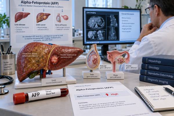
Alpha-fetoprotein, or AFP, is a protein made in large amounts by the fetal liver and yolk sac but normally present only at low levels in nonpregnant adults. An AFP blood test is used mainly in two cancer-related settings: hepatocellular carcinoma, the most common primary liver cancer, and certain germ cell tumors of the testis or ovary. AFP can also rise for noncancer reasons, especially active hepatitis, cirrhosis with liver-cell regeneration, and pregnancy. That is why a high AFP result is a clue rather than a diagnosis. In people at increased risk for liver cancer, AFP is often interpreted with liver ultrasound and changes over time. In germ cell tumors, it is combined with hCG and LDH for diagnosis, staging, prognosis, and treatment monitoring. The exact meaning depends on the concentration, trend, clinical setting, liver function, imaging, and whether AFP was elevated before treatment.
- Normal adult AFP is usually low: many laboratories use an upper reference limit around 8–10 ng/mL, but the lab’s own range should be used.
- AFP can rise in liver cancer and germ cell tumors: especially hepatocellular carcinoma, yolk sac tumor, embryonal carcinoma, and mixed nonseminomatous germ cell tumors.
- A high AFP does not automatically mean cancer: active hepatitis, cirrhosis, liver regeneration, pregnancy, and some rare disorders can also increase it.
- An AFP of 20 ng/mL or more can trigger liver-cancer evaluation in high-risk patients: current liver guidance uses AFP trends and imaging rather than AFP alone to diagnose hepatocellular carcinoma.
- AFP falls with an approximate half-life of 5–7 days: after successful treatment of an AFP-producing germ cell tumor, a slower-than-expected decline can suggest persistent disease.
Table of Contents
- What AFP Is and Why It Is Measured
- Normal Range and Test Preparation
- High AFP in Liver Disease and Liver Cancer
- AFP in Germ Cell Tumors
- How to Interpret AFP Trends
- False Positives, Limitations, and Related Tests
- What to Do After an Abnormal AFP Result
What AFP Is and Why It Is Measured
AFP is a fetal plasma protein with functions similar in some respects to adult albumin. During pregnancy, it is produced mainly by the fetal yolk sac and liver. After birth, AFP levels fall rapidly and are usually very low by later infancy. In a healthy nonpregnant adult, substantial AFP production is unusual.
Cancer cells can sometimes reactivate fetal protein programs. This occurs most notably in hepatocellular carcinoma and in germ cell tumors that contain yolk sac or embryonal elements. The result is measurable AFP in serum.
AFP is not a universal cancer marker. Most common cancers do not produce it, and even cancers known to produce AFP may not do so in every patient. A person can therefore have hepatocellular carcinoma with a normal AFP. The reverse is also true: a person can have a high AFP without cancer.
Clinicians use AFP for different purposes depending on context:
- Liver-cancer surveillance: AFP may be added to ultrasound in people at high risk for hepatocellular carcinoma.
- Diagnostic evaluation: a rising AFP can strengthen concern when liver imaging or symptoms are abnormal.
- Treatment monitoring: falling AFP after surgery, ablation, chemotherapy, or other treatment can support response when the tumor was AFP-producing.
- Germ cell tumor assessment: AFP is part of the standard serum tumor-marker set with hCG and LDH.
- Prognosis and staging: in testicular nonseminomatous germ cell tumors, marker levels after orchiectomy contribute to risk classification.
AFP is often discussed with other liver biomarkers. Des-gamma-carboxy prothrombin, also called DCP, and AFP-L3 are used in some settings to add information about hepatocellular carcinoma risk or biology. These tests complement rather than simply replace AFP.
Normal Range and Test Preparation
Most nonpregnant adults have AFP concentrations below about 8–10 ng/mL, although the upper limit varies by assay and laboratory. Some reports use slightly different reference intervals, so the number printed beside the result is the correct comparison point.
AFP does not usually require fasting. A standard blood sample is collected from a vein. The main preparation issue is not food; it is giving the clinician enough context to interpret the number correctly.
Important information includes:
- pregnancy status;
- chronic hepatitis B or C;
- cirrhosis or other chronic liver disease;
- recent liver inflammation or injury;
- a history of testicular, ovarian, liver, or germ cell tumor treatment;
- the date of surgery or chemotherapy if AFP is being followed over time.
Pregnancy is a major exception to the usual adult reference range. AFP rises physiologically during pregnancy and has a separate role in prenatal screening. A value that would be abnormal for a nonpregnant adult can be expected during pregnancy, so cancer interpretation must use obstetric context.
A single small elevation just above the reference range is rarely enough to define a diagnosis. The trend often matters more. For example, an AFP that remains stable around 12 ng/mL in chronic liver disease may have a different meaning from a value that rises from 8 to 20 to 60 ng/mL over sequential tests.
Laboratories can use different assay platforms. For long-term tumor monitoring, using the same laboratory or at least the same method when practical reduces noise from inter-assay differences.
High AFP in Liver Disease and Liver Cancer
AFP is most closely associated with hepatocellular carcinoma, but it is not specific enough to diagnose HCC by itself. This is especially important because the people most likely to be tested often already have chronic liver disease, which can raise AFP without cancer.
Active hepatitis can stimulate hepatocyte injury and regeneration. Regenerating liver cells may produce more AFP, creating mild or moderate elevations. Cirrhosis can do the same, particularly during inflammatory flares. For this reason, clinicians often look at liver enzymes, bilirubin, viral hepatitis activity, and imaging before deciding how much weight to give an AFP result.
For people at increased HCC risk, current liver guidance commonly recommends surveillance every six months with ultrasound plus AFP. The combination can detect more early cancers than ultrasound alone, although neither test is perfect. AFP is most useful when interpreted dynamically rather than as a one-time cutoff.
A commonly used clinical trigger is AFP at or above about 20 ng/mL, or a rising pattern on serial testing, particularly if the patient has cirrhosis or chronic hepatitis B. That does not mean 20 ng/mL equals cancer. It means the probability is high enough in an at-risk person to justify diagnostic imaging such as multiphase CT or contrast-enhanced MRI.
Very high AFP levels increase concern for HCC, but there is no concentration at which imaging and clinical confirmation become unnecessary. Some HCCs produce little AFP, while benign liver inflammation can occasionally cause striking elevations. Imaging features, tumor size, liver background, and clinical history remain essential.
AFP can also carry prognostic information after HCC has been diagnosed. Higher values are often associated with larger tumor burden or more aggressive tumor biology, although there is substantial overlap between patients. AFP is used in some transplant-selection and post-treatment models because it adds information beyond tumor size and number alone.
When an AFP-producing liver tumor is treated, a substantial fall can support response. A later rise may precede radiographic recurrence in some patients, but imaging is still needed because AFP alone cannot locate disease or prove progression.
Related liver markers include PIVKA-II, another name used for abnormal prothrombin associated with HCC. Multi-marker models such as GALAD combine age, sex, AFP, AFP-L3, and DCP to improve risk estimation, although availability and guideline adoption vary.
AFP in Germ Cell Tumors
AFP has a particularly important role in germ cell tumors because its biology can reveal tumor type. It is produced by yolk sac tumor and can be elevated in embryonal carcinoma and mixed nonseminomatous germ cell tumors. It is not expected to be elevated in pure seminoma.
That distinction has direct clinical value. If a testicular mass has pathology that appears to be pure seminoma but AFP is clearly elevated, clinicians consider whether an occult nonseminomatous component was missed or whether another cause of AFP elevation is present. The tumor is not managed as uncomplicated pure seminoma until that discrepancy is resolved.
Before orchiectomy for suspected testicular cancer, standard serum markers include AFP, total hCG, and LDH. The beta-hCG tumor marker is complementary because seminoma can produce hCG, while AFP elevation points toward nonseminomatous elements. LDH is less specific but can reflect tumor burden.
After orchiectomy, AFP is repeated. The expected biological half-life is roughly 5–7 days. If all AFP-producing tumor tissue has been removed and there is no metastatic disease, the level should fall in a pattern consistent with that half-life. A plateau or slower-than-expected decline raises concern for persistent tumor, although liver disease and assay issues must also be considered.
In metastatic nonseminomatous germ cell tumor, post-orchiectomy marker levels help assign prognostic risk groups. AFP is also measured during chemotherapy and follow-up when it was elevated initially. A falling marker supports treatment response; a rising marker can signal resistant or recurrent disease.
Ovarian germ cell tumors can also produce AFP, particularly yolk sac tumors and mixed tumors. In younger patients with an ovarian mass, AFP may therefore be combined with hCG, LDH, and imaging. The pattern of markers helps guide the differential diagnosis and gives a baseline for treatment monitoring.
The broader hCG tumor marker test is often paired with AFP because neither marker covers all germ cell tumor biology. Normal AFP and hCG do not exclude a germ cell tumor, especially a pure seminoma or a tumor with marker-negative components.
How to Interpret AFP Trends
Serial AFP values are often more informative than a single measurement. The direction, speed, and clinical timing of change can distinguish a meaningful tumor signal from background variation.
Rising AFP
A persistent rise is more concerning than one isolated high value. In chronic liver disease, a pattern of increasing AFP can trigger diagnostic imaging even if the absolute value is not extremely high. In a treated germ cell tumor or HCC, a confirmed upward trend may prompt evaluation for residual or recurrent disease.
Falling AFP
A decline after effective treatment is reassuring when AFP was elevated before therapy. In germ cell tumors, the expected half-life provides a rough biological clock. A rapid fall after tumor removal or chemotherapy is different from a plateau that remains well above normal.
In HCC, AFP kinetics can be slower and less predictable because liver inflammation may contribute to the baseline concentration. A fall should be interpreted with imaging and liver-function changes.
Stable mildly elevated AFP
Persistent mild elevation can occur in chronic liver disease and, rarely, as a benign familial trait. Stability over a long period lowers the likelihood that the number alone represents an aggressively growing cancer, but it does not eliminate risk in someone who already qualifies for HCC surveillance.
A useful way to review AFP is to place values on a timeline with major events: hepatitis flare, imaging, surgery, chemotherapy cycles, or liver-directed treatment. This prevents overinterpreting a change that occurred during acute liver injury or too soon after therapy for the marker to have cleared.
Do not calculate tumor doubling time directly from AFP unless the treating specialist specifically uses a validated approach. AFP production per tumor cell can change, and the marker does not translate directly into tumor volume.
False Positives, Limitations, and Related Tests
The main limitation of AFP is imperfect sensitivity and specificity. Some cancers do not produce it, and some noncancer conditions do.
Noncancer causes of elevated AFP include:
- pregnancy;
- active viral hepatitis;
- cirrhosis with hepatocyte regeneration;
- acute or chronic liver injury;
- rare hereditary persistence of AFP.
Cancer-related causes extend beyond classic HCC and germ cell tumors. Hepatoblastoma, a liver cancer of early childhood, often produces AFP. Rare gastric or other gastrointestinal cancers can show hepatoid differentiation and secrete AFP as well.
The result must therefore be matched to the patient’s age, sex, pregnancy status, organ findings, and imaging. A tumor marker panel can help when the diagnosis is uncertain, but panels should be targeted rather than used as broad cancer screens. The blood tumor marker panel article explains why AFP, CEA, CA-125, CA 19-9, and PSA are not interchangeable screening tests.
For HCC, AFP-L3 measures a glycoform of AFP more closely associated with malignant liver cells, while DCP/PIVKA-II reflects abnormal prothrombin production. These markers may increase specificity or add prognostic information, but no marker removes the need for appropriate imaging.
A normal AFP should never be used to dismiss a suspicious liver lesion. A substantial proportion of HCCs are AFP-negative. Likewise, normal AFP cannot rule out seminoma or all nonseminomatous germ cell tumors.
Another limitation is that the same numeric value can carry different weight at different times. An AFP of 25 ng/mL during an acute hepatitis flare may fall as liver inflammation improves, while a rise from 6 to 25 ng/mL over several surveillance visits may prompt closer investigation even though the final number is identical. This is why clinicians often review AFP together with alanine aminotransferase, aspartate aminotransferase, bilirubin, prior imaging, and the direction of change.
In cancer follow-up, laboratories should avoid switching assay platforms unnecessarily. Small method-related shifts can look like biological change when values are near a decision threshold. When a change is unexpected and would alter treatment, repeating the test before acting may be appropriate unless the clinical picture already requires urgent imaging or intervention.
What to Do After an Abnormal AFP Result
The right next step depends on why the test was ordered. The same AFP value can mean very different things in a person with cirrhosis, a young man with a testicular mass, a pregnant patient, or someone recovering from chemotherapy.
If AFP is elevated in a person at risk for HCC, the clinician will usually review the trend, liver tests, and the most recent surveillance imaging. Rising AFP or a result at a clinically important threshold may lead to contrast-enhanced MRI or multiphase CT.
If AFP is elevated with a testicular or ovarian mass, evaluation is more urgent because the marker can affect tumor classification and staging. Imaging and specialist referral should not be delayed while waiting for the number to normalize spontaneously.
If the result is only mildly high and there is no known cancer, repeating AFP after reviewing liver inflammation, pregnancy status, and medications may be reasonable. The repeat should use the same laboratory when practical.
After cancer treatment, ask whether AFP was a reliable marker at baseline. If it was normal before treatment, serial AFP is unlikely to become the best stand-alone surveillance tool. If it was clearly elevated, the post-treatment decline and later trend can be highly useful.
Seek prompt medical evaluation for jaundice, rapidly increasing abdominal swelling, severe right-upper-abdominal pain, a new testicular mass, acute testicular pain, or other concerning symptoms. Tumor markers are supportive tests; symptoms and imaging often determine urgency.
References
- Hepatocellular Carcinoma: The Search for an Optimal Screening Test 2025 (Review)
- Alpha-fetoprotein: Past, present, and future 2024 (Review)
- Emerging and Clinically Accepted Biomarkers for Hepatocellular Carcinoma 2024 (Review)
- French AFU Cancer Committee Guidelines – Update 2024-2026: Testicular germ cell cancer. 2024 (Guideline)
- AASLD Practice Guidance on prevention, diagnosis, and treatment of hepatocellular carcinoma 2023 (Guideline)
Disclaimer
AFP is not a stand-alone cancer diagnosis and should not be interpreted without the clinical setting, liver status, imaging, and other tumor markers when relevant. Reference ranges and decision thresholds vary by laboratory and indication. Prompt medical assessment is appropriate for a new testicular mass, jaundice, rapidly worsening abdominal symptoms, or other concerning findings.