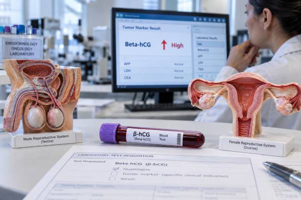
Beta-human chorionic gonadotropin, or beta-hCG, is best known as a pregnancy hormone, but it is also an important blood tumor marker in germ cell cancers. In testicular cancer, hCG is measured with alpha-fetoprotein (AFP) and lactate dehydrogenase (LDH) before surgery, after orchiectomy, during treatment, and in follow-up when appropriate. Some nonseminomatous germ cell tumors produce large amounts of hCG, while a minority of pure seminomas also raise it. In ovarian germ cell tumors, beta-hCG can help identify choriocarcinoma or mixed tumors containing hCG-producing tissue. However, an elevated result is not automatically cancer. Normal pregnancy, recent pregnancy, pituitary hCG, laboratory interference, and some non-germ-cell cancers can also produce positive results. Interpretation therefore depends on the patient’s sex, pregnancy status, tumor type, imaging, pathology, other markers, and how quickly hCG changes over time.
- In nonpregnant adults, beta-hCG is usually very low or undetectable: many laboratories use a reference limit near 5 IU/L, but assay-specific ranges apply.
- Beta-hCG is a core testicular germ cell tumor marker: it is interpreted with AFP and LDH before and after orchiectomy.
- Both seminoma and nonseminoma can raise hCG: a normal result does not exclude testicular cancer.
- Ovarian choriocarcinoma and mixed germ cell tumors may produce hCG: pregnancy must be excluded before attributing an elevation to cancer.
- Trends matter more than one isolated value: persistent or rising hCG after treatment can indicate residual or recurrent disease, but false-positive results must be considered.
Table of Contents
- What Beta-hCG Measures
- Normal Range and Causes of High Beta-hCG
- Beta-hCG in Testicular Cancer
- Beta-hCG in Ovarian Germ Cell Tumors
- How Beta-hCG Is Used for Staging and Monitoring
- False-Positive and Misleading Results
- What to Do After an Abnormal Result
What Beta-hCG Measures
Human chorionic gonadotropin is a glycoprotein hormone made of alpha and beta subunits. The alpha subunit resembles the alpha subunits of luteinizing hormone, follicle-stimulating hormone, and thyroid-stimulating hormone. The beta subunit gives hCG much of its biological and laboratory specificity.
During pregnancy, placental trophoblast cells produce hCG to support the corpus luteum and early pregnancy. Some cancers can also produce intact hCG, free beta subunit, or related molecular forms. Germ cell tumors are the classic example because certain malignant germ cells can differentiate toward trophoblastic tissue.
Laboratories may report total hCG, intact hCG, or beta-hCG, depending on the assay. These measurements overlap but are not analytically identical. For routine germ cell tumor management, clinicians generally follow the test offered by the same laboratory so serial results are comparable.
The hCG tumor marker is useful because a clearly abnormal concentration can support diagnosis, contribute to risk classification, and provide a measurable signal that should fall when hCG-producing cancer is successfully treated. It does not replace pathology or imaging.
Beta-hCG is especially useful when combined with AFP testing. The pattern of the two markers can suggest which germ cell components may be present. For example, pure seminoma should not produce AFP, whereas hCG can be elevated in seminoma because syncytiotrophoblastic cells may produce it. Nonseminomatous tumors can elevate one or both markers depending on histology.
LDH is usually measured as well. It is much less specific but can reflect tumor burden and is included in germ cell tumor risk assessment. The three markers answer different questions and are most informative as a group.
Normal Range and Causes of High Beta-hCG
In nonpregnant adults, many laboratories consider serum hCG below about 5 IU/L, numerically equivalent to 5 mIU/mL, to be negative. Some laboratories use a small indeterminate interval before a result is considered clearly positive. Always use the reference interval and units on the actual report.
For women or people who can become pregnant, pregnancy is the first explanation to consider for a positive hCG. Values rise rapidly in early pregnancy, so a tumor-marker interpretation should never be made without considering pregnancy history, menstrual timing, fertility treatment, recent delivery, miscarriage, or pregnancy termination.
High hCG can occur in several settings:
- normal intrauterine or ectopic pregnancy;
- gestational trophoblastic disease;
- testicular germ cell tumors, especially choriocarcinoma, embryonal carcinoma, and mixed nonseminomatous tumors;
- some seminomas with syncytiotrophoblastic cells;
- ovarian germ cell tumors containing choriocarcinoma or other hCG-producing components;
- selected nongerm-cell malignancies that ectopically produce hCG or free beta subunit;
- low-level pituitary hCG, particularly around or after menopause;
- laboratory interference, including heterophile antibodies.
The height of the value can be informative but is not diagnostic by itself. Germ cell choriocarcinoma can produce extremely high concentrations, while seminoma more often causes modest elevations. Yet there is substantial overlap, and pregnancy can produce very high values as well.
A low positive result deserves careful confirmation before anyone labels it malignant. In a person with no cancer findings, repeating the test, checking a urine hCG, testing with another assay platform, or evaluating pituitary hCG may be more appropriate than immediately pursuing cancer treatment.
Beta-hCG in Testicular Cancer
Current testicular cancer guidance recommends measuring serum hCG, AFP, and LDH before orchiectomy when a testicular germ cell tumor is suspected. These markers support diagnosis, help establish a baseline, and can provide clues about histology.
Up to roughly 90% of nonseminomatous germ cell tumors have an elevation of AFP or beta-hCG at diagnosis. HCG can also be elevated in a minority of pure seminomas, reported in guidelines as up to about 30%. This is why an elevated hCG does not automatically mean the tumor is nonseminomatous.
AFP behaves differently. A significant AFP elevation in a tumor otherwise called seminoma raises concern that an occult nonseminomatous component is present. Clinicians therefore interpret the beta-hCG tumor marker alongside AFP rather than trying to identify histology from hCG alone.
The marker is repeated after orchiectomy. If the testicle contained the only hCG-producing tumor, the concentration should decline according to the hormone’s serum half-life. Guidelines generally describe an hCG half-life of about one to three days. A value that falls appropriately toward normal supports removal of the source. A plateau or rise can indicate persistent metastatic disease, although an unexpected low-level result should still be checked for noncancer causes.
Normal markers do not exclude testicular cancer. Teratoma commonly produces neither AFP nor hCG, and many seminomas are marker-negative. A testicular mass is evaluated primarily with examination and scrotal ultrasound, followed by appropriate surgery and pathology rather than being ruled in or out by blood tests.
For metastatic nonseminomatous germ cell cancer, pretreatment marker levels are part of established prognostic classification. Very high hCG is associated with a less favorable risk group, but classification also considers AFP, LDH, the primary tumor site, and where metastases are present. It is a formal oncology tool, not a general interpretation table for patients reading a lab report.
During chemotherapy, serial hCG can provide rapid evidence of response because its expected biologic decline is relatively fast. Failure to fall as expected may prompt closer assessment of disease response, but treatment decisions integrate scans, other tumor markers, pathology, and the specific clinical situation.
Beta-hCG in Ovarian Germ Cell Tumors
Malignant ovarian germ cell tumors are rare and occur most often in children, adolescents, and young adults. They include dysgerminoma, yolk sac tumor, immature teratoma, embryonal carcinoma, choriocarcinoma, and mixed germ cell tumors. The marker pattern varies with the tissue components present.
Beta-hCG is particularly associated with choriocarcinoma and tumors that contain trophoblastic differentiation. Mixed ovarian germ cell tumors may therefore raise both beta-hCG and AFP. Yolk sac tumor is classically associated with AFP. Dysgerminoma usually does not produce AFP, though some cases can produce modest hCG when syncytiotrophoblastic giant cells are present.
This makes a marker panel useful at diagnosis. In a young patient with an ovarian mass, clinicians may measure beta-hCG, AFP, LDH, and sometimes other markers according to the differential diagnosis. The results can help narrow the possibilities, but surgery and histopathology establish the definitive tumor type.
Pregnancy is a particularly important confounder in this population because ovarian germ cell tumors commonly affect people of reproductive age. A positive beta-hCG cannot simply be assigned to an ovarian mass until pregnancy has been assessed. Imaging findings, serial hCG behavior, clinical history, and pathology help separate these possibilities.
If an ovarian germ cell tumor produced hCG before treatment, serial measurements can be valuable afterward. A rapid fall toward normal after surgery and chemotherapy is reassuring. A plateau, new rise, or reappearance after normalization can suggest persistent or recurrent hCG-producing disease and usually triggers clinical and imaging reassessment.
Not every ovarian germ cell tumor has a useful serum marker. Immature teratoma, for example, may have no specific marker signal, and an elevated AFP can sometimes reflect associated yolk sac elements. The absence of hCG therefore cannot be used to exclude an ovarian germ cell malignancy.
Because these cancers are uncommon and often highly curable, management by gynecologic oncology and germ cell tumor specialists is important. Marker interpretation should support fertility-conscious, evidence-based treatment rather than drive treatment in isolation.
How Beta-hCG Is Used for Staging and Monitoring
A tumor marker is most powerful when it is used at the correct clinical time point. Beta-hCG has several distinct roles.
Before treatment
A pretreatment value establishes whether the tumor secretes hCG and provides a baseline. In suspected testicular cancer, it is drawn with AFP and LDH before orchiectomy whenever possible. In an ovarian mass, pregnancy must be considered at the same time.
After surgery
The expected decline helps show whether all hCG-producing tissue may have been removed. Because hCG normally clears over days, clinicians do not expect an immediate zero result. They examine the direction and rate of change.
During chemotherapy
Falling concentrations generally support treatment response. A plateau or repeated rise is more concerning than one small fluctuation. The most useful comparisons use the same laboratory and assay because different methods can produce somewhat different absolute numbers.
During follow-up
For a tumor that was hCG-positive at diagnosis, a new confirmed rise can be an early sign of recurrence. It should be interpreted with symptoms, examination, imaging, AFP, and sometimes LDH. Treating a laboratory number without confirming the clinical picture risks unnecessary therapy when the result is false positive.
One reason serial hCG is valuable is that it changes faster than many structural findings. A biochemical rise may appear before a mass is obvious on imaging. The opposite can also occur: a marker-negative tumor such as teratoma may progress while hCG remains normal. Marker surveillance therefore complements rather than replaces imaging and clinical follow-up.
Marker kinetics after treatment
The speed of decline is sometimes as important as the absolute result. After removal of an hCG-producing testicular tumor, clinicians expect serum hCG to fall over several days because its typical biologic half-life is roughly one to three days. They may plot repeated results rather than focusing on whether the first postoperative value is still above the laboratory limit. A steadily falling series can be compatible with normal clearance, while a plateau or renewed increase raises concern that viable hCG-producing tumor remains.
The calculation is not perfectly mechanical. The starting concentration may be extremely high, tests may be drawn at different intervals, and chemotherapy can begin before a marker has fully normalized. Kidney function, assay changes, and small low-level false positives can also distort the pattern. For these reasons, germ cell tumor teams interpret kinetics within established staging and treatment protocols rather than applying a homemade doubling-time rule.
The same principle applies to ovarian germ cell tumors. If beta-hCG was clearly elevated at diagnosis, normalization provides a useful biochemical endpoint. If it was normal from the start, however, it cannot serve as a reliable surveillance marker for that patient’s tumor. In that setting, follow-up must rely more heavily on the marker that was abnormal, if any, plus imaging, examination, and symptoms.
False-Positive and Misleading Results
False or misleading hCG results matter because germ cell tumors are treated with intensive chemotherapy, and no one should receive treatment solely because of an unverified low positive value.
Heterophile antibodies are a classic cause of so-called phantom hCG. These human antibodies can interact with animal antibodies used in immunoassays and produce a persistent serum signal even though true hCG is absent. A urine hCG may be negative because heterophile antibodies are not filtered into urine in the same way. Repeating the sample on another laboratory platform or performing interference studies can resolve the discrepancy.
Pituitary hCG can produce low concentrations in perimenopausal and postmenopausal patients. Loss of ovarian estrogen feedback raises gonadotropins, and the pituitary may produce small amounts of hCG. FSH, age, menopausal status, and suppression testing can help clarify the source when clinically necessary.
Recent pregnancy can keep hCG detectable after delivery, miscarriage, or termination until the hormone has cleared. Fertility medications containing hCG can also temporarily raise measured concentrations.
Assay differences matter when following trends. One laboratory may measure total hCG while another has different recognition of intact hormone and free beta subunit. A sudden small change after switching laboratories may be analytical rather than biological.
Kidney dysfunction can complicate low-level hCG interpretation in some patients because clearance and hormone metabolism change. The broader review of blood tumor markers is relevant here: benign disease, physiologic states, and analytical interference can affect many markers, so specificity comes from context rather than from the word “tumor” in the test name.
When a result is unexpected, confirmation is safer than assumption. The clinician may repeat serum testing, obtain urine hCG, compare another assay, review pregnancy history and medications, and correlate with imaging before concluding that cancer is active.
What to Do After an Abnormal Result
The next step depends on who was tested and why.
For a person with a testicular mass, an elevated beta-hCG supports a germ cell tumor diagnosis but does not replace scrotal ultrasound and urologic evaluation. AFP and LDH should be checked, and the marker should be repeated after orchiectomy to establish the post-surgical trend.
For a reproductive-age patient with an ovarian mass, pregnancy should be assessed immediately. If pregnancy is excluded and beta-hCG remains elevated, gynecologic evaluation, pelvic imaging, AFP and LDH, and specialist review help determine whether a germ cell tumor or another cause is present.
For someone already treated for an hCG-producing germ cell tumor, the most important finding is a confirmed trend. A persistent rise should be discussed promptly with the oncology team. A single low positive result that conflicts with imaging and other markers should be verified for assay interference before a major treatment decision.
Seek urgent care for severe abdominal or pelvic pain, fainting, heavy bleeding, sudden testicular pain or swelling, shortness of breath, neurologic symptoms, or other acute symptoms. These can reflect conditions that need immediate evaluation regardless of the tumor marker level.
Beta-hCG is one of the most useful serum markers in germ cell oncology because it can change quickly and carry diagnostic, prognostic, and monitoring information. Its value is greatest when clinicians respect its limitations: pregnancy and benign sources exist, not all germ cell tumors produce hCG, and the correct interpretation comes from the full pattern of markers, pathology, imaging, and serial change.
References
- Diagnostic Evaluation – EAU Guidelines on Testicular Cancer – Uroweb 2026 (Guideline)
- Is Human Chorionic Gonadotropin a Reliable Marker for Testicular Germ Cell Tumor? New Perspectives for a More Accurate Diagnosis 2025 (Review)
- French AFU Cancer Committee Guidelines – Update 2024-2026: Testicular germ cell cancer. 2024 (Practice Guideline)
- Malignant ovarian and testicular germ cell tumors: Common characteristics but different prognoses 2024 (Review)
- A Comprehensive Review of Current Trends in the Diagnosis and Treatment of Ovarian Germ Cell Tumors. 2024 (Review)
- Factors influencing blood tumor marker concentrations in the absence of neoplasia 2024 (Review)
Disclaimer
Beta-hCG is not a stand-alone cancer test. Pregnancy, recent pregnancy, pituitary production, assay interference, and other conditions can raise hCG, while some germ cell tumors produce no hCG at all. Results should be interpreted by a qualified clinician with AFP, LDH, imaging, pathology, pregnancy status, and serial measurements.