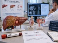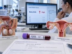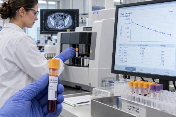
Human chorionic gonadotropin (hCG) is best known as the hormone detected in pregnancy tests, but it is also an important blood tumor marker for certain germ cell tumors and gestational trophoblastic diseases. In testicular germ cell cancer, hCG is usually interpreted with alpha-fetoprotein (AFP) and lactate dehydrogenase (LDH) before and after orchiectomy, during treatment, and in follow-up. Some ovarian and extragonadal germ cell tumors can also produce hCG. A high result does not automatically mean cancer: pregnancy, pituitary production, laboratory interference, and some other malignancies can cause detectable or elevated hCG. The trend is often more important than one number. Because hCG has a short blood half-life of roughly 24–36 hours, clinicians expect it to fall in a predictable pattern after successful removal or treatment of an hCG-producing tumor. Persistent or rising values require careful confirmation and evaluation.
- hCG is a key tumor marker for choriocarcinoma and some other testicular, ovarian, and extragonadal germ cell tumors.
- In testicular cancer, hCG is interpreted with AFP and LDH; normal markers do not rule out a germ cell tumor.
- hCG has a blood half-life of about 24–36 hours, so the rate of decline after treatment can provide important information.
- Low-level hCG can come from the pituitary or assay interference, so an unexpected borderline result should be confirmed before major treatment decisions.
- Pregnancy must be considered whenever biologically possible because normal pregnancy produces far more hCG than most nonpregnant reference ranges allow.
Table of Contents
- What the hCG Tumor Marker Test Measures
- Which Tumors Can Produce hCG
- What High hCG Levels Mean
- hCG in Testicular Cancer Staging and Risk
- How hCG Is Used for Treatment Monitoring
- False-Positive and Non-Cancer hCG Results
- Testing, Follow-Up, and Questions to Ask
What the hCG Tumor Marker Test Measures
Human chorionic gonadotropin is a glycoprotein hormone made of two protein subunits, called alpha and beta. The alpha subunit resembles parts of several pituitary hormones, while the beta subunit gives hCG much of its biological and immunologic specificity.
During pregnancy, placental syncytiotrophoblast cells produce large amounts of hCG. Certain tumors can develop similar trophoblastic cells or pathways and release intact hCG, free beta-hCG, or related molecular forms into the blood.
This creates an important laboratory detail: not every hCG assay measures exactly the same molecular forms. Some laboratories report total hCG, others beta-hCG, and different commercial methods can give somewhat different results from the same specimen. For tumor monitoring, clinicians prefer serial testing with the same assay whenever possible.
Results are commonly reported in IU/L or mIU/mL. Numerically, 1 IU/L equals 1 mIU/mL. The upper reference limit for a nonpregnant person varies by assay, age, and sex. A value around 5 IU/L is often used as a general nonpregnant cutoff, but some laboratories use lower limits in men and different limits in peri- or postmenopausal women. The laboratory’s own reference interval should be used.
The tumor-marker hCG test is closely related to the beta-hCG tumor marker test. In practice, names can overlap because many oncology assays target the beta portion or detect intact hCG plus beta-related forms. When comparing results, check the actual assay rather than assuming two tests labeled “hCG” are interchangeable.
hCG is not useful as a stand-alone screening test for the general population. Its greatest value comes when a germ cell tumor is already suspected or diagnosed and the result is interpreted with imaging, pathology, examination, and other serum markers.
Which Tumors Can Produce hCG
hCG is most strongly associated with tumors that contain trophoblastic differentiation. The amount produced varies widely by tumor type and by the amount of hCG-secreting tissue.
Testicular germ cell tumors
In testicular cancer, hCG is one of three traditional serum tumor markers. Choriocarcinoma characteristically produces hCG, often at very high levels. Embryonal carcinoma and mixed nonseminomatous germ cell tumors may also produce it.
Pure seminoma can sometimes cause a modest hCG elevation when syncytiotrophoblastic giant cells are present. This is an important exception because seminoma does not usually produce AFP. A significant AFP elevation in a tumor otherwise called seminoma raises concern that a nonseminomatous component is present.
The companion markers are AFP and LDH. AFP is particularly associated with yolk sac and embryonal components, while LDH is less specific and often reflects tumor burden or tissue turnover.
Ovarian germ cell tumors
Some ovarian germ cell tumors also produce hCG. Ovarian choriocarcinoma and tumors containing trophoblastic elements can produce marked elevations. Dysgerminoma may occasionally be associated with hCG when syncytiotrophoblastic cells are present. Other ovarian germ cell tumors may be better tracked with AFP or LDH depending on histology.
For this reason, younger patients with an ovarian mass may have a tailored marker panel rather than CA-125 alone. The beta-hCG test for ovarian germ cell tumors is interpreted with age, pregnancy status, imaging, AFP, LDH, and pathology.
Extragonadal germ cell tumors
Germ cell tumors can rarely arise outside the ovaries or testes, including in the mediastinum or retroperitoneum. hCG can support the diagnosis and help monitor treatment when these tumors contain hCG-producing components.
Gestational trophoblastic disease
hCG is central to the diagnosis and follow-up of hydatidiform mole and gestational trophoblastic neoplasia, including choriocarcinoma. Here the biology is pregnancy-related rather than a gonadal germ cell cancer. Serial hCG measurement after uterine evacuation is essential for identifying persistent disease. A dedicated gestational trophoblastic disease hCG test pathway uses its own diagnostic criteria and monitoring schedules.
What High hCG Levels Mean
A high hCG result means that the assay detected more hCG than expected for the laboratory’s reference population. The number does not identify the cause by itself.
In a person with a known hCG-producing germ cell tumor, the result may reflect tumor burden and biological activity. Very high concentrations are particularly characteristic of choriocarcinoma, but there is no single level that identifies a specific histology in every patient.
In someone with a new testicular mass, an elevated hCG can support the diagnosis of a germ cell tumor, but the test is not sensitive enough to exclude disease when normal. Many localized germ cell tumors have normal conventional serum markers.
In a person who can become pregnant, pregnancy is the first explanation that must be considered. Normal pregnancy hCG changes rapidly with gestational age and can overlap or greatly exceed tumor-marker levels. Clinical history, repeat quantitative testing, and ultrasound distinguish pregnancy-related patterns from oncologic ones.
The direction of change often matters more than the absolute value. If hCG was 20,000 IU/L before tumor removal and then falls rapidly, that pattern is very different from a value that remains flat or begins rising.
A small isolated elevation deserves special caution. Low-level hCG can come from the pituitary, especially when normal gonadal hormone feedback is reduced. Heterophile antibodies and other assay interferences can also create a positive serum result even when true circulating hCG is absent.
This is why clinicians often repeat an unexpected borderline result before changing cancer therapy. They may test urine, use a different assay platform, evaluate luteinizing hormone and gonadal function, or ask the laboratory to investigate antibody interference.
| Pattern | Possible interpretation | Typical next step |
|---|---|---|
| Normal hCG with suspicious mass | Germ cell tumor still possible | Use imaging, AFP/LDH, and pathology |
| Elevated and falling appropriately after treatment | Supports reduction of hCG-producing tumor | Continue scheduled serial monitoring |
| Persistent or rising hCG | Residual, progressive, or recurrent disease possible | Confirm result and reassess clinically |
| Stable low-level unexpected hCG | Pituitary or assay interference possible | Investigate before assuming relapse |
hCG in Testicular Cancer Staging and Risk
In testicular germ cell cancer, serum markers are used for more than diagnosis. AFP, hCG, and LDH contribute to staging and prognostic classification, especially after orchiectomy and before chemotherapy.
Markers should be measured before orchiectomy to establish a baseline. They are measured again afterward because the testicle may have contained the main source of the marker. If hCG falls as expected and normalizes, that supports removal of the marker-producing tumor. Persistent elevation may indicate remaining disease, although false-positive and non-cancer causes must be considered when the elevation is small or inconsistent.
The TNM staging system includes an S category based on serum markers. For hCG, the commonly used prechemotherapy thresholds are:
| S category | hCG component | Important context |
|---|---|---|
| S0 | Within normal limits | AFP and LDH also considered |
| S1 | <5,000 mIU/mL | Also requires AFP <1,000 ng/mL and LDH <1.5× upper limit |
| S2 | 5,000–50,000 mIU/mL | Any one marker meeting S2 can determine the category |
| S3 | >50,000 mIU/mL | Represents a high serum-marker category |
These are staging thresholds, not normal ranges. An hCG of 2,000 mIU/mL is dramatically abnormal even though it falls within the S1 band. Also, the final S category depends on all three markers, not hCG alone.
Marker levels are also part of the International Germ Cell Cancer Collaborative Group risk classification for metastatic disease. Site of the primary tumor, location of metastases, and marker levels together help define prognosis and guide treatment intensity.
A high hCG may prompt additional imaging in advanced disease. For example, current testicular cancer guidance gives special attention to brain imaging in patients with poor-prognosis features, multiple lung metastases, neurologic symptoms, or markedly elevated hCG.
How hCG Is Used for Treatment Monitoring
hCG is particularly useful for monitoring because it changes relatively quickly. Its serum half-life is about 24–36 hours, meaning that if production stops completely, the concentration should fall by roughly half over that interval.
After orchiectomy, clinicians compare serial results with the expected decline. A high preoperative value will not become normal immediately; it takes several half-lives. The rate of fall helps distinguish expected clearance from persistent tumor production.
During chemotherapy, AFP and hCG are measured at defined intervals, often around treatment cycles. A consistent decline supports treatment response. A slow decline, plateau, or renewed rise can suggest resistant or progressive disease and may influence prognosis or the need for further assessment.
The calculation is not as simple as checking whether each value is lower than the previous one. Clinicians consider the starting concentration, timing of samples, biological half-life, treatment stage, imaging, and the possibility of temporary fluctuations. Tumor lysis early in chemotherapy can occasionally alter marker values before the overall decline becomes clear.
After successful treatment, serial hCG may help detect relapse in marker-producing tumors. Surveillance schedules vary by tumor type, stage, treatment, and guideline. A relapse can sometimes be signaled by a rising marker before symptoms appear, but not all recurrent germ cell tumors secrete hCG.
For gestational trophoblastic disease, hCG monitoring is even more central because the marker directly reflects trophoblastic activity in most cases. The required duration and frequency of follow-up depend on the type of molar pregnancy or trophoblastic neoplasia and the pattern of hCG regression.
Whenever serial monitoring matters, use the same laboratory method when practical. A study comparing multiple commercial hCG immunoassays found meaningful inter-method differences because assays recognize hCG molecular forms differently. Switching methods can create an apparent change that is partly analytical rather than biological.
False-Positive and Non-Cancer hCG Results
Unexpected hCG results deserve confirmation because acting on a false-positive tumor marker can lead to unnecessary imaging, surgery, or chemotherapy.
Pregnancy
Pregnancy is the most common physiologic source of hCG. In anyone with pregnancy potential, a positive hCG cannot be interpreted as a tumor marker until pregnancy has been appropriately considered.
Pituitary hCG
The pituitary gland can produce small amounts of hCG, particularly when sex-hormone feedback is reduced. This is most familiar in perimenopausal and postmenopausal women, but it can also occur in men with hypogonadism or after loss of testicular function.
Pituitary hCG is generally low-level and may remain relatively stable rather than showing the progressive rise expected from active tumor. Clinicians can use gonadotropin levels, hormone status, repeat testing, and in selected cases medically supervised hormone-suppression testing to clarify the source.
Heterophile antibody interference
Heterophile antibodies are human antibodies that can interact with laboratory assay antibodies and create a false serum signal. One clue is a persistent low-level serum hCG result that does not fit the clinical picture. Urine hCG may be negative because large interfering antibodies do not pass into urine in the same way as true hCG.
The laboratory can investigate by testing on another platform, performing serial dilutions, using heterophile-blocking reagents, or comparing serum and urine results.
Assay and supplement interference
Different hCG assays recognize different molecular forms, so small discrepancies between platforms are expected. Certain immunoassays can also be affected by high-dose biotin or other analytical interferences, depending on the test design. Tell the clinician and laboratory about supplements and medications when a result is unexpected.
Other cancers
Some non-germ-cell malignancies can produce hCG or free beta-hCG, including certain cancers of the lung, gastrointestinal tract, kidney, bladder, and other organs. This is uncommon compared with pregnancy and classic hCG-producing tumors, but it reinforces that hCG is not specific to one cancer.
Testing, Follow-Up, and Questions to Ask
An hCG tumor marker test uses a routine blood sample and usually requires no fasting. Preparation is mainly about providing accurate clinical context: pregnancy possibility, recent cancer treatment, current medications and supplements, testicular or ovarian surgery, and prior hCG values.
For suspected testicular cancer, AFP, hCG, and LDH should ideally be drawn before orchiectomy because the baseline helps with diagnosis and post-surgical interpretation. A testicular mass still requires proper imaging and urologic evaluation even when all three markers are normal.
If hCG is high after surgery or during surveillance, the key questions are whether the value is changing as expected and whether the result fits imaging and pathology. A persistent major elevation is very different from a stable value just above the assay cutoff.
Useful questions to ask include:
- Is this assay measuring total hCG, beta-hCG, or both?
- What is the laboratory’s reference range for my age and sex?
- Was hCG elevated before surgery or treatment?
- Is the current fall consistent with the expected 24–36-hour half-life?
- What are my AFP and LDH results, and how do all three affect staging or risk classification?
- Could pregnancy, pituitary hCG, kidney or hormonal changes, supplements, or assay interference explain a low-level result?
- If the result is unexpected, should it be repeated with the same method, checked in urine, or confirmed on another assay platform before treatment changes?
Seek prompt medical attention for a new testicular lump, rapidly increasing testicular swelling, severe abdominal or chest symptoms, coughing blood, neurologic symptoms, or heavy pregnancy-related bleeding. Tumor markers can guide evaluation, but symptoms and imaging may reveal clinically important disease even when hCG is normal.
The central principle is to use hCG as a dynamic marker. Its strongest value is not simply whether it is above a reference limit, but whether the result fits the tumor type and whether the serial pattern behaves as expected after treatment.
References
- European Association of Urology Guidelines on Testicular Cancer: Summary of the 2026 Guidelines 2026 (Guideline)
- Current and Evolving Biomarkers in the Diagnosis and Management of Testicular Germ Cell Tumors 2024 (Review)
- Is Human Chorionic Gonadotropin a Reliable Marker for Testicular Germ Cell Tumor? New Perspectives for a More Accurate Diagnosis 2025 (Review)
- Utility of Commercially Available Quantitative hCG Immunoassays as Tumor Markers in Trophoblastic and Non-Trophoblastic Disease 2023
- Practical Guidelines for the Treatment of Gestational Trophoblastic Disease: Collaboration of the European Organisation for the Treatment of Trophoblastic Disease (EOTTD)-European Society of Gynaecologic Oncology (ESGO)-Gynecologic Cancer InterGroup (GCIG)-International Society for the Study of Trophoblastic Diseases (ISSTD) 2025 (Guideline)
- Human chorionic gonadotropin of pituitary origin in Chinese postmenopausal women: A single-center retrospective study 2024
Disclaimer
This article is for general education and does not diagnose cancer, pregnancy, or recurrence. hCG results must be interpreted by a qualified clinician using the assay method, pregnancy status, tumor type, other markers, imaging, pathology, and the pattern over time. An unexpected low-level hCG should be confirmed before major treatment decisions whenever clinically safe.