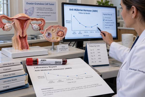
Anti-Müllerian hormone, or AMH, is best known as a blood marker of ovarian reserve, but it also has an important tumor-marker role in ovarian granulosa cell tumors. Granulosa cells normally produce AMH, so a tumor arising from these cells can release unusually high amounts into the bloodstream. In a patient with a confirmed adult-type granulosa cell tumor, AMH may help estimate tumor activity, assess response after surgery or other treatment, and detect recurrence during long-term follow-up. The test is most useful when AMH was clearly elevated at diagnosis and then falls after treatment. There is no single cancer cutoff that applies to every patient because AMH varies with age, ovarian reserve, menopause, polycystic ovary syndrome, remaining ovarian tissue, and assay method. A rising value is therefore interpreted as a trend, usually alongside inhibin B, symptoms, examination, and imaging rather than as proof of recurrence by itself.
- AMH can act as a granulosa cell tumor marker: these ovarian sex cord-stromal tumors often retain the ability to produce AMH.
- A falling AMH after treatment is generally reassuring: especially when the marker was elevated before surgery or therapy.
- A rising AMH during follow-up can suggest recurrent tumor activity: but imaging and clinical assessment are needed to confirm recurrence.
- There is no universal tumor-marker cutoff: premenopausal ovarian reserve, PCOS, menopause, ovarian surgery, and laboratory method strongly affect the expected level.
- AMH and inhibin B are often complementary: using both markers can improve follow-up when each was informative at diagnosis.
Table of Contents
- What AMH Measures
- Why Granulosa Cell Tumors Can Raise AMH
- Normal Levels and Why There Is No Tumor Cutoff
- Using AMH After Diagnosis and Treatment
- AMH Versus Inhibin A and Inhibin B
- Causes of High or Changing AMH Not Due to Recurrence
- Follow-Up and Next Steps
What AMH Measures
AMH is a glycoprotein hormone in the transforming growth factor-beta family. In females, it is produced by granulosa cells surrounding small growing ovarian follicles. In routine reproductive medicine, serum AMH is used as an indirect marker of the remaining pool of recruitable follicles. Higher levels generally indicate more small follicles, while levels decline with age and become very low after menopause.
That same biological origin explains its use as a tumor marker. Granulosa cell tumors arise from sex cord-stromal cells of the ovary and often preserve hormone-producing functions of normal granulosa cells. A tumor may therefore release AMH, inhibins, estrogen, or a combination of these hormones.
The AMH blood test does not distinguish normal granulosa cells from tumor granulosa cells. It simply measures circulating hormone. In a young premenopausal patient with two functioning ovaries, a moderately high AMH can reflect normal ovarian reserve or polycystic ovary syndrome rather than cancer. In a postmenopausal patient, or after both ovaries have been removed, detectable or rising AMH can be more suspicious because normal ovarian production should be minimal or absent.
AMH is also different from a tissue marker. Pathologists can use morphology, inhibin, calretinin, FOXL2-related findings, and other features to establish a granulosa cell tumor diagnosis. Serum AMH is primarily a monitoring marker once the clinical context is known.
Adult-type granulosa cell tumors account for most granulosa cell tumors and frequently carry a characteristic FOXL2 mutation. They often behave more slowly than epithelial ovarian cancers but are notable for late recurrences. This long natural history makes a repeatable blood marker especially attractive for follow-up.
Why Granulosa Cell Tumors Can Raise AMH
Granulosa cell tumors can raise AMH because the neoplastic cells originate from the same cell lineage that normally produces the hormone. The amount released varies widely. Some tumors produce very high levels, while others produce little enough that AMH is not useful for that individual.
Research comparing AMH with inhibin B has shown that both markers can be elevated in primary and recurrent adult-type granulosa cell tumors and that concentrations may correlate with tumor size. In a long-term cohort, AMH showed high diagnostic performance for macroscopic disease, and combining AMH with inhibin B improved detection compared with either marker used alone.
This does not mean AMH should be used to screen the general population for ovarian cancer. Granulosa cell tumors are rare, and AMH is influenced by ordinary reproductive physiology. In an asymptomatic premenopausal person, a high AMH has a much more likely explanation in ovarian reserve or PCOS than in a granulosa cell tumor.
AMH becomes clinically valuable when the prior probability is already high. Examples include:
- a pathology-confirmed granulosa cell tumor;
- a suspicious ovarian sex cord-stromal mass with hormone-related symptoms;
- follow-up after surgery for an AMH-producing granulosa cell tumor;
- investigation of possible recurrence when AMH previously tracked disease activity.
Granulosa cell tumors can also produce estrogen. Excess estrogen may cause abnormal uterine bleeding, endometrial thickening, early puberty in children with juvenile tumors, or breast tenderness. In postmenopausal patients, new vaginal bleeding is particularly important and deserves direct evaluation rather than waiting for a tumor-marker result.
The marker should therefore be viewed as one piece of a hormone-producing tumor profile. A high AMH can support the biological picture, but pathology establishes the diagnosis and imaging determines where disease is located.
Normal Levels and Why There Is No Tumor Cutoff
There is no single AMH value that separates granulosa cell tumor from normal ovarian function. Unlike markers with a broadly used upper limit such as CA-125, expected AMH changes dramatically with reproductive age and ovarian physiology.
AMH is usually highest in adolescence and young adulthood, then gradually declines as the follicle pool falls. Levels become very low or undetectable after natural menopause. Ovarian surgery, chemotherapy, radiation, and some medical treatments can reduce AMH by damaging follicles. PCOS can raise AMH because affected ovaries often contain many small follicles that produce the hormone.
Laboratories also use different assays and units, commonly ng/mL or pmol/L. Conversion is possible, but assay calibration is not perfectly interchangeable. A value considered high on one platform may not map exactly to another.
For tumor monitoring, the patient’s personal baseline and post-treatment nadir are often more useful than a generic cutoff. Consider three examples:
- A 28-year-old with both ovaries and AMH of 5 ng/mL may simply have a high-normal ovarian reserve.
- A 55-year-old several years after menopause with a clearly measurable and rising AMH has a different reference context.
- A patient who had bilateral oophorectomy for granulosa cell tumor and whose AMH fell from 40 ng/mL to undetectable, then later becomes repeatedly measurable, has a trend that deserves investigation even if the absolute number is modest.
The same principle applies after fertility-sparing surgery. If one ovary remains, normal follicles continue to make AMH. Complete suppression to an undetectable level is not expected in every patient, so surveillance must focus on changes from the individual’s established baseline.
It is also useful to separate reference intervals from decision limits. A reproductive laboratory may flag a result as high or low for age, but that flag is designed for ovarian-reserve interpretation, not for cancer recurrence. In oncology follow-up, a value can be clinically important even if it still sits inside the laboratory’s broad reproductive range, particularly when it has risen repeatedly from a much lower post-treatment baseline. The reverse can also happen: a value flagged as high may be expected in a younger patient with PCOS and no evidence of active tumor.
Small fluctuations should be interpreted cautiously because analytical and biological variation exists. When a result would trigger imaging, biopsy, or a major treatment change, clinicians often confirm that the rise is reproducible. This is especially sensible when the increase is modest, the patient feels well, and other markers remain stable. A clearly progressive increase accompanied by symptoms or a rising inhibin B carries more weight than one isolated borderline value.
Because of these variables, the laboratory’s age-related reference interval is useful for reproductive interpretation but is not a complete cancer rule. Tumor-marker interpretation should be anchored to prior values, menopausal status, ovarian anatomy, and whether AMH tracked the original tumor.
Using AMH After Diagnosis and Treatment
AMH is most useful when it is measured before treatment and shown to be elevated. That creates a baseline against which later results can be compared.
After complete surgical removal of an AMH-producing granulosa cell tumor, the level should fall. The exact speed depends on the starting concentration, residual normal ovarian tissue, assay, and treatment context. A substantial decline supports removal of hormone-producing tumor tissue, but imaging and pathology determine whether the surgery was complete.
During follow-up, clinicians may repeat AMH at scheduled visits. A stable low level is reassuring. A confirmed upward trend can raise concern for recurrence, sometimes before a mass produces obvious symptoms. Older longitudinal studies found that AMH or inhibin elevations could precede clinically apparent recurrence, which is one reason these markers remain useful despite the rarity of the disease.
A single small increase is usually not enough to declare recurrence. Before acting, the care team may:
- repeat the test using the same laboratory;
- review menopausal status and whether ovarian tissue remains;
- compare inhibin B and other previously informative markers;
- assess symptoms and perform pelvic examination;
- obtain ultrasound, CT, or MRI if the rise is persistent or clinically concerning.
The marker can also help during treatment of recurrent disease. If AMH was elevated at relapse, a falling level during surgery, chemotherapy, or hormonal therapy can support response. A plateau or renewed rise may prompt earlier imaging, but treatment should not be changed solely because of one lab value.
Granulosa cell tumors can recur many years after initial treatment. Long-term surveillance is therefore important even after a prolonged disease-free interval. The exact schedule depends on tumor stage, treatment, age, fertility-preserving decisions, and specialist guidance.
AMH Versus Inhibin A and Inhibin B
AMH is not the only serum marker associated with granulosa cell tumors. Inhibins are also produced by granulosa cells and are commonly used in diagnosis and follow-up.
Inhibin B is often considered one of the most useful circulating markers for adult-type granulosa cell tumors. Inhibin A can also be informative, although its sensitivity varies and reproductive physiology affects interpretation.
AMH and inhibin B have several practical similarities:
| Feature | AMH | Inhibin B |
|---|---|---|
| Produced by granulosa cells | Yes | Yes |
| Can rise in granulosa cell tumors | Yes | Yes |
| Useful for serial follow-up | Yes, if elevated at baseline | Yes, if elevated at baseline |
| Affected by normal ovarian physiology | Yes | Yes |
| Universal tumor cutoff | No | No |
| Can be combined with the other marker | Yes | Yes |
The best marker is often the one that was clearly abnormal at diagnosis and became low after successful treatment. Some tumors produce both. Others produce one more reliably than the other. Measuring both can increase the chance that biochemical recurrence is detected, but more tests are not automatically better if neither marker was informative initially.
Estradiol is less reliable as a tumor marker because many common physiological and treatment-related factors change estrogen levels. It can still be clinically relevant when the tumor is strongly estrogen-producing, especially in patients with uterine bleeding or endometrial effects.
The key is consistency. Longitudinal follow-up should use the marker or combination that demonstrated a relationship with that patient’s disease rather than ordering every ovarian tumor marker at each visit.
Causes of High or Changing AMH Not Due to Recurrence
A changing AMH result does not always mean the tumor has changed. Normal ovarian biology and laboratory factors can alter the number.
Common non-tumor explanations include:
- Age and ovarian reserve: younger patients naturally have higher AMH.
- PCOS: a higher number of small follicles can produce elevated AMH.
- Remaining ovarian tissue: fertility-sparing surgery leaves a normal source of AMH.
- Menopause transition: AMH declines as follicle number falls, which can change the surveillance baseline over time.
- Chemotherapy or ovarian surgery: treatment can lower AMH independently of tumor response.
- Assay differences: changing laboratories or platforms can create apparent shifts.
Pregnancy and hormonal contraception can also affect reproductive hormone interpretation, although their impact on AMH is generally smaller and more variable than their impact on some other ovarian hormones. All reproductive medications should still be reported.
Another important distinction is between serum AMH used for monitoring and AMH staining in a tumor specimen. A tumor can express AMH in tissue without producing a dramatic serum elevation. Conversely, serum AMH should not be used to identify the exact location of recurrent disease.
If a result changes unexpectedly, repeat testing is often appropriate before concluding that disease has returned, provided there are no urgent symptoms or imaging findings. Repeating the assay under the same conditions helps separate true biological movement from analytic variation.
Follow-Up and Next Steps
Granulosa cell tumor follow-up needs a long time horizon because recurrence can occur years or even decades after initial treatment. AMH can make this surveillance more sensitive in patients whose tumor secreted it, but it should remain part of a broader plan.
A practical follow-up visit may include symptom review, physical and pelvic examination, AMH and inhibin B when appropriate, and imaging based on stage, prior treatment, symptoms, and marker trends. There is no reason to obtain intensive imaging after every minor laboratory fluctuation if the overall picture is stable, but a persistent upward trend deserves attention.
Contact the treating team promptly for new or recurrent abdominal or pelvic pain, increasing abdominal girth, a pelvic mass sensation, early satiety, unexplained weight change, or abnormal uterine bleeding. Postmenopausal bleeding should be evaluated directly because estrogen-producing granulosa cell tumors can stimulate the endometrium.
If AMH begins to rise, ask four questions: Was AMH elevated with the original tumor? Is the increase confirmed on repeat testing? Does normal ovarian tissue remain? Is inhibin B or imaging changing in the same direction? Those questions turn a potentially alarming number into a more useful clinical signal.
For patients who underwent fertility-sparing surgery, reproductive goals also matter. AMH may simultaneously carry information about ovarian reserve and tumor follow-up, but the same value cannot be interpreted identically for both purposes. A gynecologic oncologist and reproductive specialist may need to interpret it together.
The strongest use of AMH as a tumor marker is individualized longitudinal monitoring: establish whether the original tumor produced AMH, document the post-treatment baseline, keep the assay method consistent when possible, and investigate confirmed changes in combination with inhibin markers, symptoms, and imaging.
Keeping copies of prior laboratory reports can be surprisingly helpful. They show the exact assay, units, reference interval, and trend over years, which is important in a disease known for late recurrence. Long-term records also help a new specialist distinguish a meaningful rise from a harmless difference between laboratories, especially when follow-up spans many years and several health systems.
References
- Anti-Müllerian hormone: biology and role in endocrinology and cancers 2024 (Review)
- Immunohistochemical markers of prognosis in adult granulosa cell tumors of the ovary – a review. 2023 (Review)
- Anti-Müllerian hormone for screening, diagnosis, evaluation, and prediction: A systematic review and expert opinions. 2023 (Systematic Review)
- Adult-type granulosa cell tumor of the ovary 2022 (Review)
- The clinical utility of serum anti-Müllerian hormone in the follow-up of ovarian adult-type granulosa cell tumors–A comparative study with inhibin B 2015
Disclaimer
AMH cannot diagnose or confirm recurrence of a granulosa cell tumor by itself. Results should be interpreted by a gynecologic oncology team using age, menopausal status, remaining ovarian tissue, prior tumor-marker behavior, inhibin levels, symptoms, and imaging. New postmenopausal bleeding, severe pelvic pain, or other concerning symptoms should receive direct medical evaluation.