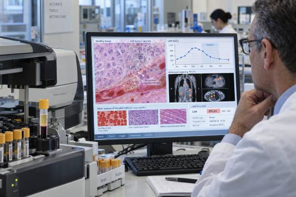
Lactate dehydrogenase, or LDH, is an enzyme found in nearly every tissue in the body. A blood LDH test is sometimes called a tumor marker test because many cancers can raise LDH, especially when disease is extensive, rapidly growing, or causing tissue injury. However, LDH is not cancer-specific. Liver disease, hemolysis, muscle injury, infection, anemia, low oxygen states, and many other conditions can also increase it. In oncology, LDH is most useful as a broad marker of cell turnover, tissue damage, metabolic activity, and prognosis rather than as a stand-alone diagnostic test. It is incorporated into risk assessment for several cancers, including some lymphomas and other hematologic malignancies, and it can help provide context when monitoring advanced disease. A high result should be interpreted against the laboratory’s upper limit of normal, the patient’s diagnosis, symptoms, other blood tests, and trends over time. A normal LDH does not rule out cancer, and an isolated high LDH does not prove that cancer is present.
- What it measures: LDH measures the activity of an enzyme released from many tissues when cells are metabolically active, stressed, injured, or destroyed.
- Typical adult range: One current laboratory uses 122–222 U/L for adults, but reference intervals differ by method and laboratory.
- High LDH is nonspecific: Cancer can raise LDH, but so can hemolysis, liver disease, muscle injury, infection, anemia, lung injury, and other conditions.
- Cancer meaning: In some cancers, an LDH above the laboratory upper limit is associated with greater disease burden or worse prognosis, but the cutoff and importance are cancer-specific.
- Common testing problem: Hemolysis during or after the blood draw can falsely increase LDH because red blood cells contain large amounts of the enzyme.
Table of Contents
- What LDH Measures
- Why LDH Is Used in Cancer Care
- LDH Reference Range and High Levels
- LDH, Cancer Burden, and Prognosis
- Non-Cancer Causes of High LDH
- How the LDH Test Is Done
- How to Interpret LDH Trends and Next Steps
What LDH Measures
LDH is an enzyme that helps cells convert lactate to pyruvate and pyruvate to lactate as part of energy metabolism. Because this pathway is used throughout the body, LDH is present in many tissues, with particularly high activity in red blood cells, liver, heart, skeletal muscle, kidneys, lungs, and some cancers.
The standard serum or plasma LDH test measures total LDH activity in the blood. It does not identify which tissue released the enzyme. That is the main reason LDH is sensitive to many kinds of cell injury but poor at identifying a specific disease.
Cancer cells often rely heavily on glycolysis and lactate metabolism, even when oxygen is available. Rapid tumor growth, hypoxia within a tumor, cell death, inflammation, and high tumor turnover can all contribute to increased circulating LDH. This biological link explains why LDH often tracks with aggressive or extensive disease in certain cancers.
At the same time, a high LDH can be completely unrelated to cancer. If red blood cells rupture in the collection tube, for example, LDH can spill into the sample and create a misleadingly high value. This is called in vitro hemolysis and is one of the most common practical reasons to repeat an unexpected LDH result.
LDH should therefore be viewed as a context marker. It can answer “Is there evidence of increased cell turnover or tissue injury?” but usually cannot answer “Which organ is responsible?” or “Does this person have cancer?” by itself.
Why LDH Is Used in Cancer Care
LDH is used in oncology because it is inexpensive, widely available, reproducible, and associated with clinically important outcomes in several malignancies. Its role depends on the cancer type.
In some lymphomas, LDH is incorporated into validated prognostic scoring systems. An LDH above the laboratory upper limit of normal can contribute to a higher-risk category when combined with age, stage, performance status, and other disease features. In leukemia and other rapidly proliferating blood cancers, LDH may reflect high cell turnover and can rise substantially when disease burden is large.
LDH is also used in selected solid tumors, including melanoma, lung cancer, germ cell tumors, and others. The test may contribute to staging, risk classification, baseline assessment, or treatment monitoring depending on the disease and guideline.
A related marker such as the beta-2 microglobulin blood test is used in some hematologic cancers for prognosis, but it reflects different biology and is strongly influenced by kidney function. The two tests are not interchangeable.
LDH is not usually ordered to screen healthy people for cancer. The lack of specificity would create many false alarms because benign conditions frequently raise it. It also misses many cancers, especially early-stage tumors that do not cause enough cell turnover or tissue injury to elevate the enzyme.
What LDH can and cannot tell you
LDH may help indicate:
- Increased cell turnover or tissue damage
- High disease burden in some cancers
- Higher-risk biology in certain validated prognostic models
- A change in disease activity when followed serially in the right setting
LDH cannot reliably determine:
- The exact cancer type
- The location of a tumor
- Whether a mass is benign or malignant
- Whether treatment should change based on one value alone
- Whether cancer is absent when LDH is normal
LDH Reference Range and High Levels
LDH is commonly reported in units per liter, or U/L. The reference interval depends on age, specimen type, analyzer, and laboratory method. One current reference interval for adults is 122–222 U/L, but many laboratories use different ranges.
The laboratory report’s own upper limit of normal, often abbreviated ULN, is more useful than comparing the number with a range found elsewhere. Cancer studies and prognostic scores often define elevation relative to the ULN rather than using one universal number.
| Result pattern | General interpretation | What usually matters next |
|---|---|---|
| Within reference range | No measurable LDH elevation | Does not exclude cancer or tissue disease |
| Mildly above ULN | Common and nonspecific | Check hemolysis, liver tests, blood count, symptoms, and trend |
| Clearly elevated | More substantial cell turnover or tissue injury may be present | Interpret with diagnosis and other laboratory findings |
| Markedly elevated | Can occur with aggressive malignancy, severe hemolysis, major tissue injury, hypoxia, or other serious illness | Prompt clinical evaluation is usually warranted |
A result two or three times the upper limit may carry different implications from a result only a few units above it. Still, there is no single LDH level that proves cancer. Even very high values have a broad differential diagnosis.
What about low LDH?
A low LDH is generally not used as a tumor marker and is usually less clinically important than a high value. Laboratories may not flag mildly low results. Rare inherited LDH deficiencies exist, but they are not the usual concern in cancer testing.
LDH, Cancer Burden, and Prognosis
High LDH is often associated with a worse prognosis in cancer, but the reason is broader than simple tumor size. LDH can reflect rapid proliferation, tumor hypoxia, metabolic reprogramming, cell destruction, inflammation, and involvement of organs that release the enzyme.
Large meta-analyses across solid tumors have found that elevated LDH is associated with shorter survival on average. More recent reviews also describe LDH as a marker connected to tumor metabolism and the immune microenvironment. These population-level associations do not mean an individual patient’s outcome can be predicted from one LDH result.
The clinical meaning is strongest when LDH is part of a validated disease-specific model. For example, lymphoma prognostic systems may explicitly score LDH as normal or above normal. In advanced melanoma and several other cancers, LDH has historically helped define prognosis and, in some settings, staging or trial stratification.
In multiple myeloma, LDH can provide prognostic context, but diagnosis and staging depend on a broader set of findings. A multiple myeloma test panel evaluates monoclonal proteins, blood counts, kidney function, calcium, bone marrow findings, and other disease markers rather than treating LDH as a diagnostic test.
Is LDH a measure of tumor burden?
Sometimes, but not perfectly. A rising LDH can accompany increasing tumor burden, and a falling LDH can occur with treatment response. However, the relationship is indirect. A small but highly aggressive tumor could generate more LDH than a larger indolent tumor, and a benign liver or muscle problem could raise LDH even when cancer is stable.
For that reason, oncologists compare LDH with imaging, symptoms, physical examination, and more specific tumor markers. A trend that matches those other data is more informative than LDH alone.
LDH can also be useful as a rate-of-change marker when the disease is known to affect it. A single value taken during dehydration, infection, liver injury, or a difficult blood draw may be misleading. Repeated measurements obtained under similar conditions are more useful. A consistent decline over several treatment cycles that parallels shrinking tumors is stronger evidence of response than one isolated normal value. Conversely, a sudden increase deserves confirmation and a search for competing explanations before it is labeled progression.
Historically, laboratories could separate LDH into five isoenzymes, called LDH-1 through LDH-5, which are distributed differently across tissues. Isoenzyme testing is now much less common because modern organ-specific tests and imaging usually provide better information. Total serum LDH remains widely available because it is inexpensive and sensitive to tissue injury, but that broad sensitivity is also its main weakness.
The degree of LDH elevation should also be interpreted against the laboratory’s upper limit of normal rather than a memorized universal number. Cancer studies often report LDH as normal versus elevated, or as a multiple of the upper limit, because assay reference intervals differ. That approach is more transferable than comparing raw U/L values from different laboratories.
Non-Cancer Causes of High LDH
The most important fact about a high LDH is that many non-cancer conditions can cause it. The result should not be interpreted as evidence of malignancy until common alternatives have been considered.
Frequent causes include:
- Hemolysis in the sample: Red blood cell rupture during blood collection or processing can falsely elevate LDH.
- Hemolytic anemia: Red blood cells breaking down inside the body release LDH and can cause substantial elevations.
- Liver disease: Hepatitis, ischemic liver injury, and other liver disorders can increase LDH, usually alongside other liver tests.
- Muscle injury: Strenuous exercise, trauma, inflammation, and muscle disease can contribute.
- Lung and oxygen-related injury: Pulmonary infarction, severe hypoxia, shock, and some pulmonary conditions can increase LDH.
- Infection and inflammation: Tissue injury from significant infections can raise the level.
- Megaloblastic or pernicious anemia: Ineffective blood-cell production can produce striking LDH elevations.
- Kidney or multisystem illness: Severe systemic disease can release LDH from several tissues at once.
A clinician trying to explain an unexpected result may order or review a complete blood count, bilirubin, haptoglobin, AST, ALT, alkaline phosphatase, creatine kinase, kidney tests, and other studies based on symptoms. The laboratory may also report a hemolysis index or note that the specimen was hemolyzed.
If the sample is visibly hemolyzed and the patient is otherwise stable, repeating the blood draw can prevent unnecessary investigation of a false elevation.
How the LDH Test Is Done
LDH is measured from a routine blood draw, usually using serum or plasma. No special preparation is required for LDH itself in most cases. If the test is part of a larger panel that requires fasting, follow those instructions instead.
The collection technique matters because red blood cells contain abundant LDH. Difficult venipuncture, prolonged tourniquet use, vigorous shaking, improper handling, or delayed processing can contribute to hemolysis. Laboratories use quality checks to identify many affected samples, but minor hemolysis can still complicate interpretation.
Exercise may also influence LDH through muscle activity, especially after unusually intense exertion. If an isolated mild elevation is unexpected, the clinician may ask about recent exercise, injury, illness, or procedures.
Medications do not usually require routine discontinuation before LDH testing. Never stop cancer treatment or another prescription medicine solely to prepare for an LDH test unless the treating clinician instructs you to do so.
How to Interpret LDH Trends and Next Steps
The best way to interpret LDH is to ask why it was measured. A mildly high result on a routine chemistry panel is a different problem from a rising LDH in a patient with known aggressive lymphoma.
When LDH is unexpectedly high, clinicians commonly consider these questions:
- Was the sample hemolyzed? A repeat specimen may be enough to clarify the result.
- How high is the value relative to the ULN? The degree of elevation helps set the urgency and differential diagnosis.
- Are other tests abnormal? Anemia, high bilirubin, abnormal liver enzymes, or high creatine kinase can point toward a non-cancer source.
- Is there a known cancer for which LDH is prognostically meaningful? Disease-specific interpretation is more reliable than general interpretation.
- What is the trend? A sustained rise, stable plateau, or fall can provide more information than one isolated measurement.
During cancer treatment, a falling LDH can be reassuring when it parallels radiographic and clinical response. A rising value may raise concern for progression, but treatment toxicity, infection, hemolysis, liver injury, or another complication can produce the same pattern. The result should be checked against the full clinical picture before changing therapy.
Seek prompt medical evaluation when a markedly abnormal LDH accompanies severe weakness, jaundice, shortness of breath, chest pain, confusion, reduced urine output, rapidly worsening anemia, or other significant symptoms. LDH itself does not cause symptoms; the underlying condition does.
For patients reviewing an oncology result, the most useful questions are: What is this laboratory’s ULN? Is the sample hemolyzed? Is LDH part of my cancer’s validated staging or prognostic model? Are my scans and other markers changing in the same direction? Those answers usually matter more than the raw number alone.
Another useful distinction is prognosis versus prediction. A prognostic marker describes how disease tends to behave in groups of patients, while a predictive marker helps identify whether a particular treatment is likely to work. LDH is often prognostic, but it is not generally a stand-alone treatment-selection test. An elevated value may identify a higher-risk clinical picture without telling the oncologist which drug will be effective.
That distinction also explains why an LDH result may remain on an oncology report even when it does not directly change treatment. It can add context to disease severity, help track a previously elevated baseline, and flag new tissue injury. Its value comes from being inexpensive, repeatable, and easy to trend—not from being specific to one cancer.
References
- LD – Overview: Lactate Dehydrogenase (LDH), Serum 2026
- The multiple roles of LDH in cancer 2022 (Review)
- Lactate Dehydrogenase and its clinical significance in pancreatic and thoracic cancers 2022 (Review)
- A Medicinal Chemistry Perspective on Lactate Dehydrogenase: Current Status and Future Directions 2025 (Review)
- Deciphering the potential of the lactate dehydrogenase-to-albumin ratio as a prognostic biomarker in malignancy: a systematic review and meta-analysis. 2025 (Systematic Review)
- Prognostic role of lactate dehydrogenase in solid tumors: a systematic review and meta-analysis of 76 studies 2015 (Systematic Review)
Disclaimer
This article provides general information about LDH testing and is not a diagnosis or a substitute for medical evaluation. LDH is highly nonspecific, and elevated results can come from cancer, blood-cell breakdown, liver or muscle injury, infection, hypoxia, or sample hemolysis. A markedly abnormal value or significant new symptoms should be reviewed promptly by a clinician.