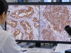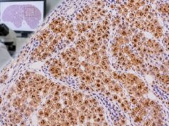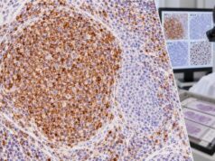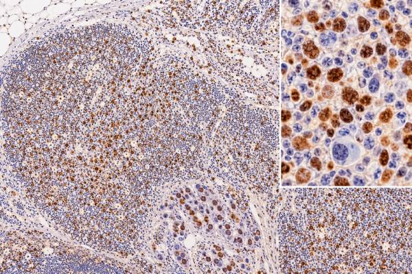
MUM1 immunohistochemistry (IHC) detects the transcription factor IRF4, also called multiple myeloma oncogene 1. In normal lymphoid tissue, MUM1 is most strongly associated with late B-cell activation and plasma-cell differentiation, although activated T cells and other immune cells can also express it. In hematopathology, a positive nuclear MUM1 stain can support plasmacytic differentiation and helps classify several lymphomas, including diffuse large B-cell lymphoma (DLBCL), plasmablastic lymphoma, plasma-cell neoplasms, classic Hodgkin lymphoma, and selected T-cell lymphomas. MUM1 positivity is not specific for one lymphoma and does not by itself prove malignancy. Reactive plasma cells are normally positive, and many unrelated lymphoid neoplasms can express IRF4. In DLBCL, MUM1 is often interpreted with CD10 and BCL6 in the Hans IHC algorithm as a practical surrogate for cell-of-origin classification, but molecular gene-expression methods are more precise. The final diagnosis always depends on morphology, other markers, clinical findings, and sometimes flow cytometry, FISH, or molecular testing.
- MUM1 and IRF4 refer to the same protein in routine pathology: positive staining is mainly nuclear and often reflects activated or late-stage B-cell differentiation.
- Plasma cells are normally MUM1 positive: a positive stain alone does not distinguish reactive plasma cells from myeloma or plasmacytoma.
- In DLBCL, MUM1 is used with CD10 and BCL6 in the Hans algorithm, commonly with a 30% positive-cell cutoff.
- MUM1 positivity is broad across lymphoma types: it can occur in DLBCL, classic Hodgkin lymphoma, plasmablastic lymphoma, plasma-cell neoplasms, and some T-cell lymphomas.
- No special patient preparation is needed: MUM1 IHC is performed on lymph node, bone marrow, mass biopsy, or other tissue submitted to pathology.
Table of Contents
- What MUM1 IHC Detects
- What Positive MUM1 Staining Means
- MUM1 in Diffuse Large B-Cell Lymphoma
- MUM1 in Plasma-Cell and Plasmablastic Neoplasms
- MUM1 in Other Lymphomas
- Limitations and Diagnostic Pitfalls
- How to Read a MUM1 Pathology Result
What MUM1 IHC Detects
MUM1 IHC detects IRF4, a nuclear transcription factor involved in immune-cell activation and the transition of B cells toward plasma-cell differentiation. The name “MUM1” comes from the gene’s historical identification in multiple myeloma, but modern terminology recognizes the protein as IRF4.
In a normal lymph node, MUM1 is not uniformly present in all B cells. It becomes prominent in a subset of late germinal-center and post-germinal-center B cells and is strongly expressed in plasma cells. Activated T cells can also express it. This normal distribution is important because reactive immune cells often provide internal positive staining around a lymphoma.
A pathologist may order MUM1 when trying to determine:
- whether a B-cell neoplasm shows a post-germinal-center or activated phenotype;
- whether large atypical cells have plasmacytic or plasmablastic differentiation;
- how a DLBCL should be classified by an IHC cell-of-origin algorithm;
- whether an unusual lymphoma fits a known immunophenotypic pattern;
- whether an IRF4-associated large B-cell lymphoma should be considered and investigated further.
The stain is generally interpreted in the nuclei of the cells of interest. Weak cytoplasmic background is not equivalent to true positivity. Depending on the diagnostic setting, the pathologist may report a simple positive/negative result or estimate the percentage of positive tumor cells.
MUM1 IHC is not the same as testing for an IRF4 gene rearrangement. A tumor can express MUM1 protein without having an IRF4 rearrangement, and a rearrangement requires a separate genetic method such as fluorescence in situ hybridization. Protein expression and gene alteration answer different questions.
What Positive MUM1 Staining Means
Positive MUM1 staining means that the nuclei of the cells being evaluated contain detectable IRF4 protein. In lymphoid tissue, that finding often suggests activation, post-germinal-center differentiation, or plasma-cell differentiation, but it is not specific enough to name a disease by itself.
A common misunderstanding is that “MUM1 positive” means multiple myeloma. It does not. Normal plasma cells, reactive plasmacytosis, plasmacytoma, multiple myeloma, plasmablastic lymphoma, and several other lymphomas can all be positive. To identify a plasma-cell neoplasm, the pathologist also evaluates morphology, CD138 and CD38, light-chain restriction, cyclin D1 or other markers when relevant, bone-marrow findings, serum or urine studies, and clinical evidence of organ involvement.
Likewise, MUM1 positivity does not automatically mean a lymphoma is “non-germinal center.” That classification depends on the full algorithm. CD10 and BCL6 results are required, and modern molecular classification can disagree with IHC surrogates.
A negative result can be useful when the suspected diagnosis usually expresses MUM1, but it does not exclude every entity. Expression varies by lymphoma subtype and even among cases within the same subtype. Small samples may also contain too few viable tumor cells for confident interpretation.
How much staining counts as positive?
There is no single MUM1 cutoff for every disease. In the Hans algorithm for DLBCL, a commonly used threshold is 30% of tumor cells. This threshold is part of a specific classification scheme and should not be applied automatically to plasma-cell neoplasms, Hodgkin lymphoma, or other tumors.
A pathologist must also separate tumor nuclei from reactive plasma cells and lymphocytes. In an inflamed biopsy, abundant benign MUM1-positive cells can make a lesion look diffusely positive at low magnification even when the malignant population is negative.
MUM1 in Diffuse Large B-Cell Lymphoma
One of the best-known uses of MUM1 is as part of the Hans algorithm for DLBCL cell-of-origin classification. DLBCL is biologically heterogeneous. Gene-expression profiling separates many cases into germinal-center B-cell-like and activated B-cell-like groups, with additional molecular complexity beyond those broad categories.
Because gene-expression testing has not always been available, the Hans algorithm uses three IHC markers—CD10, BCL6, and MUM1—to classify cases as germinal-center B-cell-like (GCB) or non-GCB. In simplified form, CD10-positive cases are generally classified as GCB. CD10-negative cases are then evaluated with BCL6 and MUM1; MUM1 positivity can shift an appropriate CD10-negative/BCL6-positive case into the non-GCB group.
| Marker | Biologic association | Role in the Hans algorithm |
|---|---|---|
| CD10 | Germinal-center phenotype | Strongly supports GCB classification when positive above the algorithm cutoff |
| BCL6 | Germinal-center transcription factor | Used mainly in CD10-negative cases |
| MUM1/IRF4 | Late germinal-center/post-germinal-center activation | Helps separate a subset of CD10-negative cases into non-GCB |
The Hans algorithm is practical, but it is a surrogate, not a direct measurement of gene-expression biology. Concordance with molecular cell-of-origin methods is imperfect. Tumors can also be heterogeneous, and cell-of-origin classification may differ between separate sites or between diagnosis and relapse.
A potentially confusing pattern is simultaneous expression of CD10, BCL6, and MUM1. These markers describe overlapping differentiation programs rather than mutually exclusive switches, so a “triple-positive” phenotype can occur. The Hans algorithm still applies its predefined sequence and thresholds, but the combination may deserve closer morphologic and genetic review. In a young patient with a large B-cell lymphoma in Waldeyer ring or another head-and-neck site, strong MUM1 together with germinal-center markers can raise consideration of large B-cell lymphoma with IRF4 rearrangement. That diagnosis requires demonstration of the rearrangement; the stain itself is only a clue. Conversely, most MUM1-positive DLBCLs do not have an IRF4 rearrangement.
Percentage estimates also have practical limitations. A result near a cutoff can be affected by which viable tumor area is scored, the amount of necrosis or crush artifact, and variation in staining intensity. If the specimen contains mixed regions, the pathologist may comment on heterogeneity rather than forcing the biology into a single simple category. This is one reason a pathology report may state both the individual marker percentages and the resulting Hans classification.
MUM1 status should therefore not be treated as a stand-alone treatment-selection biomarker. Modern DLBCL management increasingly incorporates morphology, clinical risk, FISH for MYC/BCL2/BCL6 in appropriate cases, and molecular features when available. A single MUM1 result does not replace those studies.
MUM1 in Plasma-Cell and Plasmablastic Neoplasms
Strong MUM1 expression is common in plasma cells because IRF4 is central to terminal B-cell differentiation. This makes MUM1 a useful marker when the neoplastic cells have plasmacytic or plasmablastic morphology, but it is not specific for myeloma.
In plasma-cell myeloma and plasmacytoma, tumor cells typically express MUM1 along with plasma-cell markers such as CD138 and CD38. Demonstration of light-chain restriction supports clonality. Additional markers can vary: CD20 may be absent or present in subsets, cyclin D1 can be expressed in tumors with certain genetic backgrounds, and CD56 is common but not universal.
Plasmablastic lymphoma can also show strong MUM1, often with CD138 and other plasma-cell-associated markers, while conventional B-cell markers such as CD20 and PAX5 may be weak or absent. This creates an important differential diagnosis with plasmablastic plasma-cell myeloma. Clinical setting, EBV testing, serum studies, marrow findings, lytic bone disease, immunophenotype, and genetics may all be needed.
The phrase “plasma-cell differentiation” therefore describes a phenotype, not one diagnosis. MUM1 is useful because it recognizes that phenotype, but the pathologist must determine which disease is producing it.
MUM1 also answers a different question from common companion stains. CD138 and CD38 highlight plasma-cell differentiation through surface or membrane-associated proteins, while MUM1 identifies a nuclear transcriptional program. PAX5 and CD20 assess conventional B-cell differentiation and may be reduced as cells become plasmacytic. A tumor that is MUM1 positive, CD138 positive, and PAX5 weak or negative can therefore look convincingly plasmablastic, yet that pattern still spans more than one disease. In marrow biopsies, admixed normal plasma cells can create the same broad immunophenotype, making light-chain restriction and correlation with aspirate flow cytometry especially important.
MUM1 in Other Lymphomas
MUM1 expression occurs in a wide spectrum of lymphoid malignancies, so its value often comes from a characteristic combination with other markers.
Classic Hodgkin lymphoma frequently expresses MUM1 in Hodgkin/Reed-Sternberg cells. Those cells are typically assessed with CD30, PAX5, CD15, EBER in selected cases, and markers used to exclude mimics. MUM1 can support the activated B-cell program of the tumor but is not specific enough to diagnose classic Hodgkin lymphoma alone.
Primary effusion lymphoma and other plasmablastic or immunoblastic lymphomas often express MUM1 because their phenotype is post-germinal-center and activation-associated. Some anaplastic large cell lymphomas and other T-cell lymphomas can also be positive, reflecting the fact that IRF4 is not restricted to B cells.
Large B-cell lymphoma with IRF4 rearrangement is a distinct diagnostic consideration, often occurring in younger patients and frequently involving Waldeyer ring or head-and-neck sites. These tumors commonly show strong MUM1/IRF4 expression and can coexpress germinal-center markers such as CD10 and BCL6. The unusual combination can prompt FISH or other molecular testing for an IRF4 rearrangement. Again, MUM1 protein positivity alone does not establish the rearrangement.
This breadth of expression is why MUM1 is best understood as a differentiation and activation marker rather than a disease-specific antibody.
Limitations and Diagnostic Pitfalls
The biggest MUM1 pitfall is mistaking a common activation marker for a lymphoma-specific marker. Reactive cells can be strongly positive, and multiple unrelated neoplasms share the same phenotype.
Important limitations include:
- Reactive plasma cells: benign plasma cells can surround or infiltrate a tumor and must be separated from the malignant population.
- Broad lymphoma expression: positive staining spans B-cell, plasma-cell, Hodgkin, and selected T-cell neoplasms.
- Algorithm dependence: the 30% cutoff is meaningful in the Hans DLBCL algorithm, not as a universal rule.
- Imperfect cell-of-origin classification: IHC surrogates do not fully reproduce gene-expression profiling.
- Tumor heterogeneity: different regions or disease sites can show different marker expression.
- Protein versus genetics: MUM1 positivity does not prove an IRF4 rearrangement.
- Technical quality: fixation, decalcification, tissue damage, and scant tumor can affect staining.
Bone-marrow specimens deserve special caution because plasma cells are naturally MUM1 positive. A marrow containing many MUM1-positive cells may represent reactive plasmacytosis or a clonal plasma-cell process. Light-chain studies, flow cytometry, morphology, and the clinical workup are needed to distinguish them.
The stain should also be interpreted in the correct cellular compartment. True MUM1 expression is nuclear. Nonspecific cytoplasmic or background staining should not be scored as tumor positivity.
Control cells matter as well. Reactive plasma cells and activated lymphocytes can demonstrate that the stain worked, but they can also obscure a sparse neoplastic population. The pathologist therefore correlates the MUM1 slide with the hematoxylin-and-eosin section and, when necessary, adjacent IHC stains to make sure the percentage estimate is based on tumor cells rather than bystanders.
How to Read a MUM1 Pathology Result
Read a MUM1 result by identifying which cells are positive, why the stain was ordered, and what the rest of the panel shows. A line such as “MUM1: positive” is not a diagnosis on its own.
A practical reading sequence is:
- Confirm the final diagnosis and specimen. Lymph node, marrow, extranodal mass, and effusion specimens have different differential diagnoses.
- Check the staining location. Convincing nuclear staining is the relevant pattern.
- Look for a percentage if DLBCL is being classified. The Hans algorithm commonly uses 30% cutoffs.
- Review CD10 and BCL6 in DLBCL. MUM1 cannot assign cell of origin alone.
- Review plasma-cell markers and clonality studies when plasmacytic differentiation is suspected. CD138, light-chain restriction, and flow or molecular data may be more decisive.
- Ask whether IRF4 rearrangement testing is needed. Strong MUM1 with an unusual germinal-center phenotype can sometimes raise that question.
If the report calls a DLBCL “non-GCB by Hans,” that wording means an IHC surrogate classification, not necessarily the same thing as molecular activated B-cell-like DLBCL. If the distinction will affect a clinical trial or targeted strategy, a molecular assay may be required.
For patients with a plasma-cell neoplasm, MUM1 positivity does not measure tumor burden or response. Those questions are answered by the percentage of clonal plasma cells, monoclonal protein studies, serum free light chains, imaging, marrow assessment, and disease-specific response criteria.
The key takeaway is that MUM1/IRF4 is a powerful marker of late B-cell activation and plasma-cell differentiation, but it is intentionally broad rather than disease-specific. Positive staining can help place a tumor within a differentiation pathway and can contribute to lymphoma classification, yet the final interpretation requires morphology, a full immunophenotype, and genetic testing when indicated.
Several interpretation details are worth keeping separate. MUM1 is read primarily as a nuclear stain, and reactive plasma cells or activated lymphocytes can provide an internal positive control. Their presence also illustrates why a positive result is not proof of lymphoma. The pathologist must show that the atypical population—not merely background immune cells—is the population expressing MUM1.
In diffuse large B-cell lymphoma, MUM1 is often used in the Hans immunohistochemical algorithm together with CD10 and BCL6. The familiar percentage thresholds in that algorithm belong to that specific classification method; they are not a universal definition of MUM1 positivity for every lymphoma. Even within DLBCL, an IHC cell-of-origin assignment is an approximation and is not identical to gene-expression profiling.
MUM1 protein expression should also not be confused with an IRF4 gene rearrangement. Large B-cell lymphoma with IRF4 rearrangement is a genetically defined entity that requires appropriate molecular confirmation. Likewise, strong MUM1 in plasmablastic lymphoma or a plasma-cell neoplasm supports terminal B-cell/plasma-cell differentiation but does not establish clonality or distinguish those diseases by itself. CD138, CD38, light-chain studies, B-cell markers, EBV testing, MYC studies, clinical site, and bone-marrow findings may all become important. The stain is most useful when it narrows a differential diagnosis and directs the next test rather than when it is treated as a stand-alone label.
References
- IRF4 (MUM1) in B-cell malignancies: molecular mechanisms, biological functions, and clinical implications 2026 (Review)
- Updates in Immunohistochemistry for Hematopoietic and Lymphoid Neoplasms 2024 (Review)
- An immunohistochemical study of diffuse large B-cell lymphoma with molecular subtyping based on Hans algorithm 2024
- Plasmablastic Lymphoma. A State-of-the-Art Review: Part 1-Epidemiology, Pathogenesis, Clinicopathologic Characteristics, Differential Diagnosis, Prognostic Factors, and Special Populations 2024 (Review)
- Clinicopathological spectrum of CD10, BCL6, MUM1 triple-positive diffuse large B-cell lymphoma and large B-cell lymphoma with IRF4 rearrangement 2026
Disclaimer
MUM1/IRF4 IHC must be interpreted by a qualified hematopathologist together with tissue morphology, the full immunophenotype, clinical findings, and genetic studies when appropriate. A positive or negative MUM1 result alone cannot diagnose a lymphoma, distinguish reactive from neoplastic plasma cells, or determine treatment. This article is for general education and does not replace individualized medical or pathology advice.