
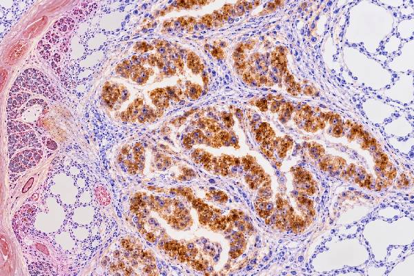
Napsin A immunohistochemistry (IHC) is a tissue stain commonly used to support a diagnosis of lung adenocarcinoma and to help evaluate the likely origin of an adenocarcinoma found in the lung or at a metastatic site. Napsin A is an aspartic protease normally expressed in type II pneumocytes and renal tubular epithelium. In tumor cells, the typical positive pattern is coarse or granular cytoplasmic staining. Many nonmucinous pulmonary adenocarcinomas are positive, and the stain is frequently paired with TTF-1 because concordant TTF-1 and Napsin A expression provides stronger evidence for pulmonary adenocarcinoma than either marker alone. However, Napsin A is not completely lung specific. Renal carcinomas and clear cell carcinomas of the ovary or endometrium can express it, and additional uncommon extrapulmonary tumors can be positive. Mucinous lung adenocarcinomas may also be negative. For those reasons, a Napsin A result must be interpreted with morphology, the complete IHC panel, imaging, clinical history, and the site of the biopsy.
- Napsin A positivity is usually granular and cytoplasmic: staining must be identified in the tumor cells, not just background pneumocytes or macrophages.
- It is a useful lung adenocarcinoma marker: especially when interpreted together with TTF-1.
- It is not lung specific: renal and gynecologic clear cell carcinomas are important positive mimics.
- A negative stain does not exclude lung origin: mucinous and some poorly differentiated pulmonary adenocarcinomas can lack Napsin A.
- Napsin A is diagnostic, not a treatment biomarker: it does not identify EGFR, ALK, ROS1, KRAS, PD-L1, or other predictive alterations.
Table of Contents
- What Napsin A IHC Detects
- What Positive Napsin A Staining Means
- Napsin A in Lung Adenocarcinoma
- Napsin A and TTF-1 for Tumor Origin
- Napsin A-Positive Tumors Outside the Lung
- Limitations and Diagnostic Pitfalls
- How to Read a Napsin A Pathology Result
What Napsin A IHC Detects
Napsin A IHC detects an intracellular aspartic protease involved in protein processing, including surfactant-related functions in type II pneumocytes. Normal expression is prominent in lung pneumocytes and kidney tubular epithelium, which explains both the marker’s usefulness and some of its diagnostic limitations.
Unlike nuclear markers such as TTF-1, Napsin A produces a cytoplasmic granular pattern. In a lung adenocarcinoma, the tumor-cell cytoplasm can show fine-to-coarse granules ranging from focal to diffuse. The pathologist determines whether the positive granules are actually within malignant epithelial cells rather than surrounding benign lung tissue.
That distinction matters because normal type II pneumocytes can be strongly positive. Alveolar macrophages may also label, and both cell types can become trapped within or adjacent to a tumor. On a small biopsy or cytology cell block, those benign positive cells can create a false impression that a Napsin A-negative carcinoma is positive unless the stained slide is carefully matched to the tumor morphology.
Napsin A is usually ordered when an adenocarcinoma is suspected but its lineage or site of origin is uncertain. Common settings include a primary lung mass, a metastatic adenocarcinoma in a lymph node, a pleural specimen, or a tumor in the lung of a patient with a previous cancer elsewhere. The stain may be part of a small, tissue-conserving panel because lung biopsies often need to preserve material for molecular and PD-L1 testing.
The marker is not used to determine whether a lesion is malignant in the first place. It can stain normal cells and selected nonpulmonary tumors. Its value comes from how the staining pattern fits a differential diagnosis that has already been built from histology, clinical data, and imaging.
What Positive Napsin A Staining Means
A positive Napsin A result means that the tumor cells express a protein commonly associated with pulmonary adenocarcinoma, but the result does not prove lung origin by itself. The strength of the inference depends on the anatomic setting and the other markers in the panel.
When a nonmucinous adenocarcinoma in the lung is positive for both Napsin A and TTF-1, pulmonary origin is strongly supported. If the tumor is also negative for markers expected from competing sites, the overall interpretation becomes more convincing. However, a pathologist does not treat the Napsin A stain as a one-marker identity test because known extrapulmonary carcinomas can express the same protein.
Reports may describe Napsin A as focal, patchy, or diffuse and may mention weak, moderate, or strong granular cytoplasmic staining. There is no single universal percentage cutoff that makes a tumor pulmonary. The diagnostic question is usually qualitative: is there convincing tumor-cell staining, and does it fit the panel?
A negative result is also not definitive. Some primary lung adenocarcinomas are Napsin A negative. The likelihood of negativity is particularly important in mucinous pulmonary adenocarcinoma, where both Napsin A and TTF-1 can be absent. Poor differentiation, limited viable tissue, and technical factors can also reduce sensitivity.
Napsin A does not provide the same type of information as predictive biomarkers. A positive stain does not indicate that the tumor has an EGFR mutation, ALK or ROS1 rearrangement, KRAS G12C mutation, or any other drug target. It also does not substitute for PD-L1 testing. Those studies answer separate therapeutic questions after the diagnosis and tumor type have been established.
Napsin A in Lung Adenocarcinoma
Napsin A is most useful in lung pathology as a marker of adenocarcinoma differentiation, particularly in nonmucinous tumors. Large tissue studies have consistently shown expression in a substantial majority of pulmonary adenocarcinomas, while conventional lung squamous cell carcinomas are generally negative in the malignant cells.
This distinction is clinically important in small non-small cell lung cancer biopsies. If morphology is clear, extensive IHC may not be necessary. When the tumor is poorly differentiated, pathologists often use a limited panel such as TTF-1 or Napsin A for adenocarcinoma and p40 for squamous differentiation. The goal is to classify the cancer accurately while preserving tissue for molecular testing.
Adenocarcinoma is not a uniform entity. Acinar, papillary, lepidic, solid, micropapillary, and other nonmucinous patterns may show Napsin A, but expression can vary. Invasive mucinous adenocarcinoma is an important exception because pulmonary mucinous tumors show lower rates of Napsin A expression. Therefore, a Napsin A-negative mucinous tumor in the lung cannot simply be labeled metastatic on that basis.
The same caution applies to metastases from a known lung adenocarcinoma. A metastatic deposit may retain the primary tumor’s Napsin A expression, making the stain useful at distant sites, but loss of expression can occur. If previous pathology is available, comparing the original tumor’s morphology and immunophenotype can be more informative than interpreting the new stain in isolation.
Napsin A may also help when TTF-1 is negative. A subset of pulmonary adenocarcinomas can be TTF-1 negative but Napsin A positive. Conversely, some are TTF-1 positive and Napsin A negative. This complementary behavior is one reason the two stains are often considered together rather than as competitors.
Napsin A and TTF-1 for Tumor Origin
The combination of Napsin A and TTF-1 is more informative for pulmonary adenocarcinoma origin than either stain alone. A large contemporary tissue-microarray study found that combining the two increased specificity for pulmonary adenocarcinoma, while also demonstrating that neither marker is absolutely restricted to lung tumors.
| IHC pattern | Typical interpretation | Main caution |
|---|---|---|
| TTF-1 positive / Napsin A positive | Strong support for pulmonary adenocarcinoma in the appropriate setting | Rare extrapulmonary tumors can share part or all of this phenotype |
| TTF-1 positive / Napsin A negative | Still compatible with lung adenocarcinoma | TTF-1 also occurs in thyroid and selected other tumors |
| TTF-1 negative / Napsin A positive | Can support lung adenocarcinoma | Renal and gynecologic clear cell carcinomas must be considered |
| TTF-1 negative / Napsin A negative | Does not exclude pulmonary origin | Mucinous lung adenocarcinoma is a major setting for double negativity |
Tumor origin panels are built around the actual differential diagnosis. A metastatic clear cell tumor might require PAX8 and organ-specific markers because Napsin A can be positive in both lung and renal/gynecologic cancers. A suspected gastrointestinal metastasis may require CDX2, SATB2, or other markers. A possible breast primary may prompt GATA3 and hormone receptors. The exact panel should be targeted rather than indiscriminately broad.
TTF-1 itself also has limitations. It is strongly associated with lung and thyroid differentiation but can appear in other tumor categories depending on antibody clone and context. Recent large-scale data reinforce that the best specificity comes from combined patterns plus morphology, not from declaring either TTF-1 or Napsin A perfectly site specific.
Napsin A-Positive Tumors Outside the Lung
The most important nonpulmonary Napsin A-positive tumors are renal carcinomas and clear cell carcinomas of the gynecologic tract. Awareness of these entities prevents a metastatic tumor from being incorrectly assigned to the lung.
Kidney tissue normally expresses Napsin A, and renal cell carcinomas—particularly papillary and some clear cell-related subtypes—can be positive. If a patient has a renal mass or a metastatic clear cell carcinoma, PAX8 and other renal-lineage markers can help place the Napsin A finding in context.
Ovarian clear cell carcinoma is another major pitfall. Multiple studies have shown frequent Napsin A expression in this histotype, and the marker is actually useful within gynecologic pathology for supporting clear cell differentiation. Endometrial clear cell carcinoma can also express Napsin A. Consequently, a Napsin A-positive pelvic tumor or metastasis in a woman cannot be assumed to be pulmonary without considering PAX8, gynecologic markers, imaging, and morphology.
Broad tumor surveys have also identified lower-frequency expression in additional neoplasms, including selected thyroid and endometrial tumors. More recently, Napsin A and TTF-1 expression has been documented as a potential pitfall in gastric adenocarcinomas arising in the setting of autoimmune metaplastic atrophic gastritis. These uncommon examples reinforce a general principle: organ-of-origin IHC is probabilistic, not absolute.
The site of biopsy changes the prior probability. Napsin A positivity in a classic peripheral lung adenocarcinoma has a different meaning from the same stain in an ovarian mass or renal lesion. Pathologists incorporate radiologic distribution, previous cancer history, and the rest of the immunophenotype before assigning a primary site.
Limitations and Diagnostic Pitfalls
The largest Napsin A pitfalls are mistaking benign positive cells for tumor, assuming lung specificity, and overinterpreting a negative result in a mucinous carcinoma. These errors are avoidable when the stain is read morphologically and as part of a panel.
Important limitations include:
- Entrapped type II pneumocytes: benign pneumocytes can be strongly positive within or around a lung tumor.
- Alveolar macrophage staining: macrophages can show granular cytoplasmic Napsin A and may mimic tumor cells on small specimens.
- Extrapulmonary positivity: renal and ovarian/endometrial clear cell carcinomas are established positive mimics.
- Mucinous lung adenocarcinoma: this subtype often lacks Napsin A, reducing the marker’s value for separating primary from metastatic mucinous tumors.
- Antibody and platform effects: monoclonal versus polyclonal reagents and laboratory-specific validation can influence sensitivity and nonspecific staining.
- Limited tissue: crushed or scant biopsies and paucicellular cell blocks can make cell identification difficult.
- No predictive information: Napsin A does not replace molecular profiling or PD-L1 assessment for lung cancer therapy.
The historical literature also shows why antibody selection matters. Polyclonal Napsin A has produced broader, potentially misleading staining in some extrapulmonary mucinous tumors. Modern laboratories validate their chosen clone and platform, and pathologists interpret results against appropriate controls rather than assuming all Napsin A assays behave identically.
In lung squamous carcinoma, apparent Napsin A positivity can result from entrapped pneumocytes or macrophages rather than the malignant squamous cells. Careful review at high magnification and comparison with the routine slide can resolve many such cases. If the tumor truly has conflicting glandular and squamous markers, adenosquamous carcinoma or another diagnosis may need consideration rather than forcing the case into one category.
How to Read a Napsin A Pathology Result
Read a Napsin A result by identifying the stained cell population, noting the granular cytoplasmic pattern, and then checking what TTF-1 and site-specific companion markers show. A single positive line is not a complete origin diagnosis.
A practical sequence is:
- Confirm what tissue was biopsied. Lung, lymph node, pleura, bone, liver, ovary, kidney, and other sites create different differential diagnoses.
- Confirm that tumor cells—not pneumocytes or macrophages—are positive. This is a central interpretation step in lung specimens.
- Review the staining pattern and extent. Convincing granular cytoplasmic staining supports a true result.
- Check TTF-1. Dual positivity strongly supports lung adenocarcinoma in the appropriate clinical setting.
- Look for markers of competing primaries. PAX8, CDX2, SATB2, GATA3, ER, and other stains may be used depending on morphology and history.
- Keep mucinous histology in mind. Negative Napsin A and TTF-1 do not exclude a primary pulmonary mucinous adenocarcinoma.
If a report says “Napsin A and TTF-1 positive, consistent with lung primary,” that wording means the pathologist has integrated the immunophenotype with the specimen and clinical context. If the report says only “Napsin A positive,” the result should not be converted by itself into a statement of tumor origin.
For a confirmed lung adenocarcinoma, the next clinically important results are usually separate predictive tests. Depending on stage and local guidelines, these may include broad molecular profiling for actionable alterations and PD-L1 testing. Napsin A helps classify the cancer; it does not determine which targeted drug or immunotherapy is appropriate.
The central takeaway is that Napsin A is a highly useful granular cytoplasmic marker for pulmonary adenocarcinoma, especially when paired with TTF-1, but it is neither perfectly sensitive nor perfectly lung specific. Correct interpretation depends on identifying the right cells, recognizing extrapulmonary positive tumors, and integrating the full pathology and clinical picture.
Several microscopic details help prevent false attribution of tumor origin. Normal type II pneumocytes can show strong granular cytoplasmic Napsin A staining, and alveolar macrophages may also contain granular signal. In a small lung biopsy, those background cells can sit next to a Napsin A-negative carcinoma. The stain should therefore be matched carefully to the tumor on the routine slide rather than judged from the field as a whole.
When the tumor is outside the lung, Napsin A positivity changes the differential but does not settle it. Renal and gynecologic clear-cell tumors are important examples of extrapulmonary expression. A metastatic clear-cell tumor may require PAX8 and other renal or Müllerian markers, while a suspected pulmonary primary is usually evaluated with TTF-1 and the clinical-radiologic pattern. This panel-based approach is especially important when a patient has more than one prior cancer.
Negative staining also has limits. Pulmonary mucinous adenocarcinoma often lacks both Napsin A and TTF-1, so a negative pair cannot exclude lung origin when the morphology and imaging fit. Conversely, dual positivity in a nonmucinous adenocarcinoma strongly supports pulmonary differentiation but is not mathematically specific to the lung. Finally, Napsin A is a diagnostic marker, not a predictive biomarker. It does not substitute for EGFR, ALK, ROS1, KRAS, other genomic testing, or PD-L1 assessment when those studies are indicated for treatment planning.
References
- Immunohistochemistry as a Cornerstone in Lung Cancer Diagnosis: Subtyping Based on the 2021 World Health Organization Classification and Its Practical Application 2026
- Pulmonary immunohistochemical markers may be positive in gastric adenocarcinomas associated with autoimmune metaplastic atrophic gastritis 2026
- TTF-1 is a highly sensitive but not fully specific marker for pulmonary and thyroidal cancer: a tissue microarray study evaluating more than 17,000 tumors from 152 different tumor entities 2024
- Diagnostic and Predictive Immunocytochemistry in Lung Cancer 2025 (Review)
- Napsin A Expression in Human Tumors and Normal Tissues 2021
- Napsin A expression in primary mucin-producing adenocarcinomas of the lung: an immunohistochemical study 2013
Disclaimer
This article is for general educational purposes and cannot determine the origin of an individual tumor. Napsin A IHC must be interpreted by a qualified pathologist with the tumor morphology, TTF-1 and other relevant stains, imaging, clinical history, and molecular testing when indicated. Lung cancer treatment decisions should be based on the complete diagnosis, stage, and validated predictive biomarkers rather than Napsin A staining alone.





