
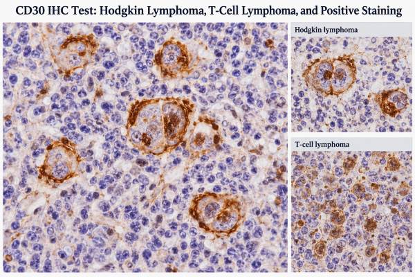
CD30 immunohistochemistry (IHC) is a tissue test that shows whether cells in a biopsy express the CD30 protein. Pathologists use the staining pattern together with cell shape, tissue architecture, clinical information, and other markers to classify lymphomas. Strong CD30 staining is a hallmark of classic Hodgkin lymphoma and anaplastic large cell lymphoma (ALCL), but CD30 is not specific to either disease. It can also appear in other T-cell and B-cell lymphomas and in activated, noncancerous immune cells.
A “CD30-positive” report therefore does not mean one diagnosis by itself. The location of staining, the percentage and intensity of positive tumor cells, and the results of markers such as CD15, PAX5, ALK, CD3, CD20, and others often determine what the finding means. CD30 expression may also matter for treatment because several lymphomas can be treated with CD30-directed therapy, although treatment eligibility is not based on one universal staining cutoff for every lymphoma type.
- CD30 IHC measures CD30 protein in tissue, usually in a lymph node, skin, bone marrow, or other biopsy specimen.
- Strong membrane and Golgi-area staining in abnormal large cells supports certain lymphomas, especially classic Hodgkin lymphoma and ALCL, when the rest of the findings fit.
- CD30 positivity is not cancer-specific; activated lymphocytes and several infections or inflammatory conditions can contain CD30-positive cells.
- There is no single universal “normal range” or positive cutoff for all diseases; diagnosis depends on pattern, cell type, and the full IHC panel.
- A positive result may affect therapy, but the pathologic diagnosis and treatment plan should be based on the complete biopsy and clinical context.
Table of Contents
- What the CD30 IHC Test Shows
- How CD30 Staining Is Performed and Reported
- CD30 in Hodgkin Lymphoma
- CD30 in T-Cell Lymphomas and ALCL
- Other Causes of CD30 Positivity
- How to Interpret a CD30 IHC Result
- What Happens After CD30 Testing
What the CD30 IHC Test Shows
CD30, also called TNFRSF8, is a cell-surface receptor in the tumor necrosis factor receptor family. Normal resting lymphocytes usually show little or no CD30, while activated B cells and T cells can express it. Several lymphomas exploit the same activation pathway and show much stronger or more extensive expression.
IHC does not measure CD30 in the bloodstream. Instead, a laboratory applies an antibody to a thin section of preserved tissue. If CD30 protein is present, the antibody produces a visible stain that the pathologist evaluates under a microscope. This makes CD30 different from a blood tumor marker with a numeric reference range.
The test can answer several related questions:
- Are the abnormal large cells in a suspicious biopsy CD30 positive?
- Does the staining pattern fit classic Hodgkin lymphoma, ALCL, or another CD30-expressing lymphoma?
- Which cells are positive: the tumor cells, background immune cells, or both?
- Is CD30 expression widespread or limited to a small subset of cells?
- Could CD30 expression be relevant to a CD30-targeted treatment?
CD30 is almost always interpreted as part of a broader tumor immunohistochemistry panel. A pathologist first asks what the tissue looks like and which cell population is abnormal. The marker panel then tests competing possibilities rather than treating each stain as an isolated yes-or-no result.
That distinction matters because the same positive stain can mean different things in different settings. Large CD30-positive cells scattered in an inflammatory background may represent Hodgkin/Reed-Sternberg cells, activated immunoblasts, or another lymphoma. Sheets of strongly CD30-positive anaplastic cells raise a different differential diagnosis. The final interpretation depends on morphology, site, clinical presentation, and companion markers.
How CD30 Staining Is Performed and Reported
CD30 IHC is most often performed on formalin-fixed, paraffin-embedded tissue from a core biopsy, excisional biopsy, skin biopsy, or other surgical specimen. No special preparation is needed from the patient after the tissue has already been collected. The important variables are largely laboratory factors: fixation quality, tissue processing, antibody performance, staining controls, and interpretation.
When CD30 is truly expressed by tumor cells, staining is usually membranous with a characteristic accentuation near the Golgi apparatus next to the nucleus. The exact appearance can vary with the cell type and assay. Pathologists also assess whether staining is strong or weak, diffuse or focal, and present in tumor cells versus reactive background cells.
A pathology report may describe CD30 in words such as “positive,” “strong and diffuse,” “variable,” or “focal.” When the result could influence therapy, expert recommendations support reporting an estimated percentage of positive tumor cells when feasible. This is more informative than simply writing positive or negative because CD30 expression can be heterogeneous within one biopsy and between different tumor sites.
There is no universally valid percentage that defines CD30 positivity for every lymphoma. Some entities have diagnostic expectations. For example, systemic ALCL typically shows strong, widespread CD30 expression, and primary cutaneous ALCL is defined within a CD30-positive lymphoproliferative spectrum. In other diseases, clinically meaningful expression may be lower or patchy. A treatment decision therefore should not be reduced to a generic cutoff borrowed from another lymphoma subtype.
Preanalytic problems can also affect results. Poor fixation, tissue crush, decalcification, extensive necrosis, or very small samples may reduce antigen detection or make the target cells hard to identify. If the morphology strongly suggests a CD30-associated lymphoma but the stain is unexpectedly absent, the pathologist may repeat the stain, use another block, review controls, or obtain additional tissue.
CD30 in Hodgkin Lymphoma
Strong CD30 expression is one of the defining immunophenotypic features of classic Hodgkin lymphoma (cHL). The malignant Hodgkin and Reed-Sternberg cells are usually intensely positive, often with both membrane staining and a paranuclear Golgi pattern. Because the cancer cells can make up only a small fraction of the biopsy, the pathologist must identify the correct large atypical cells rather than judge the slide by the total number of stained cells.
CD30 alone does not establish cHL. The typical diagnosis combines compatible architecture and cell morphology with a panel. Hodgkin/Reed-Sternberg cells are commonly positive for CD15 staining and show weak or dim PAX5 expression, reflecting their altered B-cell program. A PAX5 IHC result can be particularly helpful because cHL often shows weaker nuclear PAX5 in tumor cells than surrounding normal B cells. Classic cases usually lack strong expression of broad B-cell markers such as CD20, although a subset can be CD20 positive.
This panel helps separate cHL from look-alikes. For example, nodular lymphocyte-predominant B-cell lymphoma has a different phenotype and usually does not show the classic strong CD30 pattern. Some diffuse large B-cell lymphomas, primary mediastinal large B-cell lymphomas, Epstein-Barr virus–associated proliferations, and T-cell lymphomas can contain large CD30-positive cells that resemble Hodgkin cells.
The background also matters. cHL often contains abundant reactive T cells, histiocytes, eosinophils, plasma cells, and fibrosis around relatively sparse tumor cells. A small needle core may sample only part of that environment. When the clinical or microscopic features are discordant, an excisional lymph node biopsy may provide better architecture for classification.
Rare cHL cases can show atypical antigen patterns, including reduced or absent CD30 staining. Those situations require caution rather than automatic exclusion of the diagnosis. The pathologist may review fixation, repeat IHC, expand the panel, and consider molecular or Epstein-Barr virus testing. The diagnosis should remain an integrated one, especially when a single marker behaves unexpectedly.
CD30 in T-Cell Lymphomas and ALCL
CD30 is especially important in the workup of mature T-cell lymphomas because strong expression is central to ALCL and variable expression occurs in several other T-cell neoplasms.
Anaplastic large cell lymphoma
ALCL consists of large atypical lymphoid cells, often with horseshoe- or kidney-shaped nuclei and a prominent Golgi region. Tumor cells characteristically show strong, usually diffuse CD30 staining. The stain often highlights cell membranes and the paranuclear area so clearly that it helps the pathologist recognize the malignant population.
Systemic ALCL is divided into ALK-positive and ALK-negative forms. An ALK IHC test is therefore a key companion study. ALK-positive ALCL often occurs in children and younger adults and is driven by an ALK rearrangement, most commonly involving NPM1. ALK-negative ALCL occurs more often in older adults and includes genetically distinct subgroups. Both are strongly CD30 positive, so CD30 cannot determine ALK status by itself.
T-cell markers may be variable in ALCL. Some tumors lose several conventional T-cell antigens, creating a “null” phenotype by routine IHC despite evidence of T-cell lineage. A CD3 IHC test may be negative in the tumor cells even when the lymphoma is classified within the T-cell family. That is one reason the diagnostic panel often includes multiple T-cell markers, cytotoxic markers, epithelial membrane antigen, ALK, and molecular studies when appropriate.
Other CD30-positive T-cell lymphomas
Peripheral T-cell lymphoma, not otherwise specified, and T-follicular helper lymphomas can show CD30 in a subset of tumor cells. Expression may be patchy or heterogeneous rather than the uniformly strong pattern expected in ALCL. Cutaneous CD30-positive lymphoproliferative disorders include primary cutaneous ALCL and lymphomatoid papulosis, which can look alarming under the microscope despite very different clinical behavior.
Clinical site is crucial in separating primary cutaneous ALCL from systemic ALCL involving skin. A skin-limited lesion, staging results, ALK status, and other pathologic features all contribute. Breast implant–associated ALCL is another distinct CD30-positive entity that usually presents with a late peri-implant fluid collection, capsular mass, or both. Its diagnosis requires the appropriate clinical setting and cytologic or tissue evaluation.
Because CD30-targeted therapy is available for several lymphomas, the pathologist may quantify expression more carefully when treatment selection is being considered. Still, CD30 percentage does not replace disease classification. The same percentage can carry different diagnostic and therapeutic implications in ALCL, cHL, cutaneous lymphoma, and other peripheral T-cell lymphomas.
Other Causes of CD30 Positivity
CD30 positivity is not synonymous with Hodgkin lymphoma or T-cell lymphoma. Activated lymphocytes can express CD30 in reactive conditions, and a range of neoplasms can contain CD30-positive cells. This is one of the most important limitations of the test.
Reactive immunoblasts may become large, atypical-looking, and CD30 positive during viral infections or other immune stimulation. Epstein-Barr virus–driven proliferations can be especially challenging because the activated B cells may resemble Hodgkin/Reed-Sternberg cells. The pathologist may use Epstein-Barr virus–encoded RNA testing, lineage markers, and the overall architecture to resolve the differential.
Some B-cell lymphomas also express CD30. These include subsets of diffuse large B-cell lymphoma and primary mediastinal large B-cell lymphoma. In that setting, a CD20 IHC test and other B-cell markers help establish lineage. Strong B-cell antigen expression in the tumor population can shift the diagnosis away from cHL even when the cells are CD30 positive.
Other tumors and non-lymphoid malignancies can occasionally stain with some CD30 antibodies, so morphology and broad lineage markers remain important when the biopsy is poorly differentiated. In a mass of uncertain origin, the first goal is often to determine whether the lesion is hematolymphoid, epithelial, melanocytic, or another lineage before interpreting disease-specific markers.
Common pitfalls include:
- Mistaking scattered reactive CD30-positive cells for the malignant population.
- Calling a lymphoma “Hodgkin” based on CD30 alone without a compatible phenotype.
- Assuming negative CD3 excludes a T-cell lymphoma such as ALCL.
- Treating focal expression as diagnostically equivalent to the diffuse pattern typical of ALCL.
- Ignoring tissue quality when a strongly suspected CD30-associated lymphoma stains unexpectedly weakly.
- Applying a treatment cutoff from one lymphoma type to another without disease-specific guidance.
The practical rule is simple: CD30 tells you that a population is expressing an activation-associated receptor. The rest of the biopsy tells you what that population is.
How to Interpret a CD30 IHC Result
A useful interpretation starts by identifying exactly which cells are positive. The phrase “CD30 positive” is incomplete if the slide contains several cell populations. Pathology reports may therefore state that the large atypical cells are positive, that only scattered background immunoblasts stain, or that a certain percentage of tumor cells shows expression.
| CD30 pattern | Possible interpretation | What usually clarifies it |
|---|---|---|
| Strong staining in Hodgkin/Reed-Sternberg–type cells | Supports classic Hodgkin lymphoma if morphology and phenotype fit | CD15, PAX5, CD20, CD45, EBV testing, architecture |
| Strong diffuse staining in sheets of anaplastic large cells | Raises ALCL strongly | ALK, T-cell markers, cytotoxic markers, clinical site, genetics |
| Variable staining in another T-cell lymphoma | May support activation and can have therapeutic relevance | Full T-cell panel, subtype-defining markers, molecular studies |
| Scattered positive immunoblasts in reactive tissue | May be noncancerous activation | Morphology, clonality when needed, EBV studies, clinical history |
| Positive large B cells | Can occur in B-cell lymphoma or EBV-related proliferation | CD20/PAX5 and other B-cell markers, EBV, genetic studies |
“Positive” does not automatically mean “high,” and “high” is not a standardized diagnostic category across all lymphoma types. Some laboratories report the estimated percentage of tumor cells with membrane staining, while others use descriptive categories. If a report gives a percentage, it should be read with the diagnosis and laboratory method rather than compared with a generic internet reference range.
The significance of intensity is also contextual. Very strong, diffuse staining can be characteristic in cHL and ALCL, but intensity alone cannot separate the two. Conversely, weaker staining does not prove a benign process. Tissue preservation, heterogeneous biology, and treatment effects can all change antigen expression.
For patients, the most useful questions are often: What diagnosis does the pathologist favor? Which cells were CD30 positive? What other markers support that diagnosis? Was expression quantified because it may affect treatment? Is more tissue or molecular testing needed? Those questions are more informative than asking whether CD30 is simply “good” or “bad.”
What Happens After CD30 Testing
After CD30 IHC, the next step depends on why the stain was ordered. If the biopsy is being evaluated for lymphoma, the pathologist typically completes a panel that defines lineage and subtype. Flow cytometry, in situ hybridization, fluorescence in situ hybridization, or sequencing may be added when they answer a specific diagnostic question.
For suspected classic Hodgkin lymphoma, the final report integrates morphology with CD30, CD15, PAX5, B-cell and T-cell markers, CD45, and sometimes Epstein-Barr virus studies. For suspected ALCL, ALK testing is essential for systemic disease classification, and additional studies may look for DUSP22 or TP63 rearrangements in selected ALK-negative cases. Staging then uses clinical examination, imaging, and other tests appropriate to the diagnosed lymphoma.
CD30 can also have treatment implications. Brentuximab vedotin is an antibody-drug conjugate that binds CD30 and is used in several CD30-expressing lymphomas. However, the relationship between the percentage of CD30-positive cells and treatment response is not identical across diseases, and low or heterogeneous expression can complicate interpretation. The oncology team uses the approved indication, lymphoma subtype, prior therapy, patient factors, and the pathology report together.
If a result seems inconsistent with the suspected disease, repeat testing may be reasonable. Pathologists can review another tissue block, confirm internal controls, repeat the antibody, or request a larger biopsy. This is especially important when the tissue is tiny, crushed, decalcified, or largely necrotic.
Patients generally do not need to act on a CD30 result before the complete pathology diagnosis is available. CD30 is one piece of a classification process, not a stand-alone screening test. A positive stain can be highly informative when it appears in the right cells and pattern, but its value comes from being interpreted with the entire biopsy.
References
- Best Practices in CD30 Immunohistochemistry Testing, Interpretation, and Reporting: An Expert Panel Consensus 2023 (Position Statement)
- The International Consensus Classification of Mature Lymphoid Neoplasms: a report from the Clinical Advisory Committee 2022 (Consensus Classification)
- Fifth Edition of the World Health Organization Classification of Tumors of the Hematopoietic and Lymphoid Tissues: Mature T-Cell, NK-Cell, and Stroma-Derived Neoplasms of Lymphoid Tissues 2024 (Review)
- Classic Hodgkin lymphoma: An illustrative review of select diagnostic limitations and immunomorphological challenges 2025 (Review)
- Anti-CD30 antibody-drug conjugate therapy in lymphoma: current knowledge, remaining controversies, and future perspectives 2023 (Review)
Disclaimer
CD30 IHC is a pathology test that must be interpreted with the full biopsy, other stains, and clinical findings. A positive or negative CD30 result alone cannot diagnose or exclude a specific lymphoma or determine treatment. Discuss the complete pathology report and treatment implications with the pathologist, hematologist, or oncologist involved in your care.





