CD15 immunohistochemistry (IHC) is a tissue stain best known for helping identify classic Hodgkin lymphoma. In that setting, Hodgkin and Reed-Sternberg cells often express CD15 together with strong CD30 and weak PAX5. The combination supports the diagnosis when the microscopic appearance is also typical. CD15 is not a lymphoma-specific marker, however. Normal granulocytes, some monocytes, and several non-hematologic tumors can also be positive, while a meaningful minority of classic Hodgkin lymphomas are CD15 negative.
That makes CD15 a supporting marker, not a stand-alone cancer test. Pathologists interpret the stain on the abnormal cells, compare it with CD30, PAX5, CD20, CD45, and other markers, and consider important mimics such as anaplastic large cell lymphoma, large B-cell lymphoma, Epstein-Barr virus-associated proliferations, and nodular lymphocyte-predominant Hodgkin lymphoma. A result described only as “CD15 positive” cannot determine the diagnosis, stage, or prognosis without the rest of the pathology findings.
- CD15 positivity in Hodgkin/Reed-Sternberg cells supports classic Hodgkin lymphoma, especially with strong CD30 and weak PAX5.
- CD15 is positive in many, but not all, classic Hodgkin lymphomas, so a negative result does not exclude the disease.
- The typical tumor-cell pattern is membranous and Golgi/perinuclear with cytoplasmic staining, but technical and biologic variation occurs.
- CD15 is not specific for Hodgkin lymphoma because granulocytes and several other tumors can express it.
- Tumor classification requires a panel, not CD15 alone, because multiple lymphomas can contain Hodgkin-like large cells.
Table of Contents
- What the CD15 IHC Test Detects
- How CD15 Staining Is Read
- CD15 in Classic Hodgkin Lymphoma
- CD15 With CD30, PAX5, and CD20
- Conditions That Can Mimic Hodgkin Lymphoma
- What Positive and Negative CD15 Results Mean
- What Happens After CD15 Testing
What the CD15 IHC Test Detects
CD15 is a carbohydrate antigen related to the Lewis X structure on the cell surface. It is normally expressed on mature neutrophils and eosinophils and on subsets of monocytes. In pathology, antibodies historically known as Leu-M1 recognize CD15 and can make the antigen visible in formalin-fixed tissue.
Its most familiar diagnostic use is in the workup of classic Hodgkin lymphoma (cHL). The malignant Hodgkin and Reed-Sternberg (HRS) cells in cHL frequently express CD15 despite having lost much of the normal B-cell program. This phenotype is unusual enough to be useful, especially when CD15 is combined with CD30 and weak B-cell lineage markers.
CD15 does not determine cell lineage by itself. It is not equivalent to CD20 for B cells or CD3 for T cells. Because reactive granulocytes are strongly positive, a pathologist must distinguish staining in background inflammatory cells from staining in the large atypical tumor cells. In cHL, those HRS cells may represent only a small fraction of the total cells in the biopsy, while the rest of the tissue contains abundant lymphocytes, eosinophils, plasma cells, histiocytes, and other reactive cells.
The stain is therefore most valuable in a targeted tumor IHC panel. It answers the question “Do these suspicious large cells show a phenotype compatible with classic Hodgkin lymphoma?” rather than “Does this patient have Hodgkin lymphoma?”
CD15 also appears in some adenocarcinomas, myeloid cells and neoplasms, and occasional other lymphoid tumors. The clinical setting and morphology determine whether those alternative explanations are relevant. A positive epithelial tumor, for example, should not be mistaken for lymphoma simply because it expresses CD15.
How CD15 Staining Is Read
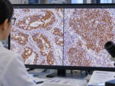
CD15 IHC is usually performed on a lymph node or other tissue biopsy that has been fixed in formalin and embedded in paraffin. The laboratory applies an antibody to CD15 and develops a visible stain. The pathologist then identifies the suspicious HRS cells and asks whether those specific cells label.
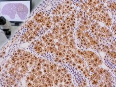
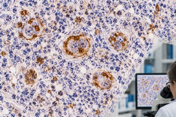
In classic Hodgkin lymphoma, convincing CD15 staining can be membranous, cytoplasmic, and often concentrated in the Golgi/perinuclear region. The distribution may be strong and diffuse or present in only a subset of tumor cells. Background neutrophils and eosinophils provide useful internal positive staining but can also create visual noise.
There is no blood-style reference range and no universal percentage cutoff that separates cHL from non-cHL. A report may state “positive in HRS cells,” “subset positive,” “focally positive,” or “negative.” That wording matters because a focal tumor-cell reaction carries a different implication from strong staining limited to background granulocytes.
Several technical issues can influence the result. Small core biopsies may contain very few diagnostic cells. Crush artifact, necrosis, poor fixation, decalcification, and exhausted tissue can make HRS cells difficult to identify or weaken antigen detection. If the morphology strongly suggests cHL but CD15 is negative, pathologists do not usually stop the workup. They assess the rest of the immunophenotype and may repeat stains on another block or obtain additional tissue if the specimen is inadequate.
A second practical issue is antibody specificity. Different CD15 antibody clones and staining platforms can produce somewhat different sensitivity. This is another reason the diagnosis rests on a coherent panel rather than on a rigid percentage threshold.
CD15 in Classic Hodgkin Lymphoma
Classic Hodgkin lymphoma is defined by characteristic HRS cells in a distinctive inflammatory background. HRS cells are derived from B cells, usually germinal center B cells, but they have profoundly altered gene expression and lose many of the markers expected on a conventional mature B-cell lymphoma.
The classic phenotype is CD30 strong positive, CD15 positive, PAX5 weak positive, CD45 negative, and CD20 negative or weaker/less extensive than in ordinary B-cell lymphoma. Real cases can deviate from that textbook pattern. CD15 is commonly positive in approximately three-quarters or more of cHL cases, depending on the series, staining method, and definition of positivity. That leaves a significant CD15-negative group.
The stain is especially persuasive when the positive cells have the right morphology. HRS cells are large, often with abundant cytoplasm and one or more striking nucleoli. Reed-Sternberg forms may be binucleated or multinucleated. Variants include mononuclear Hodgkin cells, lacunar cells, and mummified forms. A mixed background may contain small lymphocytes, eosinophils, histiocytes, plasma cells, and fibrosis, with the balance depending on the histologic subtype.
The four traditional subtypes of cHL—nodular sclerosis, mixed cellularity, lymphocyte-rich, and lymphocyte-depleted—remain morphologic categories. CD15 is not a test that assigns those subtypes on its own. In practice, the overall diagnosis and exclusion of mimics matter more than trying to infer subtype from a single immunostain.
CD15 positivity is also not a staging test. Stage is determined from the distribution of disease using clinical evaluation and imaging, with bone marrow assessment only in selected circumstances. Likewise, the intensity of CD15 staining does not provide a treatment dose or a simple numerical prognosis.
A particularly important point is that CD15 negativity does not exclude cHL. When CD30 is strong and diffuse, PAX5 is weak, CD45 is absent, morphology fits, and alternative diagnoses are excluded, a CD15-negative cHL diagnosis may be entirely appropriate. Modern diagnostic reviews emphasize recognition of these immunophenotypic variations rather than requiring every case to match the classic panel perfectly.
CD15 With CD30, PAX5, and CD20
CD15 works best as part of a pattern. In suspected classic Hodgkin lymphoma, CD30 IHC is typically the most sensitive hallmark stain. HRS cells usually show strong membranous and Golgi-region CD30 staining. CD15 adds support but is less consistently positive.
PAX5 provides a different kind of information. It is a B-cell transcription factor, and HRS cells in cHL often retain weak nuclear PAX5 expression. That weak staining can help establish their B-cell origin while distinguishing them from many strongly PAX5-positive large B-cell lymphomas. PAX5 staining should still be judged against nearby normal B cells, which serve as an internal intensity comparison.
CD20 is commonly absent or only focal/weak in cHL, but some cases show stronger expression. Conversely, the tumor cells of nodular lymphocyte-predominant Hodgkin lymphoma—called nodular lymphocyte-predominant B-cell lymphoma in the International Consensus Classification—usually retain a robust B-cell program with CD20 and PAX5 and are typically negative for CD15 and CD30. The fifth WHO edition retains the NLPHL terminology while recognizing its distinct biology.
A simplified pattern comparison is useful, but it is not a substitute for full diagnosis:
| Entity | CD15 | CD30 | B-cell markers | Key caution |
|---|---|---|---|---|
| Classic Hodgkin lymphoma | Often positive | Usually strong positive | PAX5 often weak; CD20 variable | Not every case has the classic phenotype |
| Nodular lymphocyte-predominant Hodgkin/B-cell lymphoma | Usually negative | Usually negative | CD20 and PAX5 strong | Architecture and LP-cell pattern are essential |
| Anaplastic large cell lymphoma | Usually negative | Strong positive | PAX5 usually negative | T-cell markers, ALK and cytotoxic phenotype may help |
| Large B-cell lymphoma | Usually negative | Variable | Often strong CD20/PAX5 | Some cases have Hodgkin-like cells |
The purpose of the panel is not merely to count positives. It is to decide whether all results describe the same biologic process. A CD15-positive, CD30-positive large cell that is strongly CD20/PAX5 positive may prompt a different differential from one that shows weak PAX5 and no CD45.
Conditions That Can Mimic Hodgkin Lymphoma
Large CD30-positive Hodgkin-like cells are not unique to cHL. They can appear in other lymphomas, viral infections, immune deficiency-associated proliferations, and reactive conditions. CD15 can sometimes be positive in these settings, so morphology and a broader panel are needed.
Anaplastic large cell lymphoma (ALCL) is one of the most important mimics because it can contain very large pleomorphic cells with strong CD30 expression. ALCL usually lacks PAX5 and CD15 and may show T-cell or cytotoxic markers, although pan-T-cell antigens can be lost. ALK IHC helps identify ALK-positive ALCL. ALK-negative ALCL requires other clinical, morphologic, and molecular features for classification.
Large B-cell lymphomas can also resemble cHL, particularly in the mediastinum or in Epstein-Barr virus-positive disease. Strong and broad B-cell marker expression, including CD20, PAX5, OCT2, and BOB1, favors a large B-cell lymphoma, but overlap exists. Mediastinal gray-zone lymphoma sits at a biologic and morphologic boundary and may show an intermediate phenotype.
Nodular lymphocyte-predominant Hodgkin lymphoma/NLP B-cell lymphoma is separated from cHL because its LP cells are usually CD20 and PAX5 strong, OCT2/BOB1 positive, and CD15/CD30 negative. The surrounding architecture, including follicular dendritic cell networks and rosetting T cells, helps establish the diagnosis.
Epstein-Barr virus-related reactive or neoplastic processes may contain large activated B cells that look like HRS cells. EBER in situ hybridization, B-cell markers, the overall tissue pattern, immune status, age, and clinical setting help resolve these cases. Infectious mononucleosis can be especially deceptive in limited tissue.
CD15 can also be expressed by non-lymphoid malignancies. If the biopsy contains a poorly differentiated tumor, broad cytokeratins and other lineage markers may be necessary before the lesion is classified as lymphoma. The correct question is always whether the CD15-positive cells are the tumor and whether the rest of the phenotype fits the proposed diagnosis.
What Positive and Negative CD15 Results Mean
A positive CD15 result in the suspicious large cells means those cells express the CD15 antigen. When morphology is classic and CD30 is strongly positive with weak PAX5, the finding supports cHL. It does not independently prove the diagnosis because other cells and tumors can express CD15.
A negative result means convincing CD15 was not detected in the relevant tumor cells. This reduces one piece of support for cHL but does not rule it out. The pathologist asks whether CD30, PAX5, CD45, CD20, EBV testing, T-cell markers, and other findings form a coherent alternative pattern.
A focal or subset-positive result is common enough to be meaningful if the stained cells are unequivocal HRS cells. It should not be confused with abundant CD15-positive neutrophils around the tumor. Reports that specify “HRS cells positive” are more informative than reports that simply list CD15 as positive.
CD15 results should also not be used to predict whether a patient will respond to a particular drug. Unlike CD30, which can serve as a therapeutic target in some lymphomas, routine CD15 IHC is primarily diagnostic. Historical studies have explored associations between CD15 expression and outcome, but modern clinical risk assessment and treatment decisions rely on disease type, stage, symptoms, response assessment, and established clinical factors rather than CD15 staining alone.
If the pathology findings remain discordant, the safest interpretation may be descriptive until more tissue or ancillary testing is available. This is particularly important for small biopsies, where only a few atypical cells may be present and the architecture needed to distinguish entities may be missing.
The clinical history can be unusually important in difficult CD15 cases. A mediastinal mass in a young adult raises a different set of competing diagnoses from a lymph node in an older immunosuppressed patient. Prior lymphoma, organ transplantation, HIV status, recent viral illness, and the distribution of disease can all change which Hodgkin-like mimics deserve focused testing.
Pathologists also compare the intensity of lineage markers with internal controls. For example, weak PAX5 in HRS cells is meaningful only when nearby normal B cells show the expected strong nuclear signal. This relative pattern often carries more diagnostic weight than simply marking a stain as positive or negative.
What Happens After CD15 Testing
After CD15 IHC, the pathologist integrates the full panel with the biopsy morphology. A common initial cHL panel includes CD30, CD15, PAX5, CD20, CD3, and CD45. Additional stains are chosen from the differential rather than ordered automatically.
If ALCL is possible, ALK and additional T-cell/cytotoxic markers may be added. If large B-cell lymphoma or mediastinal gray-zone lymphoma is possible, stronger B-cell transcription factors and B-cell markers such as OCT2, BOB1, CD79a, and CD20 can be helpful. If an EBV-associated process is in the differential, EBER in situ hybridization is commonly used. Clinical location and immune status can be decisive in difficult cases.
When the sample is too small to show diagnostic architecture or contains very few HRS cells, an excisional or larger core biopsy may be recommended. Fine-needle aspiration alone is often insufficient for initial lymphoma classification because lymphoma diagnosis depends on architecture as well as cytology, although flow cytometry and specialized methods can add value to small samples.
For patients, the most useful part of the report is the final integrated diagnosis, not the isolated CD15 line. Questions worth asking include whether the phenotype was typical for cHL, whether important mimics were excluded, whether EBV was assessed, and whether the tissue was adequate for classification. Once cHL is confirmed, staging and treatment planning proceed separately from CD15 interpretation.
No special preparation is needed specifically for the stain because CD15 IHC is performed on tissue already collected. The clinically important preparation concerns the biopsy itself, such as medication instructions and procedural planning. If a repeat biopsy is recommended, the reason is usually tissue adequacy or diagnostic uncertainty rather than an abnormal CD15 “level.”
CD15 is most useful when it provides one well-placed piece of a larger pattern. Strong diagnostic confidence comes from agreement among morphology, CD30, B-cell and T-cell markers, viral studies when needed, and the clinical setting.
References
- Classic Hodgkin lymphoma: An illustrative review of select diagnostic limitations and immunomorphological challenges. 2025 (Review)
- Diagnostic approach for classic Hodgkin lymphoma in small samples with an emphasis on PD-L1 expression and EBV harboring in tumor cells: a brief review from morphology to biology 2023 (Review)
- Hodgkin Lymphoma Classification-From Historical Concepts to Current Refinements 2025 (Review)
- Histopathologic Features and Differential Diagnosis in Challenging Cases of Nodular Lymphocyte Predominant B-cell Lymphoma/Nodular Lymphocyte Predominant Hodgkin Lymphoma. 2024
- American Registry of Pathology Expert Opinions: Immunohistochemical evaluation of classic Hodgkin lymphoma 2019 (Expert Review)
Disclaimer
CD15 IHC is a diagnostic tissue stain and cannot confirm or exclude Hodgkin lymphoma by itself. The result must be interpreted with biopsy morphology, a complete immunohistochemical panel, and clinical information. Individual diagnosis, staging, and treatment decisions should be discussed with the treating clinician and pathology team.