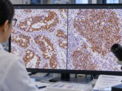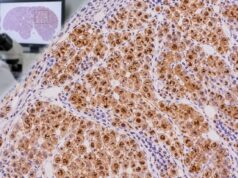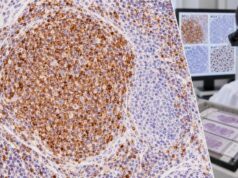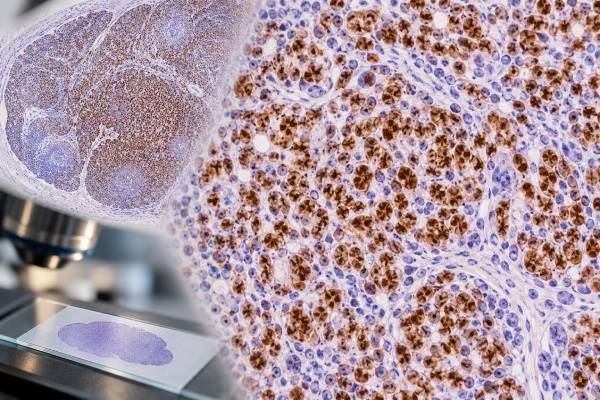
The PAX5 IHC test is a tissue stain used mainly to identify B-cell lineage in lymphomas and other hematolymphoid neoplasms. PAX5 is a nuclear transcription factor that helps commit developing lymphocytes to the B-cell program and remains expressed through much of B-cell maturation. In pathology, nuclear PAX5 staining supports B-cell differentiation, including in many tumors that may show weak or absent staining for some surface B-cell markers. It is especially useful in classical Hodgkin lymphoma, B-lymphoblastic leukemia/lymphoma, and many mature B-cell lymphomas. The result is not interpreted by itself because PAX5 can be weak, lost in plasmacytic differentiation, or rarely expressed in non-B-cell tumors. Pathologists combine PAX5 with morphology and markers such as CD20, CD79a, CD3, CD30, CD15, OCT2, BOB1, MUM1, and plasma-cell markers. The full immunophenotypic pattern, not a single stain, determines the lymphoma classification.
- PAX5 positivity usually appears as nuclear staining and supports B-cell lineage in the right context.
- Most mature B-cell lymphomas are PAX5 positive, but plasma-cell neoplasms and plasmablastic tumors often lose PAX5.
- Classical Hodgkin lymphoma usually shows weak PAX5 in Hodgkin/Reed–Sternberg cells, weaker than surrounding reactive B cells.
- PAX5 is not completely B-cell specific, so unexpected staining requires a broader panel and morphologic correlation.
- There is no blood “normal range” for PAX5 IHC; it is a qualitative or semiquantitative tissue interpretation.
Table of Contents
- What the PAX5 IHC Test Detects
- What Positive PAX5 Staining Means
- PAX5 Patterns in Major Lymphoma Types
- How PAX5 IHC Is Performed and Reported
- How PAX5 Works With Other Lymphoma Markers
- Limitations and Diagnostic Pitfalls
- What Happens After a PAX5 Result
What the PAX5 IHC Test Detects
PAX5 is a transcription factor encoded by the PAX5 gene. It plays a central role in B-cell development by activating B-cell programs and suppressing alternative lineage programs. In normal lymphoid tissue, PAX5 is expressed in B cells from relatively early stages of differentiation through mature B-cell stages, then is usually downregulated as B cells become terminally differentiated plasma cells.
Immunohistochemistry takes advantage of this biology. The diagnostically meaningful reaction is nuclear staining. If an atypical lymphoid population shows convincing nuclear PAX5, the result supports B-cell differentiation. That can be particularly valuable in a poorly differentiated tumor or a lymphoma in which CD20 expression is weak, absent, or difficult to interpret.
PAX5 is therefore considered a lineage-associated marker, not a marker of one single disease. Diffuse large B-cell lymphoma, follicular lymphoma, mantle cell lymphoma, many other mature B-cell lymphomas, and B-lymphoblastic leukemia/lymphoma are typically PAX5 positive. Classical Hodgkin lymphoma is also usually positive, but the neoplastic Hodgkin/Reed–Sternberg cells often stain only weakly.
The marker is complementary to CD20. CD20 is a membrane protein and a major therapeutic target, while PAX5 is a nuclear transcription factor. A tumor can show discordant expression, especially after anti-CD20 therapy or when the cells have plasmablastic differentiation.
PAX5 does not determine whether a B-cell neoplasm is indolent or aggressive, and it does not by itself identify the exact lymphoma subtype. The classification of lymphoid tumors uses morphology, immunophenotype, clinical setting, cytogenetics, and molecular findings according to modern WHO and International Consensus Classification frameworks.
What Positive PAX5 Staining Means
A positive PAX5 result means the nuclei of the cells being evaluated contain detectable PAX5 protein. The pathologist first determines which cells are positive. This is essential in lymph nodes because normal reactive B cells are commonly present and can stain strongly even when the tumor cells do not.
In many B-cell lymphomas, tumor-cell staining is moderate to strong and widespread. In classical Hodgkin lymphoma, the typical pattern is different: Hodgkin/Reed–Sternberg cells often show weak nuclear PAX5 staining compared with the strong staining of small background B lymphocytes. This relative weakness is diagnostically useful when combined with CD30 positivity, frequent CD15 positivity, and reduced expression of the usual B-cell program.
PAX5 positivity supports B-cell lineage, but it should not be translated as “B-cell lymphoma confirmed” without context. Reactive B-cell proliferations are also PAX5 positive. Some non-B-cell neoplasms have rare or aberrant PAX5 expression. The morphology must show a neoplastic process, and the rest of the panel must support the same interpretation.
A negative PAX5 result also has several meanings. It may argue against conventional B-cell differentiation, but plasma cells normally lose PAX5, and plasma-cell neoplasms are usually negative. Plasmablastic lymphoma and some highly differentiated or therapy-altered B-cell neoplasms can also be negative or weak. For that reason, an apparently PAX5-negative lymphoma may still require OCT2, BOB1, CD79a, MUM1, CD138, immunoglobulin light-chain studies, or molecular tests.
There is no universal percentage cutoff that turns PAX5 into a positive clinical score. The stain is interpreted in relation to the distribution and intensity expected for a particular diagnostic question.
PAX5 Patterns in Major Lymphoma Types
PAX5 is used across several lymphoma families, but the pattern differs by disease and maturation stage.
In mature B-cell lymphomas, PAX5 is commonly positive. This includes many diffuse large B-cell lymphomas, follicular lymphomas, mantle cell lymphomas, marginal zone lymphomas, and other mature B-cell neoplasms. Subtyping then depends on additional markers and genetics. For example, a germinal-center workup may use BCL6 and CD10, while mantle cell lymphoma often requires cyclin D1, SOX11, and testing for CCND1 rearrangement.
In B-lymphoblastic leukemia/lymphoma, PAX5 can be valuable because the immature cells may not express strong CD20. Other B-lineage markers such as CD79a and CD19, along with markers of immaturity such as TdT, are usually assessed. Flow cytometry and genetic testing often provide additional classification information.
In classical Hodgkin lymphoma, weak PAX5 expression in Hodgkin/Reed–Sternberg cells supports their B-cell derivation. The characteristic phenotype commonly includes strong CD30, variable CD15, weak PAX5, and reduced or absent CD20. The CD30 IHC test is therefore an important companion, but CD30 is not specific for Hodgkin lymphoma and can be positive in activated lymphocytes and other lymphomas.
In plasma-cell neoplasms, PAX5 is generally negative because terminal plasma-cell differentiation switches off much of the conventional B-cell program. CD138, MUM1, light-chain restriction, and other plasma-cell studies are more useful. Plasmablastic lymphoma can similarly show little or no PAX5 despite its B-cell origin.
These patterns illustrate why lineage markers are interpreted according to cell differentiation rather than as rigid labels. Loss of a marker does not erase the biologic lineage of a tumor.
How PAX5 IHC Is Performed and Reported
PAX5 IHC is performed on tissue that has usually been fixed in formalin and embedded in paraffin. Lymph-node biopsies, extranodal biopsies, bone marrow cores, and surgical specimens are common sources. The laboratory applies a PAX5 antibody to a thin tissue section and uses a detection system that creates visible nuclear staining where the protein is present.
The patient does not need to prepare for the stain itself. The important preanalytic issues are adequate tissue sampling, fixation, decalcification when bone marrow is involved, and preservation of morphology. Strong acid decalcification can reduce some antigens, so pathologists interpret bone marrow IHC in light of the laboratory’s processing method.
A pathology report may describe PAX5 as positive, negative, weakly positive, or positive in a subset of cells. In classical Hodgkin lymphoma, the report may specifically note weak nuclear staining in the large atypical cells. In other lymphomas, diffuse strong positivity may be expected.
The pathologist also uses internal controls. Small normal B lymphocytes in the specimen can provide a useful reference for staining strength. If background B cells are strongly positive but the tumor cells are weak, that difference can be meaningful. If all expected control cells are negative, the stain may have failed technically and should not be interpreted as a true negative tumor result.
PAX5 is one component of the broader tumor immunohistochemistry workflow. In a suspected lymphoma, the first goal is often to establish hematolymphoid lineage, then B-cell versus T-cell lineage, then maturation stage and disease-specific features. Flow cytometry, fluorescence in situ hybridization, PCR-based clonality studies, and next-generation sequencing may follow when needed.
How PAX5 Works With Other Lymphoma Markers
A PAX5 result becomes most useful when interpreted as part of a panel designed around a specific differential diagnosis. The panel is different for a large-cell lymphoma, a small B-cell lymphoma, a Hodgkin-like process, or a lymphoblastic neoplasm.
For B-cell versus T-cell lineage, PAX5 and CD20 may be compared with CD3. A population that is PAX5 positive and CD3 negative generally supports B-lineage, although mixed reactive cells must be separated from the tumor population.
For classical Hodgkin lymphoma versus anaplastic large cell lymphoma, PAX5 can be especially helpful. Classical Hodgkin lymphoma usually shows weak PAX5, whereas anaplastic large cell lymphoma is generally PAX5 negative. Both can be CD30 positive. ALK, T-cell markers, cytotoxic markers, EMA, and other studies may be needed because rare aberrant cases exist.
For mature B-cell lymphoma classification, pathologists may add CD10, BCL6, BCL2, cyclin D1, SOX11, MUM1, MYC, LEF1, and other markers based on morphology. Some diagnoses then require FISH or molecular confirmation. Modern classifications increasingly integrate genetics rather than relying only on a long IHC list.
For plasmacytic or plasmablastic lesions, loss of PAX5 and CD20 can be expected. MUM1, CD138, light-chain restriction, EBV testing, and clinical context become more informative. This is why a negative PAX5 stain cannot be used as a blanket rule-out for all B-cell-derived malignancies.
The most reliable interpretation asks whether the markers create a coherent biological story. A panel should explain both the positive and negative findings rather than simply count how many stains react.
Limitations and Diagnostic Pitfalls
The first major pitfall is mistaking reactive B cells for tumor cells. A lymph node may contain numerous PAX5-positive normal follicles or scattered B cells around a T-cell lymphoma, carcinoma, or Hodgkin lymphoma. The pathologist must map the staining to the abnormal cells seen on the routine section.
The second pitfall is assuming that PAX5 is absolutely B-cell specific. Although it is strongly associated with B-cell differentiation, aberrant expression has been reported in rare non-B-cell tumors and unusual lymphomas. The more unexpected the result, the more important it is to confirm lineage with other markers and, when necessary, molecular studies.
A third pitfall is overinterpreting negativity. Plasma-cell neoplasms, plasmablastic lymphoma, and some B-cell lymphomas with terminal differentiation may lose PAX5. Prior therapy can also alter marker expression. Anti-CD20 therapy is a common example of why CD20 may disappear while other B-lineage evidence persists; PAX5 may help, but even PAX5 is not guaranteed to remain.
Technical issues matter too. Weak fixation, decalcification, small crushed biopsies, and low tumor-cell content can complicate interpretation. The laboratory’s positive controls and internal lymphocytes help distinguish a biologic negative result from assay failure.
Finally, PAX5 cannot replace classification criteria. A diagnosis such as diffuse large B-cell lymphoma, follicular lymphoma, or classic Hodgkin lymphoma requires the appropriate morphology and supporting phenotype. In many modern lymphoma categories, cytogenetic or molecular findings are defining or clinically important.
The practical lesson is to treat PAX5 as a strong lineage clue, not as a solitary verdict.
What Happens After a PAX5 Result
After PAX5 staining, the pathologist decides whether the result resolves the lineage question or whether additional testing is needed. If a large-cell neoplasm is PAX5 positive, the next step may be to characterize the B-cell lymphoma subtype with germinal-center markers, activation markers, proliferation markers, and genetic studies. If the cells are weakly PAX5 positive and strongly CD30 positive, a Hodgkin lymphoma workup may take priority.
If PAX5 is negative in a suspected lymphoma, the pathologist does not stop. The differential may shift toward T-cell lymphoma, plasma-cell neoplasm, myeloid neoplasm, metastatic carcinoma, melanoma, or sarcoma, but additional lineage markers are required. A broad marker such as CD45 can help establish hematolymphoid origin, while lineage-specific stains refine the diagnosis.
For some B-cell lymphomas, flow cytometry or molecular testing provides information that IHC cannot. Light-chain restriction can support clonality. FISH may detect defining rearrangements. Next-generation sequencing can identify mutations important for classification, prognosis, or therapy. The current WHO and International Consensus Classification systems intentionally integrate these data with morphology and immunophenotype.
Patients reading a pathology report can ask whether PAX5 was positive in the tumor cells or only in background lymphocytes, how strong the staining was, which other B-cell or T-cell markers were tested, and whether genetic confirmation is needed. These questions are more useful than trying to infer a lymphoma subtype from PAX5 alone.
PAX5 is valuable because it follows the B-cell program through much of normal and neoplastic B-cell development. Used carefully, it helps pathologists establish lineage, recognize weak B-cell differentiation in classical Hodgkin lymphoma, and build the correct diagnostic panel for final tumor classification.
Reactive lymphoid tissue provides an important comparison for interpreting PAX5. In a normal or reactive lymph node, PAX5 labels B-cell zones while T-cell-rich areas remain negative. That organized distribution can act as an internal map for the pathologist. A lymphoma may replace or distort the normal architecture, producing sheets or clusters of PAX5-positive cells rather than the expected compartmental pattern. The distinction between a reactive population and neoplastic B cells still depends on cell shape, architecture, clonality, and the rest of the immunophenotype. PAX5 positivity alone does not prove lymphoma because normal B cells are supposed to express it.
Treatment history can also change how much weight the marker receives. Some B-cell lymphomas treated with B-cell-directed therapy can lose familiar surface markers, and highly differentiated plasma-cell neoplasms often have weak or absent PAX5 because plasma cells downregulate the B-cell transcriptional program. In a recurrent tumor with an unusual phenotype, the pathologist may therefore compare several lineage markers rather than insist that every B-cell neoplasm retain PAX5. Clinical history, prior pathology, flow cytometry, and molecular studies can become particularly valuable in that setting.
PAX5 is also useful in selected difficult Hodgkin lymphoma differentials. Classical Hodgkin lymphoma often shows weak PAX5 expression in the malignant Hodgkin/Reed-Sternberg cells, typically weaker than the surrounding small B lymphocytes. That pattern can support B-cell derivation while helping distinguish classical Hodgkin lymphoma from some large-cell mimics. It is not specific, however, and must be interpreted with CD30, CD15, EBER when indicated, broad B- and T-cell markers, and morphology. A strong diffuse PAX5 pattern in large atypical cells may instead push the differential toward a conventional B-cell lymphoma, depending on the rest of the case.
For this reason, the final lymphoma diagnosis rarely rests on a single lineage stain. Modern classification integrates morphology, immunophenotype, cytogenetics, molecular findings, and clinical setting. PAX5 answers an important question—whether the cells retain a B-lineage transcriptional program—but the exact lymphoma name comes from the combined pattern.
When tissue is limited, preserving material for flow cytometry, fluorescence in situ hybridization, and sequencing can be important. A concise IHC panel that uses PAX5 to establish B-lineage can therefore improve both diagnostic efficiency and tissue stewardship.
References
- Fifth Edition of the World Health Classification of Tumors of the Hematopoietic and Lymphoid Tissues: B-cell Neoplasms 2024 (Review)
- The International Consensus Classification of Mature Lymphoid Neoplasms: a report from the Clinical Advisory Committee 2022 (Position Statement)
- The 5th edition of the World Health Organization Classification of Haematolymphoid Tumours: Lymphoid Neoplasms 2022 (Review)
- A Diagnostic Approach in Large B-Cell Lymphomas According to the Fifth World Health Organization and International Consensus Classifications and a Practical Algorithm in Routine Practice 2024 (Review)
- Diagnostic uses of Pax5 immunohistochemistry 2007 (Review)
Disclaimer
PAX5 IHC is an ancillary pathology test that must be interpreted with morphology, other immunostains, and when appropriate flow cytometry or molecular studies. PAX5 positivity alone does not establish a specific lymphoma diagnosis, and a negative result does not exclude every B-cell-derived neoplasm. Discuss the integrated pathology report with the treating hematologist or pathologist.