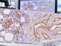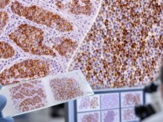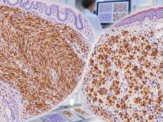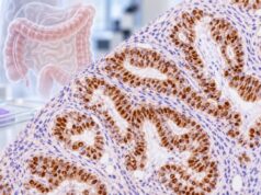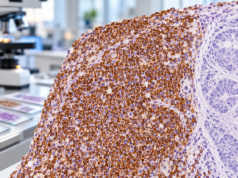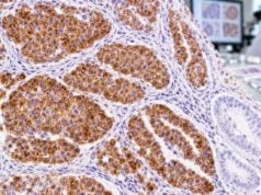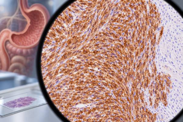
DOG1 immunohistochemistry is mainly used to help identify gastrointestinal stromal tumors (GISTs), especially when the tumor’s shape and location suggest GIST but other stains are unclear. DOG1 is a cell-membrane protein encoded by ANO1, and most GISTs show membranous and/or cytoplasmic staining for it. A positive DOG1 result strongly supports GIST in the right microscopic and clinical setting, but it does not prove GIST by itself because several non-GIST tumors can also express DOG1. Pathologists therefore interpret DOG1 with tumor morphology, KIT (CD117), CD34, smooth muscle and neural markers, and sometimes molecular testing for KIT, PDGFRA, SDH pathway abnormalities, or other drivers. The practical value of DOG1 is greatest in diagnostically difficult tumors, including a subset of KIT-negative GISTs. Its result can help classify a mass correctly, which matters because GIST treatment and risk assessment differ from those for leiomyosarcoma, schwannoma, and other mesenchymal tumors.
- DOG1-positive staining usually supports GIST, particularly in a compatible gastrointestinal spindle-cell or epithelioid tumor.
- DOG1 negativity does not completely rule out GIST, so the result must be interpreted with KIT, morphology, and molecular findings.
- Staining is typically membranous and cytoplasmic, and it may be diffuse or patchy depending on the tumor and assay.
- DOG1 is not fully specific for GIST; some salivary, smooth-muscle, epithelial, and neuroendocrine tumors can stain positive.
- A difficult or treatment-relevant case may need molecular testing even when DOG1 and KIT results are convincing.
Table of Contents
- What the DOG1 IHC Test Measures
- When DOG1 Is Used in GIST Diagnosis
- How to Interpret DOG1 Staining
- DOG1 With Other IHC Markers
- Pitfalls, False Results, and Non-GIST Positivity
- Molecular Testing, Prognosis, and Treatment Relevance
- Questions to Ask After a DOG1 Result
What the DOG1 IHC Test Measures
DOG1 IHC detects DOG1 protein in preserved tumor tissue. DOG1 stands for “discovered on GIST-1,” a name that reflects how the marker was identified rather than a claim that it appears only in GIST. The protein is also known as anoctamin-1 and TMEM16A and is encoded by the ANO1 gene. It functions as a calcium-activated chloride channel in several normal and neoplastic tissues.
The test is performed on a biopsy or surgical specimen that has been fixed, processed, and embedded in paraffin. A thin tissue section is exposed to an antibody that binds DOG1. A chemical detection system then creates visible staining where the antibody has attached. The pathologist evaluates where the staining occurs, how strong it is, how much of the tumor is involved, whether internal controls behave as expected, and whether the pattern fits the tumor’s appearance.
DOG1 is a tissue marker, not a blood tumor marker. There is no standard “normal range” such as a concentration in ng/mL. Reports instead use descriptive language such as positive, negative, focal, diffuse, weak, moderate, or strong. Some laboratories may use a semiquantitative score, but there is no single universal percentage cutoff that makes a tumor a GIST.
In a typical GIST, DOG1 staining is often membranous with cytoplasmic accentuation. Some tumors show strong circumferential membrane staining, while others show diffuse granular cytoplasmic reactivity. Pattern and intensity can vary with antibody clone, fixation, tumor subtype, and technical conditions. This is why a pathology report should be read as an integrated interpretation rather than as a stand-alone plus or minus sign.
GISTs arise from, or share differentiation with, the interstitial cells of Cajal and related precursors in the gastrointestinal tract. They most often occur in the stomach and small intestine, but they can also involve the esophagus, colon, rectum, or extra-gastrointestinal sites. Histologically, they may be spindle-cell, epithelioid, or mixed. Because several other tumors can mimic these appearances, immunohistochemistry is central to the diagnostic workup. A broader tumor immunohistochemistry panel may be used when lineage is not obvious.
When DOG1 Is Used in GIST Diagnosis
DOG1 is most useful when a pathologist is evaluating a mesenchymal tumor in or near the gastrointestinal tract and GIST is in the differential diagnosis. It is commonly ordered with KIT, also called CD117. Most conventional GISTs express one or both markers, so concordant positive staining provides strong support for GIST when the morphology also fits.
A classic example is a spindle-cell gastric mass. The microscopic differential may include GIST, leiomyoma, leiomyosarcoma, schwannoma, and other less common mesenchymal tumors. If the tumor is strongly DOG1-positive and KIT-positive, while desmin and S100-family markers do not support a competing lineage, GIST becomes much more likely. Conversely, strong diffuse desmin and smooth-muscle marker expression with absent DOG1 and KIT may favor a smooth-muscle neoplasm.
DOG1 is especially valuable in KIT-negative or weakly KIT-positive GIST. A small minority of GISTs lack convincing KIT immunoreactivity, including some tumors with PDGFRA mutations. In that setting, DOG1 can preserve diagnostic sensitivity. The complementary use of DOG1 and the CD117 (c-KIT) IHC test is one of the main reasons both stains are common in contemporary GIST workups.
Pathologists may also use DOG1 in metastatic disease. A liver, peritoneal, or soft-tissue metastasis composed of spindle or epithelioid cells may raise a broad differential. DOG1 positivity can point toward metastatic GIST, especially if the patient has a known gastrointestinal mass or prior GIST history. The final conclusion still depends on clinical imaging, morphology, other stains, and molecular data.
DOG1 can be informative on small biopsies, including endoscopic ultrasound-guided samples, but small samples introduce extra limitations. Tumor heterogeneity may mean the sampled fragment does not fully represent the lesion. Necrosis, crush artifact, poor fixation, or scant viable tumor can weaken staining. If the biopsy result conflicts with imaging and morphology, the pathology team may repeat staining, test an additional block, or recommend further tissue sampling.
The stain is not a screening test for people without a tumor. It is ordered after a mass or abnormal tissue has already been sampled. It also does not determine whether a GIST is benign or malignant. Modern GIST risk assessment relies more on tumor size, mitotic rate, anatomic site, rupture status, and selected molecular features than on DOG1 intensity.
How to Interpret DOG1 Staining
A positive DOG1 result means that tumor cells express detectable DOG1 protein. In the right context, this is strong evidence of GIST, but the amount of confidence depends on the entire case.
| Result pattern | Typical interpretation | Common next step |
|---|---|---|
| Strong, diffuse DOG1 positivity in a typical GI spindle-cell tumor | Strongly supports GIST | Correlate with KIT, morphology, risk features, and molecular testing when indicated |
| DOG1 positive, KIT negative | GIST remains possible and may be strongly favored | Review morphology and consider KIT/PDGFRA and other molecular studies |
| Focal or weak DOG1 staining | Supportive but less decisive | Check controls, other markers, tumor distribution, and competing diagnoses |
| DOG1 negative, KIT positive | GIST can still be diagnosed in an appropriate setting | Integrate morphology and molecular findings |
| DOG1 and KIT both negative | Conventional GIST is less likely but not excluded | Reassess differential; consider molecular and SDH-related testing |
The most important point is that positive does not equal malignant. DOG1 tells the pathologist about differentiation, not behavior. A small low-mitotic gastric GIST and a large metastatic GIST can both be strongly DOG1-positive. Likewise, staining intensity is not a validated substitute for recurrence risk models.
A negative result is also not absolute. Technical issues can reduce antigen detection, and biologically unusual GISTs may express little or no DOG1. The pathologist should first confirm that the slide contains viable tumor and that positive internal or external controls worked. If a tumor is morphologically convincing for GIST but DOG1 is negative, a CD34 IHC result, KIT stain, SDHB stain, and molecular profile may help resolve the case.
The percentage of stained cells may be reported, but there is no single internationally applied DOG1 percentage threshold comparable with ER scoring in breast cancer. The diagnosis depends on the pattern’s quality and specificity in the context of the tissue.
DOG1 With Other IHC Markers
DOG1 is strongest when used as part of a focused panel rather than alone. The choice of companion stains depends on the tumor’s location and appearance.
KIT (CD117) is the most common partner. KIT is a receptor tyrosine kinase and is positive in most GISTs. DOG1 can add sensitivity, particularly when KIT is absent or equivocal. Molecular KIT mutations and KIT protein staining are related concepts but are not identical: a tumor can express KIT protein without having a KIT mutation, and mutation status must be determined by molecular testing.
CD34 is positive in many GISTs, but it is less specific. It also labels vascular endothelium and several fibroblastic or other mesenchymal tumors. CD34 is therefore supportive rather than decisive.
Desmin and smooth muscle markers help separate GIST from true smooth-muscle tumors. Leiomyomas and leiomyosarcomas commonly express smooth muscle actin, desmin, and h-caldesmon. GIST can sometimes express smooth muscle actin, but diffuse strong desmin is uncommon in typical GIST. The desmin IHC test is therefore useful when the differential includes leiomyoma or leiomyosarcoma.
S100 and SOX10 are useful when schwannoma or another neural-crest tumor is considered. Gastrointestinal schwannomas usually have strong S100/SOX10 expression and lack the characteristic DOG1/KIT profile of GIST. An S100 IHC test can therefore be informative in a spindle-cell lesion.
SDHB immunohistochemistry is important in selected GISTs, especially tumors with unusual clinical features, gastric location in younger patients, multifocality, or morphology suggesting succinate dehydrogenase deficiency. Loss of SDHB staining identifies an SDH-deficient pathway and can trigger further genetic or molecular evaluation.
No panel is completely automatic. For example, DOG1 can be positive in some non-GIST tumors, smooth muscle actin can be positive in GIST, and CD34 has broad expression. The pathologist uses the combination of positive and negative findings to build a coherent diagnosis.
Pitfalls, False Results, and Non-GIST Positivity
The main interpretive pitfall is assuming that DOG1 is absolutely specific for GIST. It is not. DOG1 expression has been documented in a range of non-GIST neoplasms, including some salivary gland tumors, certain carcinomas, selected neuroendocrine neoplasms, and occasional mesenchymal tumors. Because of this overlap, DOG1 positivity outside the usual GIST setting should prompt caution rather than an automatic diagnosis.
Acinic cell carcinoma of salivary gland is a classic non-GIST example. DOG1 may stain the apical or membranous region in these tumors because normal salivary acinar cells can express DOG1. Secretory carcinoma and other salivary lesions can enter the differential, which is why tumor location and histology matter.
Technical factors also affect results. Common pre-analytic and analytic issues include:
- delayed fixation or under-fixation;
- prolonged fixation that alters antigen accessibility;
- decalcification in unusual specimens;
- necrotic or crushed tumor tissue;
- antibody-clone and platform differences;
- weak background staining that can be mistaken for true tumor reactivity;
- staining of non-neoplastic structures that is misread as tumor positivity.
Another pitfall is using DOG1 to infer a specific mutation. DOG1 IHC does not identify KIT, PDGFRA, NF1, BRAF, SDH, or other molecular alterations. It also cannot predict sensitivity to every tyrosine kinase inhibitor. Molecular testing answers those questions more directly.
Sampling is especially important in tumors with mixed morphology or after treatment. A treated GIST may show fibrosis, hyalinization, necrosis, or reduced viable tumor. Marker expression can become patchy in these settings. A negative stain on a tiny treated biopsy should be weighed against the prior diagnosis and treatment history.
When a tumor’s morphology, DOG1, and KIT results disagree, a second pathology review can be valuable. GIST mimics can have major treatment implications, so difficult cases often benefit from review by a pathologist with gastrointestinal or soft-tissue tumor expertise.
Molecular Testing, Prognosis, and Treatment Relevance
DOG1 helps establish diagnosis, while molecular testing helps explain tumor biology and guide therapy. Most adult GISTs have activating mutations in KIT or PDGFRA. Other subsets are driven by SDH deficiency, NF1 alterations, BRAF mutations, NTRK fusions, or other less common mechanisms.
Molecular testing is particularly important when systemic therapy is being considered. Different mutations can predict sensitivity or resistance to specific tyrosine kinase inhibitors. For example, some KIT-mutated GISTs respond well to imatinib, while PDGFRA D842V-mutated tumors are resistant to standard-dose imatinib and require a different targeted strategy. DOG1 positivity alone cannot make that distinction.
Molecular studies can also resolve diagnostic uncertainty. A DOG1-positive, KIT-negative gastrointestinal mesenchymal tumor with a pathogenic KIT or PDGFRA alteration strongly supports GIST. Conversely, absence of a typical GIST driver may prompt evaluation for an SDH-deficient or other wild-type GIST pathway rather than immediate rejection of the diagnosis.
Risk of recurrence after complete resection is assessed using clinicopathologic factors, especially tumor size, mitotic rate, site, and rupture. Gastric GISTs generally behave less aggressively than similarly sized and mitotically active small-bowel GISTs. DOG1 intensity is not part of standard risk models.
Treatment decisions may include surgery, observation after resection, adjuvant tyrosine kinase inhibitor therapy, or systemic treatment for unresectable or metastatic disease. The pathology report provides the diagnosis, size, mitotic count, margins where relevant, and sometimes risk category, while the molecular report adds predictive information. A DOG1 result is therefore one piece of a larger clinical decision pathway, not a stand-alone treatment test.
Questions to Ask After a DOG1 Result
If your pathology report mentions DOG1, the most useful next questions focus on the final diagnosis and what evidence supports it.
You may want to ask:
- Does the overall pathology favor GIST, or is DOG1 only one possibility-supporting finding?
- Was KIT/CD117 also tested, and was it positive or negative?
- Were smooth-muscle, neural, or epithelial markers used to exclude common mimics?
- Is the tumor considered conventional GIST, SDH-deficient GIST, or another subtype?
- Has molecular testing for KIT and PDGFRA been performed or recommended?
- If the tumor was removed, what are its size, mitotic rate, site, rupture status, and recurrence-risk category?
- Would expert gastrointestinal or sarcoma pathology review change management?
A report that says “DOG1 positive” should not be interpreted in isolation. The decisive line is usually the final diagnosis or comment section, where the pathologist explains how morphology and the full test panel fit together. If treatment depends on mutation status, ask specifically whether molecular testing has been completed because immunohistochemistry cannot substitute for it.
A pathology diagnosis may also distinguish diagnostic certainty from treatment planning. DOG1 can make GIST very likely, yet oncologists may still need the exact molecular driver before choosing a tyrosine kinase inhibitor. KIT exon 11, KIT exon 9, PDGFRA, and other molecular subgroups can differ in drug sensitivity, while some GISTs lack the common KIT or PDGFRA alterations. That is why a strongly DOG1-positive tumor can still move on to sequencing. The stain identifies a phenotype; molecular testing can define the biologic subtype that matters for therapy.
When comparing reports over time, differences in DOG1 intensity should not be treated as a reliable measure of response or recurrence. Imaging, tumor size and site, pathology from any new specimen, and molecular context are more informative. If a recurrent mass is biopsied, DOG1 may help confirm that it represents GIST, but treatment response is not graded by how dark the stain appears.
References
- Pathologic diagnosis and molecular features of gastrointestinal stromal tumors: a mini-review 2024 (Review)
- Gastrointestinal Stromal Tumors: Variants and Some Pitfalls That They Create. 2024 (Review)
- Gastrointestinal stromal tumours: ESMO-EURACAN-GENTURIS Clinical Practice Guidelines for diagnosis, treatment and follow-up 2022 (Guideline)
- [Clinical practice guideline for the pathological diagnosis of gastrointestinal stromal tumor (2022 version)]. 2022 (Guideline)
- Old and new immunohistochemical markers for the diagnosis of gastrointestinal stromal tumors 2011 (Review)
Disclaimer
DOG1 IHC is a pathology test that must be interpreted with the tumor’s microscopic appearance, clinical setting, other stains, and sometimes molecular testing. A positive or negative DOG1 result alone cannot diagnose or exclude GIST, determine cancer stage, or select treatment. Discuss the complete pathology and molecular report with your treating clinician or pathologist.