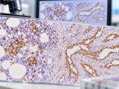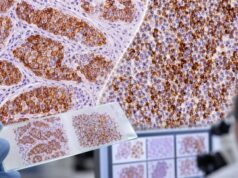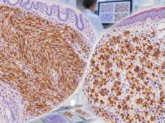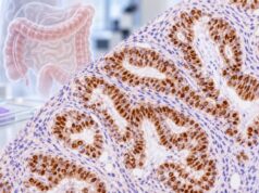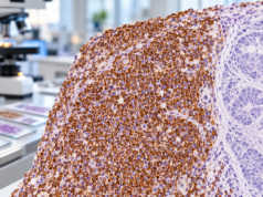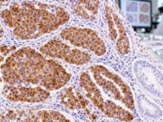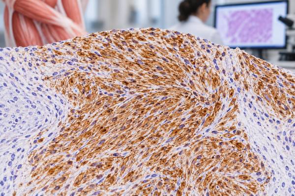
Desmin immunohistochemistry (IHC) is a tissue stain that helps pathologists identify muscle differentiation in a tumor. Desmin is an intermediate-filament protein normally found in skeletal, cardiac, and smooth muscle cells. When tumor cells show convincing cytoplasmic desmin staining, the finding can support a muscle-lineage tumor such as leiomyoma, leiomyosarcoma, or rhabdomyosarcoma. The result is not a stand-alone cancer diagnosis, however. Several non-muscle tumors can express desmin, and some true muscle tumors show weak, focal, or absent staining. Pathologists therefore interpret desmin alongside the tumor’s microscopic appearance, location, patient age, and a panel of other markers. In smooth-muscle lesions, these may include smooth muscle actin and h-caldesmon. In suspected skeletal-muscle tumors, nuclear markers such as myogenin and MyoD1 are especially important. The key question is not simply whether desmin is “positive,” but whether its pattern fits the proposed tumor type and the rest of the diagnostic evidence.
- Desmin-positive staining supports muscle differentiation, but it does not by itself prove that a tumor is a sarcoma or identify the exact subtype.
- True desmin staining is cytoplasmic, often diffuse in smooth-muscle tumors and frequently present in rhabdomyosarcoma.
- Smooth-muscle tumors need a marker panel, commonly including smooth muscle actin and h-caldesmon in addition to desmin.
- Rhabdomyosarcoma requires stronger lineage confirmation, usually with nuclear myogenin and MyoD1 rather than desmin alone.
- A negative desmin stain does not completely exclude muscle differentiation, especially in poorly differentiated, treated, or limited tumor samples.
Table of Contents
- What Desmin IHC Measures
- When Desmin Is Used in Tumor Diagnosis
- How Positive Desmin Staining Is Interpreted
- Desmin in Smooth-Muscle Tumors
- Desmin in Rhabdomyosarcoma
- Differential Diagnosis and Pitfalls
- What to Ask After a Desmin Result
What Desmin IHC Measures
Desmin IHC detects desmin protein in cells preserved in a tissue specimen. Desmin belongs to the intermediate-filament family, a group of structural proteins that helps cells maintain their shape and organize their internal architecture. In normal tissue, desmin is strongly associated with muscle cells. That makes it useful as a lineage marker when a pathologist is trying to determine what type of tissue a tumor resembles.
The test is performed on a biopsy, resection, or other pathology specimen. A thin tissue section is placed on a slide, treated with an antibody directed against desmin, and developed so antibody binding becomes visible under a microscope. The pathologist then evaluates which cells stain, where the staining is located inside the cells, and how extensive and intense it is.
Desmin is different from a blood tumor marker. There is no serum desmin “normal range,” and patients do not need to fast or adjust medications for the stain itself. Tissue quality matters far more than patient preparation. Fixation, decalcification, necrosis, prior treatment, and the amount of viable tumor can all affect how confidently the stain can be read.
A positive result means that detectable desmin protein is present in the stained cells. It does not automatically mean cancer. Normal muscle stains for desmin, and benign tumors such as leiomyomas can be strongly positive. The stain also does not tell the pathologist whether a tumor is low grade or high grade. Those questions depend on morphology, mitotic activity, necrosis, invasion, molecular findings, and other features specific to the tumor being considered.
For that reason, desmin is usually one component of a broader tumor IHC panel. Its greatest value comes from how it changes the probability of competing diagnoses when interpreted with the rest of the case.
When Desmin Is Used in Tumor Diagnosis
Pathologists most often order desmin when the microscopic appearance raises the possibility of smooth-muscle or skeletal-muscle differentiation. This can happen in a mass made of spindle cells, epithelioid cells, or small round cells. The same basic shapes occur in many unrelated tumors, so morphology usually narrows the possibilities but cannot always settle the diagnosis alone.
Common situations include:
- a uterine or soft-tissue spindle-cell tumor in which leiomyoma or leiomyosarcoma is considered;
- a pediatric or young-adult small round cell tumor that could represent rhabdomyosarcoma;
- an abdominal, retroperitoneal, or gastrointestinal mass where smooth-muscle tumor and gastrointestinal stromal tumor are competing possibilities;
- a metastatic tumor whose lineage is unclear on routine staining;
- a poorly differentiated sarcoma that needs an initial lineage-directed panel.
Desmin can be particularly useful in a spindle-cell lesion because it may move the workup toward myogenic differentiation. Yet it should not end the workup. Gastrointestinal stromal tumors, for example, are usually evaluated with markers such as DOG1 and CD117/c-KIT. A tumor can occasionally express markers outside its expected lineage, so a coherent pattern is more persuasive than a single positive stain.
The clinical setting also changes how a result is interpreted. Strong desmin staining in a classic uterine smooth-muscle lesion carries a different diagnostic meaning from focal staining in an undifferentiated retroperitoneal sarcoma. Patient age is especially important for small round cell tumors, while anatomic site can sharply alter the differential diagnosis for spindle-cell tumors.
Desmin is also useful when confirming, rather than discovering, a suspected diagnosis. Once routine histology strongly suggests a muscle tumor, IHC can document the lineage and help exclude close mimics. In modern soft-tissue pathology, this phenotypic information may then be integrated with molecular testing when a specific fusion, amplification, mutation, or other genomic event defines the entity more reliably than morphology and IHC alone.
How Positive Desmin Staining Is Interpreted
Desmin positivity is judged primarily by cytoplasmic staining in the tumor cells. Pathologists assess the pattern rather than applying one universal percentage cutoff. A report may describe staining as diffuse, patchy, focal, strong, moderate, or weak. Those terms describe what is seen but do not have one standardized numeric meaning across all tumor types and laboratories.
A useful interpretation asks four questions: Are the correct tumor cells staining? Is the staining in the expected cellular location? How much of the tumor is positive? Does the pattern fit the proposed diagnosis?
| Finding | Typical meaning | Important caution |
|---|---|---|
| Strong, diffuse cytoplasmic staining | Strong evidence of myogenic differentiation in a compatible tumor | Still not specific for one smooth- or skeletal-muscle tumor type |
| Focal cytoplasmic staining | May support muscle differentiation, depending on morphology and other markers | Focal positivity occurs in several non-myogenic tumors |
| No convincing tumor staining | Provides less support for a muscle-lineage diagnosis | Does not absolutely exclude a true muscle tumor |
| Only background or entrapped muscle stains | Shows the antibody can recognize desmin in the specimen | Must not be mistaken for positive tumor cells |
Internal controls can be valuable. Normal vascular or muscular structures in the specimen may stain and demonstrate that the assay worked in that tissue. The pathologist still has to distinguish those normal elements from the neoplastic cells being evaluated.
There is no universally meaningful “desmin score” comparable with receptor scoring systems used for some breast-cancer biomarkers. The diagnostic weight depends on the candidate tumor. For example, diffuse desmin plus convincing h-caldesmon and smooth muscle actin expression in a morphologically smooth-muscle tumor is more informative than isolated weak desmin staining in a poorly differentiated mass.
Negative results also require context. A tumor can lose lineage-associated proteins as it becomes less differentiated. Small samples can miss a positive area, and preanalytic problems can reduce staining. If morphology remains strongly suspicious, the pathologist may repeat the stain on another block, use additional antibodies, or move to molecular testing rather than treating one negative result as final.
Desmin in Smooth-Muscle Tumors
Desmin is commonly expressed in smooth-muscle tumors, including leiomyomas and leiomyosarcomas. In a classic lesion, tumor cells often show broad cytoplasmic positivity. However, desmin cannot by itself distinguish a benign leiomyoma from a malignant leiomyosarcoma. Both may be positive because both show smooth-muscle differentiation.
The benign-versus-malignant distinction relies heavily on tumor-specific morphologic criteria. Depending on the site, pathologists evaluate cytologic atypia, mitotic activity, tumor-cell necrosis, growth pattern, borders, and clinical context. In uterine smooth-muscle tumors, the combination and degree of these findings are central to classification. IHC can assist difficult cases, but it does not replace those criteria.
A typical smooth-muscle panel may include desmin, smooth muscle actin, and h-caldesmon. Smooth muscle actin is sensitive for contractile differentiation but is also found in myofibroblasts and other cell types. Desmin is more lineage-associated but is not entirely specific. H-caldesmon can add evidence for mature smooth-muscle differentiation. The pattern across these markers is more useful than any single result.
This distinction matters in the gastrointestinal tract and abdomen. True smooth-muscle tumors can resemble GIST, while GIST has its own characteristic marker profile. A pathologist comparing these possibilities may use DOG1, CD117, desmin, smooth muscle actin, and other markers selected for the site and morphology. Strong desmin with absent GIST markers may favor smooth-muscle differentiation, but unusual cases can still require molecular confirmation.
Smooth-muscle differentiation can also occur in tumors outside the uterus, including lesions arising in soft tissue, blood vessels, skin, and visceral organs. Marker sensitivity can vary by tumor location and degree of differentiation. A leiomyosarcoma that is pleomorphic or poorly differentiated may show less uniform marker expression than a well-differentiated example.
Importantly, classification of a smooth-muscle sarcoma is only one part of patient management. Grade, size, anatomic site, depth, margins, stage, and metastatic status usually carry more direct prognostic importance than whether desmin staining is strong or weak. Desmin establishes lineage; it is not a stand-alone prognosis test.
Desmin in Rhabdomyosarcoma
Rhabdomyosarcoma is a malignant tumor showing skeletal-muscle differentiation. Desmin is often positive and can be a sensitive clue, but the diagnosis generally requires more specific evidence. Nuclear myogenic transcription factors are particularly helpful because they more directly support skeletal-muscle lineage.
Two commonly used markers are myogenin and MyoD1. Unlike desmin’s cytoplasmic pattern, myogenin and MyoD1 are interpreted as nuclear stains. Convincing nuclear expression in the correct cells can strongly support rhabdomyoblastic differentiation when the morphology is compatible.
The intensity and distribution of myogenic markers can vary among rhabdomyosarcoma subtypes. Modern classification also uses molecular findings. For example, some tumors are defined or risk-stratified by characteristic gene fusions, while other subtypes have different molecular alterations. This means an IHC pattern that establishes skeletal-muscle differentiation may be the beginning of classification rather than the final step.
Desmin is especially useful in the initial evaluation of a small round blue cell tumor, a broad morphologic category that includes several pediatric and adult malignancies. A panel may be needed to separate rhabdomyosarcoma from lymphoma, Ewing sarcoma, poorly differentiated carcinoma, neuroendocrine tumors, and other round-cell sarcomas. Because some of these tumors can show unexpected focal staining, no single antibody should be allowed to override an otherwise contradictory panel.
A positive desmin stain also should not be interpreted as proof of rhabdomyosarcoma in an adult spindle-cell lesion. Smooth-muscle tumors are often desmin-positive, and other neoplasms can be positive as well. The presence of rhabdomyoblast-like cells, cross-striations in rare cases, nuclear myogenin/MyoD1, age, site, and molecular features provide the needed context.
For patients, the practical point is that “desmin positive” and “rhabdomyosarcoma” are not synonyms. The pathology report’s final diagnosis, subtype, molecular findings, and stage are much more important for treatment planning than the isolated stain result.
Differential Diagnosis and Pitfalls
Desmin has broad value precisely because muscle differentiation appears in several tumor families, but this breadth creates pitfalls. Positive staining can occur in tumors that are not conventional smooth- or skeletal-muscle sarcomas. Desmoplastic small round cell tumor, for example, often shows a distinctive perinuclear or dot-like desmin pattern rather than the diffuse pattern expected in many muscle tumors. Some myofibroblastic tumors, glomus tumors, and other mesenchymal neoplasms may also express desmin variably.
Other practical pitfalls include:
- Entrapped normal muscle. Skeletal or smooth muscle at the edge of a tumor can stain strongly and mimic tumor-cell positivity if cell identity is not carefully traced.
- Focal expression. A small positive population should be weighed less heavily than a coherent, diffuse myogenic profile when the differential is broad.
- Poorly differentiated tumors. Loss of mature lineage markers can produce false reassurance from a negative stain.
- Limited biopsy material. A core biopsy may sample only one component of a heterogeneous tumor.
- Technical effects. Delayed fixation, overfixation, decalcification, necrosis, or treatment effect can weaken antigen detection.
- Panel mismatch. Ordering only broad markers can create an ambiguous result that more specific lineage or molecular tests could resolve.
Desmin can also overlap with lesions in which epithelial, neural, vascular, or GIST markers are more decisive. Depending on morphology, a pathologist may add broad cytokeratins, S100 or SOX10, CD34, ERG, DOG1, CD117, ALK, or other markers. The goal is not to collect as many stains as possible; it is to use the smallest informative panel that separates the realistic diagnoses.
A further limitation is that IHC reflects protein expression, not necessarily the genetic alteration that defines a modern sarcoma entity. Newer entity-specific antibodies can sometimes act as reliable surrogates for molecular changes, but confirmatory sequencing, fluorescence in situ hybridization, or RNA-based fusion testing may still be appropriate. The need depends on tumor type, specimen quality, clinical consequences, and how specific the IHC pattern is.
The most reliable interpretation therefore combines morphology, a targeted panel, and molecular testing when indicated. A surprising desmin result should prompt reconciliation of all three rather than an immediate change in diagnosis.
What to Ask After a Desmin Result
A pathology report may list desmin among many stains without explaining the reasoning behind each one. The final diagnostic line and comment section usually matter more than the stain list itself. If the result affects a suspected sarcoma diagnosis, useful questions include:
- Was desmin staining diffuse or only focal, and was it clearly present in the tumor cells?
- Does the overall pattern support smooth-muscle differentiation, skeletal-muscle differentiation, or another tumor type?
- Which companion markers were tested, such as smooth muscle actin, h-caldesmon, myogenin, or MyoD1?
- Were GIST, nerve-sheath, epithelial, vascular, or other spindle-cell tumors excluded with appropriate markers?
- Is molecular testing needed to confirm the diagnosis or assign a modern sarcoma subtype?
- If the tumor is malignant, what are its grade, size, margin status, and stage?
- Would review by a pathologist who specializes in bone and soft-tissue tumors be useful for an unusual or high-impact diagnosis?
The answer may differ between a straightforward excision and a tiny biopsy. On a small sample, the pathologist may be able to establish a likely lineage but defer precise classification until more tissue or molecular data are available. That is not necessarily a problem with the stain; it reflects the limits of the specimen.
Patients should also distinguish diagnostic markers from treatment biomarkers. Desmin generally helps identify what the tumor is. It is not, by itself, a target that determines standard drug selection, and its percentage of staining is not usually followed over time like a serum tumor marker. Treatment decisions for sarcoma depend on the final tumor type, molecular profile when relevant, extent of disease, surgical options, and multidisciplinary assessment.
If a report uses terms such as “consistent with,” “supports,” or “favors,” those words often show that the diagnosis is based on combined evidence rather than one definitive stain. Asking how desmin fits with the morphology and the rest of the panel usually gives a clearer answer than asking whether the stain is simply positive or negative.
References
- Replacing Molecular Testing With Next-Generation Immunohistochemistry: I Can Diagnose That Soft Tissue Tumor With a Single Antibody! 2026 (Review)
- Diagnosis and treatment of primary soft tissue sarcomas: comprehensive review 2025 (Review)
- Update on immunohistochemistry in bone and soft tissue tumors: Cost-effectively replacing molecular testing with immunohistochemistry 2024 (Review)
- Rhabdomyosarcoma: Updates on classification and the necessity of molecular testing beyond immunohistochemistry 2024 (Review)
- Molecular-Based Immunohistochemical Algorithm for Uterine Leiomyosarcoma Diagnosis 2023
- Immunohistochemistry of soft tissue tumours – review with emphasis on 10 markers 2014 (Review)
Disclaimer
Desmin IHC must be interpreted by a qualified pathologist in the context of the full specimen, other stains, and clinical findings. A positive or negative desmin result alone cannot establish a specific sarcoma diagnosis or determine treatment. Discuss the final pathology interpretation and any need for molecular testing or specialist review with your care team.