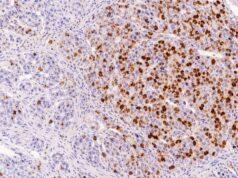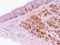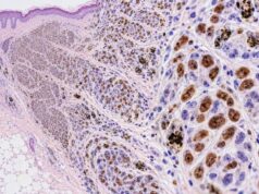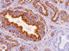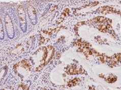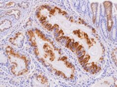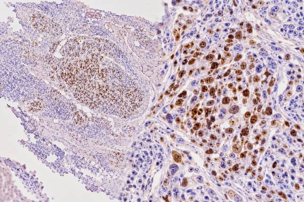
MyoD1 immunohistochemistry (IHC) is a tissue stain used to identify skeletal-muscle differentiation in tumors. MyoD1 is a myogenic regulatory transcription factor, so the diagnostically meaningful pattern is nuclear staining in the tumor cells. In surgical pathology, MyoD1 is most often used when rhabdomyosarcoma is suspected, especially in a small round cell, spindle cell, or poorly differentiated sarcoma. A positive MyoD1 result can strongly support rhabdomyoblastic differentiation when the morphology and the rest of the immunohistochemical panel agree. However, MyoD1 positivity is not the same as a diagnosis of rhabdomyosarcoma, and it is not a substitute for molecular classification. Myogenin and desmin are common companion stains, while FOXO1 fusion testing, MYOD1 mutation testing, or broader sequencing may be required in selected subtypes. An important distinction is that routine MyoD1 IHC measures protein expression; it does not determine whether a tumor carries a pathogenic MYOD1 gene mutation.
- True MyoD1 positivity is nuclear: cytoplasmic or background staining should not be interpreted as evidence of skeletal-muscle differentiation.
- MyoD1 is a key rhabdomyosarcoma marker: it is usually interpreted with myogenin, desmin, morphology, and clinical context rather than alone.
- A positive stain does not identify the RMS molecular subtype: FOXO1 fusion status and other molecular alterations require dedicated testing.
- MyoD1 IHC is not a MYOD1 mutation test: diffuse protein expression can occur without the MYOD1 p.L122R mutation.
- No special patient preparation is needed: the stain is performed on biopsy or resection tissue in the pathology laboratory.
Table of Contents
- What MyoD1 IHC Detects
- What Positive MyoD1 Staining Means
- MyoD1 in Rhabdomyosarcoma
- MyoD1 Versus Myogenin and Desmin
- MYOD1 Mutation and Spindle/Sclerosing RMS
- Limitations and Diagnostic Pitfalls
- How to Read a MyoD1 Pathology Result
What MyoD1 IHC Detects
MyoD1 IHC detects the MyoD1 protein, a nuclear transcription factor that helps commit cells to the skeletal-muscle lineage. It belongs to the group of myogenic regulatory factors that coordinate muscle differentiation. In pathology, that biology is useful because rhabdomyosarcomas recapitulate parts of skeletal-muscle development even when the tumor cells look primitive and do not form obvious muscle fibers.
The stain is applied to formalin-fixed, paraffin-embedded tissue from a biopsy or surgical specimen. When the assay is working correctly, the pathologist evaluates whether the nuclei of the tumor cells are stained, how strong the staining is, and what proportion of the neoplastic population is positive. Mature or regenerating skeletal muscle in the specimen can also show myogenic-marker expression and must not be mistaken for tumor.
MyoD1 is particularly useful in lesions that are morphologically ambiguous. Rhabdomyosarcoma can resemble Ewing sarcoma, lymphoma, neuroblastoma, poorly differentiated carcinoma, melanoma, synovial sarcoma, malignant peripheral nerve sheath tumor, and other round cell or spindle cell neoplasms. The exact differential diagnosis depends heavily on the patient’s age, anatomic site, and microscopic pattern.
A typical diagnostic panel may include MyoD1 together with myogenin and desmin, then add lineage-specific markers for the most plausible alternatives. For example, a round cell tumor may require cytokeratins, CD45, SOX10/S100, NKX2.2, or other stains depending on the morphology. Immunohistochemistry narrows the possibilities, while molecular testing can establish genetically defined entities.
The key feature is cellular localization. Because MyoD1 functions as a transcription factor, convincing nuclear labeling is the relevant finding. Cytoplasmic staining, diffuse haze, or staining confined to nonneoplastic muscle is not equivalent to tumor-cell nuclear positivity. This distinction matters because interpretation errors can create a false impression of myogenic differentiation.
What Positive MyoD1 Staining Means
A positive MyoD1 stain means that the evaluated cells are expressing a skeletal-muscle-lineage transcription factor; in the right setting, this strongly supports rhabdomyoblastic differentiation. The result is most persuasive when nuclear staining occurs in morphologically abnormal tumor cells and is supported by another myogenic marker.
Pathology reports may describe the stain as focal, patchy, multifocal, or diffuse, and as weak, moderate, or strong. There is no universal percentage cutoff that converts MyoD1 positivity into a diagnosis. Instead, the amount of staining is interpreted in context. A tiny focus of nuclear labeling in an otherwise discordant tumor warrants more caution than broad nuclear staining in a lesion with classic rhabdomyoblasts and supportive myogenin/desmin expression.
Positive MyoD1 can answer an important lineage question without answering every classification question. It may tell the pathologist that a sarcoma is showing skeletal-muscle differentiation, yet it does not determine whether the tumor is embryonal, alveolar, spindle cell/sclerosing, pleomorphic, or one of the emerging molecularly defined rhabdomyosarcoma groups. Modern RMS classification increasingly integrates histology with genomic findings.
Similarly, the stain does not provide a direct prognosis. Prognosis in rhabdomyosarcoma depends on multiple variables, including molecular subtype, primary site, tumor size, stage or metastatic status, age, resectability, and treatment response. Some molecular groups with strong MyoD1 expression have aggressive behavior, but the protein stain itself is not the causal prognostic assay.
A negative stain also needs context. Some bona fide rhabdomyosarcomas can show limited or absent MyoD1 immunoreactivity, particularly in small biopsies or technically compromised tissue. For that reason, pathologists commonly use more than one skeletal-muscle marker. A negative MyoD1 result should not automatically exclude RMS when morphology and other data remain suspicious.
MyoD1 in Rhabdomyosarcoma
MyoD1 is one of the core immunohistochemical markers used to establish skeletal-muscle differentiation in rhabdomyosarcoma. Its role is especially important because RMS spans a broad morphologic spectrum and can occur in children, adolescents, and adults.
Embryonal rhabdomyosarcoma often contains a mixture of primitive round or spindle cells and more differentiated rhabdomyoblasts. MyoD1 may be expressed in a substantial subset of tumor cells, while myogenin can be more variable and sometimes focal. The combination of nuclear myogenic markers with desmin is often diagnostically useful, but classification also depends on architecture, site, age, and molecular findings when indicated.
Alveolar rhabdomyosarcoma has historically been defined by an alveolar pattern, but contemporary risk assessment places greater emphasis on PAX3::FOXO1 or PAX7::FOXO1 fusion status. Myogenin is often strongly and diffusely expressed in fusion-positive tumors. MyoD1 may also be positive, but neither stain can establish the FOXO1 fusion. Dedicated FISH, RT-PCR, or next-generation sequencing is needed when fusion status is clinically or diagnostically important.
Spindle cell/sclerosing rhabdomyosarcoma is particularly relevant to MyoD1 because this histologic category contains distinct molecular groups. Some tumors in infants carry VGLL2-related fusions, while another clinically important group harbors a MYOD1 hotspot mutation. These molecular categories can have very different clinical behavior despite overlapping morphology. MyoD1 IHC can support the myogenic nature of the tumor but cannot reliably sort those genetic groups by itself.
Pleomorphic rhabdomyosarcoma, seen mainly in adults, can resemble other high-grade pleomorphic sarcomas. Demonstrating nuclear MyoD1 and/or myogenin in the malignant cells can be valuable, especially when desmin alone would be too nonspecific. Still, a broad differential should be considered because rhabdomyoblastic differentiation can occasionally arise as a component of other malignancies rather than representing a conventional primary RMS.
The increasingly molecular framework for RMS means that diagnosis has two layers: first, establish that the tumor truly shows rhabdomyoblastic differentiation; second, determine whether the morphology and genomic findings place it into a recognized subtype. MyoD1 contributes strongly to the first layer but only indirectly to the second.
MyoD1 Versus Myogenin and Desmin
MyoD1, myogenin, and desmin are complementary rather than interchangeable markers. Understanding what each stain represents helps explain why pathologists frequently order them together.
| Marker | Typical staining pattern | Main diagnostic contribution |
|---|---|---|
| MyoD1 | Nuclear | Supports skeletal-muscle lineage commitment and rhabdomyoblastic differentiation |
| Myogenin | Nuclear | Strong evidence of skeletal-muscle differentiation; often especially diffuse in fusion-positive alveolar RMS |
| Desmin | Cytoplasmic | Sensitive muscle/intermediate-filament marker, but less specific for skeletal-muscle differentiation |
Desmin is useful because many RMSs express it strongly, but desmin is not restricted to rhabdomyosarcoma. Smooth-muscle tumors, some myofibroblastic lesions, and other neoplasms can be desmin positive. MyoD1 and myogenin are more lineage-specific because they are nuclear myogenic regulatory proteins.
MyoD1 and myogenin can also show different distributions within the same tumor. Older immunohistochemical studies found that both are useful for RMS diagnosis but may highlight different stages of differentiation. Primitive tumor cells may show MyoD1, while more differentiated rhabdomyoblasts can demonstrate stronger myogenin. In practice, this means one nuclear marker can rescue a case in which the other is focal or weak.
The staining pattern can sometimes offer a clue to subtype but should not be overinterpreted. Diffuse myogenin is associated with many alveolar/fusion-positive RMSs, while spindle cell/sclerosing tumors—especially MYOD1-mutant examples—may show strong diffuse MyoD1 with more patchy myogenin. These are tendencies, not mutation or fusion tests.
A panel also protects against technical problems. If desmin is positive but both nuclear markers are convincingly negative, the pathologist may reconsider whether the tumor is truly rhabdomyoblastic or whether antigen preservation is poor. If MyoD1 is positive but the staining is cytoplasmic only, the result should not be counted as a true nuclear myogenic signal.
MYOD1 Mutation and Spindle/Sclerosing RMS
One of the most important interpretation points is that MyoD1 IHC and MYOD1 mutation analysis are different tests. The IHC stain detects protein. Molecular testing examines the gene for pathogenic sequence changes, most notably the p.L122R hotspot mutation described in a subset of spindle cell/sclerosing RMS.
MYOD1 p.L122R–mutant RMS is now recognized as a biologically aggressive group. Studies have shown high rates of local and distant recurrence and poor outcomes across pediatric and adult age groups. The mutation can coexist with other alterations, including PIK3CA mutations in some tumors. International molecular-testing recommendations have therefore highlighted MYOD1 p.L122R assessment as clinically important for risk stratification in appropriate spindle/sclerosing RMS cases.
Many MYOD1-mutant tumors show diffuse, strong MyoD1 protein expression, but that correlation is not sufficiently specific to infer the genotype from IHC. Wild-type RMS can also express MyoD1 strongly, and some rhabdomyosarcomas without the hotspot mutation have substantial staining. Conversely, technical factors and tumor heterogeneity can alter the observed IHC pattern.
This distinction has practical consequences. A report saying “MyoD1: diffuse positive” does not mean “MYOD1 mutation detected.” If mutation status matters, the pathology report should contain a separate molecular result, such as targeted sequencing, an NGS panel, or another validated DNA-based assay. The same principle applies to FOXO1: immunophenotype may suggest a category, but fusion status requires a molecular test.
Spindle/sclerosing RMS also illustrates why molecular classification is increasingly necessary beyond immunohistochemistry. Histologically similar tumors may fall into infantile fusion-driven, MYOD1-mutant, or other genetic groups with different implications. Recent classification reviews emphasize integrating morphology, IHC, and molecular findings rather than trying to force every tumor into a subtype by stain pattern alone.
Limitations and Diagnostic Pitfalls
MyoD1 is highly useful, but interpretation errors can occur if nuclear localization, morphology, and the broader tumor context are ignored. The most important pitfalls are practical rather than theoretical.
- Cytoplasmic staining is not the diagnostic pattern. MyoD1 is a nuclear transcription factor; nonspecific cytoplasmic signal should not be scored as true positivity.
- Normal skeletal muscle can be present in the biopsy. Entrapped or regenerating muscle fibers must be distinguished from malignant cells.
- Small biopsies can under-sample the tumor. A negative result in scant tissue may not represent the whole lesion.
- Rhabdomyoblastic differentiation can occur in other tumors. A malignant neoplasm with focal skeletal-muscle differentiation is not automatically conventional rhabdomyosarcoma.
- Desmin positivity is not enough. Desmin has a broader distribution and needs nuclear myogenic markers or other supporting evidence.
- Protein expression does not define genotype. MyoD1 IHC cannot diagnose a MYOD1 mutation, and it cannot establish FOXO1 fusion status.
- Technical factors matter. Fixation, decalcification, crush artifact, necrosis, and assay performance can reduce or distort staining.
The differential diagnosis also changes with anatomic site. A sinonasal spindle or round cell tumor, for example, may require consideration of biphenotypic sinonasal sarcoma or other genetically defined neoplasms; a bone or head-and-neck lesion can raise additional fusion-associated entities. Some unusual tumors can express epithelial or neural markers together with myogenic proteins, so a seemingly contradictory IHC panel may be a signal to pursue molecular testing rather than dismiss one result.
Age is similarly important. A spindle cell RMS in an infant has a different molecular differential from a sclerosing tumor in an adolescent or adult. The stain does not encode that clinical information. Pathologists combine the IHC result with age, site, imaging, histology, and molecular findings to avoid overcalling or undercalling RMS.
How to Read a MyoD1 Pathology Result
The most useful way to read a MyoD1 result is to ask which cells are positive, whether the staining is nuclear, and what the companion studies show. “MyoD1 positive” is a piece of diagnostic evidence, not a complete tumor diagnosis.
A practical reading sequence is:
- Confirm the specimen and final diagnosis. MyoD1 may be one stain within a much larger sarcoma workup.
- Look for nuclear staining. Nuclear labeling of the tumor cells is the relevant positive pattern.
- Check how extensive the staining is. Focal versus diffuse expression can help the pathologist judge how strongly the stain supports myogenic differentiation, although no universal percentage alone defines RMS.
- Review myogenin and desmin. Concordant nuclear myogenin and cytoplasmic desmin usually strengthen the interpretation.
- Look for molecular results. FOXO1 fusion testing, MYOD1 sequencing, or other fusion/NGS studies may be reported separately.
- Separate protein staining from mutation status. A diffuse MyoD1 stain does not prove the p.L122R alteration.
If a report says that MyoD1 and myogenin are positive and the morphology is consistent with rhabdomyosarcoma, the stains are supporting skeletal-muscle differentiation. If it additionally says “FOXO1 rearrangement detected,” that is a separate molecular result that can refine subtype and risk assessment. Likewise, “MYOD1 p.L122R mutation detected” is a genomic finding with implications that cannot be inferred from IHC alone.
Patients should also avoid treating stain intensity as a direct measure of how aggressive the cancer is. A tumor with strong MyoD1 expression is not necessarily more advanced than one with focal expression. Clinical stage, molecular subtype, primary site, metastatic disease, and treatment response are much more relevant to prognosis.
The central takeaway is that MyoD1 IHC is a powerful marker of rhabdomyoblastic differentiation when true nuclear staining is present, but it works best as part of an integrated sarcoma diagnosis. Its greatest value is establishing lineage; modern molecular testing then helps define the biologic subtype that may carry prognostic or therapeutic significance.
Slide quality and cellular localization deserve special attention with MyoD1. The diagnostically meaningful result is nuclear staining in the neoplastic cells. Cytoplasmic background or nonspecific signal should not be counted as equivalent evidence of skeletal-muscle differentiation. Adjacent normal or regenerating skeletal muscle can also be positive and may help confirm that the assay worked, but those cells must be distinguished from the tumor on the matching routine section.
A negative result on a tiny, crushed, or poorly fixed biopsy may be less informative than a negative result in a well-preserved resection. If the morphology still raises strong concern for rhabdomyosarcoma, repeating the stain on another block or adding myogenin, desmin, and molecular testing may be appropriate. The reverse is also true: focal MyoD1 positivity in an otherwise discordant spindle-cell tumor should not override morphology and the rest of the panel. These quality-control steps are particularly important because the diagnosis can lead to disease-specific chemotherapy and risk stratification.
References
- Rhabdomyosarcoma Classification: Refining Our Understanding 2025 (Review)
- Rhabdomyosarcoma: Updates on classification and the necessity of molecular testing beyond immunohistochemistry 2024 (Review)
- Biological Role and Clinical Implications of MYOD1L122R Mutation in Rhabdomyosarcoma 2023 (Review)
- Molecular testing of rhabdomyosarcoma in clinical trials to improve risk stratification and outcome: A consensus view from European paediatric Soft tissue sarcoma Study Group, Children’s Oncology Group and Cooperative Weichteilsarkom-Studiengruppe 2022 (Position Statement)
- Molecular profile of head and neck rhabdomyosarcomas: A systematic review and meta-analysis 2022 (Systematic Review)
- Are myogenin and myoD1 expression specific for rhabdomyosarcoma? A study of 150 cases, with emphasis on spindle cell mimics 2001
Disclaimer
This article is for general educational purposes and does not interpret an individual pathology specimen. MyoD1 staining must be assessed by a qualified pathologist together with tissue morphology, companion immunostains, clinical findings, and any indicated molecular studies. Treatment and prognosis for rhabdomyosarcoma should be discussed with the patient’s oncology and pathology teams using the complete diagnosis and stage.