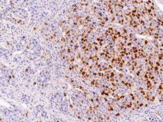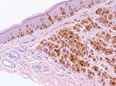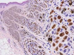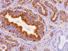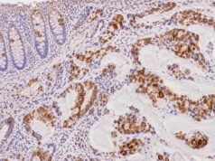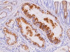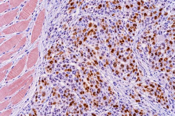
Myogenin immunohistochemistry (IHC) is a nuclear tissue stain used to demonstrate skeletal-muscle differentiation in tumors. The myogenin protein, encoded by the MYOG gene, is a myogenic regulatory transcription factor expressed as cells progress toward skeletal-muscle differentiation. In diagnostic pathology, convincing nuclear myogenin staining in tumor cells is strong evidence of rhabdomyoblastic differentiation and is especially useful when rhabdomyosarcoma is being considered. Myogenin is commonly interpreted with MyoD1 and desmin because no single stain should be used in isolation. The extent of staining can vary across rhabdomyosarcoma subtypes: fusion-positive alveolar rhabdomyosarcoma often shows widespread strong nuclear expression, whereas embryonal and some spindle cell/sclerosing tumors may be more focal. Even so, the stain cannot determine FOXO1 fusion status, a MYOD1 mutation, or the complete molecular subtype. A positive result must be matched to the abnormal cells on the biopsy and integrated with morphology, age, tumor site, other immunostains, and any required molecular studies.
- Myogenin is interpreted as a nuclear stain: nuclear labeling of the tumor cells is the meaningful positive pattern.
- Positive myogenin strongly supports skeletal-muscle differentiation: it is a core marker in the diagnosis of rhabdomyosarcoma.
- Diffuse staining can suggest, but not prove, a fusion-positive alveolar phenotype: FOXO1 status requires molecular testing.
- Focal positivity can still be important: embryonal and spindle cell/sclerosing rhabdomyosarcomas may stain less extensively.
- No special preparation is required from the patient: myogenin IHC is performed by the pathology laboratory on tissue from a biopsy or resection.
Table of Contents
- What Myogenin IHC Detects
- What Positive Myogenin Staining Means
- Myogenin in Rhabdomyosarcoma Subtypes
- Myogenin Versus MyoD1 and Desmin
- Myogenin and Molecular Testing
- Limitations and Diagnostic Pitfalls
- How to Read a Myogenin Pathology Result
What Myogenin IHC Detects
Myogenin IHC detects a transcription factor involved in skeletal-muscle differentiation, so the diagnostically relevant signal is located in cell nuclei. The protein participates in the developmental program that converts myogenic precursor cells into differentiated skeletal muscle. Rhabdomyosarcoma can activate this program even when the tumor is composed almost entirely of primitive cells.
That feature makes myogenin valuable on small biopsies. A pediatric or young-adult soft tissue mass may consist of sheets of undifferentiated round cells with few obvious clues to lineage. In other cases, a spindle cell or sclerosing tumor may resemble a non-rhabdomyoblastic sarcoma. Nuclear myogenin provides direct evidence that at least part of the neoplasm is following a skeletal-muscle differentiation pathway.
The stain is usually performed on formalin-fixed, paraffin-embedded tissue. The pathologist first identifies the neoplastic cells on the routine hematoxylin-and-eosin section and then determines whether those same cells contain nuclear labeling. Percentage, intensity, and distribution matter, but they are interpreted qualitatively rather than through one universal diagnostic cutoff.
Normal or regenerating skeletal muscle can also express myogenic regulatory proteins. Therefore, a positive cell at the edge of a biopsy is not automatically malignant. In a tumor infiltrating muscle, the pathologist must distinguish entrapped myocytes or regenerative nuclei from the atypical tumor population. The architecture and morphology on corresponding sections are essential for that distinction.
Myogenin is particularly useful because its nuclear pattern is generally more specific for skeletal-muscle differentiation than broad muscle markers such as desmin. However, “skeletal-muscle differentiation” is a biologic finding, not always a complete disease name. Some mixed or unusual neoplasms can contain a rhabdomyoblastic component, so the final diagnosis depends on the whole tumor rather than the stain alone.
What Positive Myogenin Staining Means
A positive myogenin result means that the cells being evaluated express a nuclear myogenic differentiation program. When those cells are the malignant population and the morphology is compatible, the result strongly supports rhabdomyosarcoma or another tumor with genuine rhabdomyoblastic differentiation.
Pathology reports may describe myogenin as rare, focal, patchy, multifocal, or diffuse. They may also provide an estimated percentage of positive tumor cells. The significance of those words depends on the diagnostic setting. Diffuse strong nuclear staining in a classic small round cell tumor is highly supportive, while rare positive nuclei in a complex spindle cell lesion require careful confirmation that the labeled cells are actually neoplastic.
A common misunderstanding is that more staining automatically means a more aggressive cancer. That is not how myogenin is used. The amount of expression can correlate with subtype—particularly the tendency for fusion-positive alveolar RMS to show extensive myogenin—but stain percentage is not a substitute for stage, molecular risk group, primary site, tumor size, or response to treatment.
A negative myogenin stain is also not an absolute exclusion test. Some rhabdomyosarcomas, especially particular spindle cell/sclerosing or poorly sampled tumors, can have limited expression. Technical issues such as fixation, decalcification, necrosis, or very scant viable tissue can further reduce staining. Pathologists therefore often pair myogenin with MyoD1 and desmin rather than relying on one negative result.
The strongest interpretation occurs when the stain fits a coherent pattern: malignant cells with rhabdomyoblastic morphology show nuclear myogenin, another myogenic marker is supportive, and markers for major mimics are absent or otherwise explained. If the immunophenotype is contradictory, molecular testing may be needed to resolve the diagnosis.
Myogenin in Rhabdomyosarcoma Subtypes
Myogenin is expressed across rhabdomyosarcoma, but the distribution of positive cells often differs by subtype and molecular background. This is one reason the stain contributes both to diagnosis and, more cautiously, to classification clues.
Embryonal rhabdomyosarcoma can show heterogeneous myogenin expression. Some tumors contain only scattered positive nuclei, while others have broader labeling. This patchiness fits the variable degree of differentiation often visible microscopically, from primitive cells to cells with more abundant eosinophilic cytoplasm. A focal result can still be diagnostically meaningful if the positive cells clearly belong to the tumor.
Alveolar rhabdomyosarcoma classically shows stronger and more extensive myogenin expression. Older large immunohistochemical studies demonstrated a marked difference between alveolar and embryonal patterns, with many alveolar tumors labeling in a high proportion of nuclei. Modern classification, however, has shifted emphasis from alveolar architecture alone to PAX3::FOXO1 or PAX7::FOXO1 fusion status because fusion status better captures biologic risk.
That change is crucial when interpreting a stain. Diffuse myogenin may raise suspicion for a fusion-positive RMS, but it cannot prove the fusion. Conversely, a tumor should not be denied appropriate molecular testing because its myogenin is only focal. The genomic result is a separate piece of evidence.
Spindle cell/sclerosing rhabdomyosarcoma can show a different relationship between myogenin and MyoD1. MYOD1-mutant tumors are often strongly MyoD1 positive but can show patchier myogenin. Infantile spindle cell tumors with fusion-driven biology constitute another group. Because these categories differ in behavior, modern reviews emphasize molecular evaluation beyond IHC when the morphology fits this spectrum.
Pleomorphic rhabdomyosarcoma, usually encountered in adults, may have highly atypical cells in which skeletal-muscle differentiation is difficult to recognize. Nuclear myogenin can help demonstrate rhabdomyoblastic differentiation, but the pathologist must still exclude other pleomorphic sarcomas and malignancies that have acquired a heterologous rhabdomyoblastic component.
Myogenin Versus MyoD1 and Desmin
Myogenin, MyoD1, and desmin answer related but different questions, so their combined pattern is more informative than any single result. Myogenin and MyoD1 are nuclear transcription factors; desmin is a cytoplasmic intermediate-filament protein.
| Marker | Positive pattern | How it helps |
|---|---|---|
| Myogenin | Nuclear | Strong evidence of skeletal-muscle differentiation; often extensive in fusion-positive alveolar RMS |
| MyoD1 | Nuclear | Supports myogenic lineage and may be especially prominent in some spindle/sclerosing RMS |
| Desmin | Cytoplasmic | Sensitive muscle-lineage support but less specific because other muscle and nonmuscle tumors can express it |
Myogenin is often easier to interpret than a nonspecific cytoplasmic stain because true positive nuclei can be mapped directly to malignant cells. It is also more specific for rhabdomyoblastic differentiation than desmin. Desmin remains useful because it is highly sensitive in many RMSs and can highlight tumor distribution, but desmin positivity alone does not establish skeletal-muscle differentiation.
MyoD1 and myogenin can complement each other when the tumor is heterogeneous. Experimental and diagnostic studies suggest that the two proteins can be expressed at different phases of myogenic differentiation. A primitive population may be more conspicuous with MyoD1, whereas cells showing greater morphologic differentiation can have stronger myogenin. This is not an absolute rule, but it explains why a dual nuclear-marker approach improves confidence.
When the stains disagree, the answer is not simply to choose the positive one. The pathologist checks whether the positive staining is in the tumor, whether internal and external controls worked, and whether the morphology supports the claimed lineage. A broad panel may then exclude lymphoma, Ewing sarcoma, neuroblastoma, carcinoma, melanoma, or other sarcoma types.
Myogenin and Molecular Testing
Myogenin IHC establishes a phenotype; molecular testing can establish a genotype. The two forms of evidence increasingly work together in modern rhabdomyosarcoma diagnosis.
For tumors in the alveolar/fusion-positive spectrum, the central molecular question is often whether a PAX3::FOXO1 or PAX7::FOXO1 fusion is present. Fusion-positive RMS has distinct biologic and prognostic significance, and current cooperative-group risk stratification uses fusion status rather than relying only on microscopic alveolar pattern. A myogenin stain cannot identify the fusion partner or confirm a rearrangement.
For spindle cell/sclerosing RMS, the relevant molecular questions are different. A subset contains the MYOD1 p.L122R mutation and behaves aggressively, while infantile cases can harbor VGLL2-related fusions. Other newly characterized RMS groups are defined by different recurrent genomic changes. These distinctions cannot be made reliably from myogenin intensity.
There is also an important terminology point: myogenin IHC is not a MYOG sequencing test. Routine pathology generally uses the antibody because the protein is a useful differentiation marker. A report of “myogenin positive” does not imply that the MYOG gene is mutated, rearranged, or amplified. In most cases, the stain is being used for lineage rather than to identify a genetic abnormality in MYOG itself.
Molecular testing may use FISH, RT-PCR, targeted DNA/RNA sequencing, or broader NGS depending on the institution and the suspected entity. The choice depends on morphology, age, site, available tissue, and whether the result affects classification or clinical risk. Recent pathology reviews specifically stress the necessity of molecular methods beyond immunohistochemistry for many RMS cases.
Limitations and Diagnostic Pitfalls
The most important myogenin pitfall is assuming that any positive nucleus in a biopsy proves conventional rhabdomyosarcoma. The pathologist must establish that the stained cells belong to the malignant population and that the tumor as a whole fits the diagnosis.
Important limitations include:
- Reactive or regenerating skeletal muscle can be positive. Entrapped muscle at the edge of a mass is not evidence that the tumor itself is rhabdomyosarcoma.
- Focal positivity is context dependent. Rare positive tumor nuclei may be meaningful, but rare nonneoplastic nuclei can also occur.
- Rhabdomyoblastic differentiation is not exclusive to conventional RMS. Other malignancies can contain heterologous skeletal-muscle differentiation.
- Negative staining does not absolutely exclude RMS. Sampling, subtype, and technical factors can reduce sensitivity.
- Staining extent is not a fusion assay. Diffuse myogenin may suggest an alveolar/fusion-positive phenotype but cannot establish FOXO1 rearrangement.
- Fixation and tissue processing matter. Poor preservation, decalcification, necrosis, or crush artifact can impair nuclear staining.
- Small biopsies may miss heterogeneity. A limited sample may not represent all morphologic or immunophenotypic areas of the tumor.
Specific diagnostic mimics depend on the tumor’s appearance. In a small round cell neoplasm, the panel may need to evaluate hematolymphoid, neural, epithelial, melanocytic, and other sarcoma lineages. In a spindle cell lesion, smooth-muscle and nerve-sheath tumors may enter the differential. Some genetically defined sarcomas can have mixed immunophenotypes, so unexpected marker combinations should prompt correlation rather than automatic exclusion.
The distinction between tumor cells and background cells is particularly important in scant needle biopsies. A few myogenin-positive nuclei adjacent to necrosis or normal muscle should be interpreted on the corresponding morphology. Proper controls, an adequate viable sample, and complementary stains reduce the risk of false-positive or false-negative conclusions.
How to Read a Myogenin Pathology Result
Read a myogenin result by focusing on nuclear staining in the malignant cells and then placing that finding within the complete sarcoma workup. The line “myogenin: positive” does not by itself specify the exact RMS subtype or prognosis.
A practical sequence is:
- Identify the final diagnosis and specimen type. A small biopsy may carry more sampling limitations than a resection.
- Confirm nuclear localization. The diagnostically meaningful signal is nuclear labeling of the target tumor cells.
- Check the extent of positivity. Focal and diffuse patterns can both be significant, but they have different levels of support and different subtype associations.
- Review MyoD1 and desmin. A coherent myogenic panel strengthens the interpretation.
- Look for the molecular classification. FOXO1 fusion status, MYOD1 mutation status, or other sequencing results may appear elsewhere in the report.
- Do not equate staining intensity with cancer stage. Stage and prognosis depend on clinical and molecular factors, not simply the darkness of the stain.
If the report describes diffuse nuclear myogenin together with compatible morphology and a confirmed PAX::FOXO1 fusion, the fusion result—not the IHC alone—establishes the molecularly defined category. If myogenin is focal but MyoD1 and desmin are positive in a spindle/sclerosing tumor, the pathologist may pursue MYOD1 mutation or fusion testing rather than dismiss rhabdomyosarcoma because the myogenin percentage is low.
For patients, the most useful question is therefore not “Is myogenin good or bad?” but “What diagnosis did the whole pathology workup establish, and were molecular tests needed to define the subtype?” Myogenin has enormous diagnostic value because it can show skeletal-muscle differentiation in otherwise primitive tumors. Its role is to help identify lineage accurately, while morphology and molecular findings complete the classification.
The key takeaway is that positive nuclear myogenin is one of the strongest routine IHC findings supporting rhabdomyoblastic differentiation, but the result must be integrated rather than read in isolation. Its pattern can guide the next diagnostic step, yet modern RMS classification depends increasingly on genetic data that the stain itself cannot provide.
The distribution of myogenin staining can provide useful context without functioning as a stand-alone subtype test. Some fusion-positive rhabdomyosarcomas show widespread nuclear labeling, while embryonal, spindle-cell, or sclerosing tumors may be more variable. A patchy result can still be diagnostically meaningful if the positive nuclei clearly belong to the malignant population and MyoD1 or desmin supports the same line of differentiation.
Technical and sampling factors can also produce misleading extremes. Crush artifact, necrosis, decalcification, scant viable tumor, or prolonged fixation may reduce staining. At the other end, normal or regenerating skeletal muscle near a tumor can contain myogenic regulatory proteins and should not be mistaken for a positive sarcoma. Correlation with the routine slide and appropriate controls is therefore essential.
Once rhabdomyoblastic differentiation is established, molecular classification may become more important than adding additional muscle markers. Testing for PAX3::FOXO1 or PAX7::FOXO1 fusions, MYOD1 alterations, and other relevant genetic events can separate biologically distinct groups that have different risk implications. Myogenin cannot substitute for those assays. Its greatest strength is answering the lineage question early and efficiently so that the pathologist can choose the most informative confirmatory and molecular tests.
References
- Rhabdomyosarcoma Classification: Refining Our Understanding 2025 (Review)
- Rhabdomyosarcoma: Updates on classification and the necessity of molecular testing beyond immunohistochemistry 2024 (Review)
- An Integrative Morphologic and Molecular Approach for Diagnosis and Subclassification of Rhabdomyosarcoma 2022 (Review)
- Molecular testing of rhabdomyosarcoma in clinical trials to improve risk stratification and outcome: A consensus view from European paediatric Soft tissue sarcoma Study Group, Children’s Oncology Group and Cooperative Weichteilsarkom-Studiengruppe 2022 (Position Statement)
- Myogenin is a specific marker for rhabdomyosarcoma: an immunohistochemical study in paraffin-embedded tissues 2000
Disclaimer
This article provides general educational information and cannot interpret an individual tumor or pathology report. Myogenin IHC should be evaluated by a qualified pathologist together with morphology, other immunostains, clinical findings, and molecular tests when indicated. Decisions about rhabdomyosarcoma treatment and prognosis require the complete pathologic diagnosis, molecular subtype, stage, and multidisciplinary oncology assessment.