
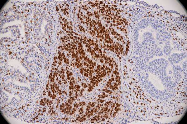
The p53 immunohistochemistry (IHC) test is a tissue stain that shows the pattern of p53 protein expression in tumor cells. Pathologists often use that pattern as a practical surrogate for abnormalities in TP53, a tumor-suppressor gene altered in many cancers. The key is not simply whether p53 is “positive.” A normal, or wild-type, pattern usually contains a variable mixture of weak, moderate, and stronger nuclear staining with some tumor nuclei unstained. In contrast, certain abnormal patterns—such as diffuse strong nuclear overexpression, complete absence of tumor-cell staining with intact internal controls, or convincing cytoplasmic staining—can support a mutation-type pattern. The exact meaning depends on the cancer type, antibody method, and morphology. p53 IHC is especially important in settings such as endometrial carcinoma classification, but it is useful in many other tumors as well. Because staining and DNA results are highly concordant rather than identical, difficult or high-stakes cases may still require molecular testing.
- What p53 IHC measures: it shows how p53 protein is distributed in tumor cells; it does not directly sequence the TP53 gene.
- What “abnormal” usually means: diffuse overexpression, complete null staining with valid controls, or abnormal cytoplasmic localization may indicate a TP53-altered pattern.
- What a wild-type pattern looks like: variable nuclear staining with a mixture of positive and negative tumor-cell nuclei usually supports normal regulation.
- Why context matters: valid interpretation differs by tumor type, specimen quality, antibody, and the specific diagnostic question.
- When sequencing may help: molecular testing can resolve discordant, subclonal, equivocal, or clinically important results.
Table of Contents
- What the p53 IHC Test Detects
- Normal and Abnormal p53 Staining Patterns
- How p53 IHC Relates to TP53 Mutations
- How p53 Helps Classify Cancer
- How to Read a p53 Pathology Result
- Limitations and Diagnostic Pitfalls
- What Happens After an Abnormal p53 Result
What the p53 IHC Test Detects
p53 is a protein encoded by the TP53 gene. It helps cells respond to DNA damage and other stresses by regulating cell-cycle arrest, DNA repair, senescence, or cell death. Because this pathway limits the survival of genetically damaged cells, TP53 is commonly described as a tumor-suppressor gene.
IHC does not measure TP53 DNA directly. Instead, an antibody binds p53 protein in a thin section of tumor tissue, and the pathologist studies the location and distribution of the staining. This distinction explains why “p53 positive” is often an incomplete description. Some normal-functioning tissue contains p53-positive nuclei, and several very different abnormal patterns can correspond to TP53 alterations.
The most informative feature is usually the pattern across the tumor. A wild-type pattern is heterogeneous: some nuclei stain, others do not, and intensity varies. Mutation-type patterns can be strikingly uniform or strikingly absent. The stain therefore behaves differently from a blood tumor marker that yields a numeric concentration.
Technical controls are essential, particularly when the tumor appears completely negative. Normal stromal cells, lymphocytes, or other non-neoplastic cells on the same slide should retain appropriate staining. If every cell on the slide is negative, the pathologist cannot confidently call the tumor “null”; the assay may simply have failed.
p53 IHC is often ordered as part of a larger classification panel rather than as a standalone test. The pathologist may pair it with lineage markers, mismatch-repair proteins, hormone receptors, or other tumor-specific markers depending on the organ and suspected diagnosis. A broader tumor IHC panel can be particularly useful when the tissue is poorly differentiated or the primary site is uncertain.
Normal and Abnormal p53 Staining Patterns
Pathologists commonly separate p53 staining into wild-type and abnormal, or mutation-type, patterns. The exact criteria are validated for particular disease settings, so the descriptions below are principles rather than universal numeric cutoffs.
A wild-type pattern shows variable nuclear staining. Different tumor nuclei have different intensities, and a meaningful fraction are unstained. This patchwork pattern reflects normal regulation in which p53 protein levels rise and fall rather than accumulating uniformly.
A classic overexpression pattern shows very strong nuclear staining in a large, often near-uniform population of tumor cells. Many missense TP53 mutations create an abnormal protein that persists in the nucleus, producing this “block-like” appearance. A common mistake is to assume that any high percentage of positive nuclei equals a mutation. The distribution and intensity matter, and thresholds can differ by tumor type and protocol.
A null pattern is the opposite: tumor-cell nuclei show complete absence of p53 staining. This can correspond to mutations that prevent stable protein production. The diagnosis is valid only when non-neoplastic internal controls are positive. Without those controls, a null-looking slide may represent technical failure.
A third abnormal pattern is cytoplasmic staining, in which convincing tumor-cell cytoplasmic labeling occurs with an inappropriate nuclear pattern. Certain TP53 alterations can disturb protein localization. Because background cytoplasmic staining can occur, this pattern requires careful morphologic correlation and assay experience.
Some tumors show subclonal abnormality. One geographic area may have a mutation-type pattern while another retains wild-type staining. This can reflect tumor evolution and heterogeneity rather than a simple technical problem. In endometrial carcinoma, recognizing subclonal p53 abnormality can be relevant to molecular classification, and the pathologist may correlate it with other markers or sequencing.
The central lesson is that p53 IHC is pattern-based. “Positive” and “negative” are not sufficient categories, because both strong positivity and complete absence can be abnormal.
How p53 IHC Relates to TP53 Mutations
p53 IHC is widely used because the staining pattern often predicts TP53 mutation status, but it is a surrogate, not a perfect replacement for sequencing. Studies in several cancers have reported high agreement when standardized pattern-based interpretation is used.
For example, a colorectal cancer study published in 2025 reported about 91% concordance between p53 IHC pattern and TP53 sequencing. In the PORTEC-3 endometrial cancer cohort, a pattern-based algorithm also showed high agreement and strong diagnostic performance. These results support routine IHC as a fast and practical first-line tool while also showing why occasional discordance is expected.
Different mutations affect the protein differently. Many missense variants stabilize abnormal p53 protein and can produce diffuse nuclear overexpression. Nonsense, frameshift, splice, or deletion-type alterations may produce no stable protein, resulting in a null pattern. Other mutations can produce less typical patterns. This biology explains why a single percentage cutoff cannot capture every TP53 alteration.
Not every abnormal-looking stain is caused by a pathogenic TP53 mutation, and not every TP53 mutation generates an unmistakable abnormal pattern. Preanalytic factors, antibody performance, tumor heterogeneity, low tumor cellularity, and interpretation all affect concordance. Sequencing also has its own limitations, including tumor purity, assay coverage, variant classification, and detection thresholds.
Molecular confirmation is most useful when the answer will change classification or management and the IHC is equivocal, technically questionable, subclonal, or inconsistent with morphology. It may also be needed in research protocols or tumor types where TP53 mutation status itself has a defined clinical role.
Patients should therefore avoid translating “p53 abnormal” into “a TP53 mutation was genetically proven” unless molecular testing actually demonstrated the variant. The IHC result is often highly informative, but the report should make clear whether it is a protein-expression interpretation or a DNA result.
How p53 Helps Classify Cancer
p53 contributes to tumor classification in several organs because TP53 alterations cluster in specific tumor types and molecular subgroups. Its value is strongest when the staining pattern is interpreted with morphology and other biomarkers.
Endometrial carcinoma is a major example. Modern molecular classification divides endometrial cancers into clinically meaningful groups using POLE mutation status, mismatch-repair status, p53 status, and other features. An abnormal p53 pattern can serve as a surrogate for the copy-number-high/TP53-abnormal molecular group when the case is evaluated using an established algorithm. This information can affect risk assessment and, together with stage and histology, treatment planning.
In high-grade serous tubo-ovarian carcinoma, TP53 alteration is extremely common, so a mutation-type p53 pattern is expected in a morphologically typical tumor. A wild-type pattern in an alleged high-grade serous carcinoma can prompt the pathologist to reconsider the diagnosis, specimen quality, or staining interpretation rather than simply ignoring the discrepancy. Other Müllerian markers, including PAX8 IHC, help establish lineage but answer a different question.
In colorectal cancer, p53 can support characterization of tumor biology and research classification, although it is not a standalone routine subtype test in the same way as mismatch-repair proteins. In lung cancer, abnormal p53 expression may correlate with TP53 mutation and has been studied for prognosis, but it does not replace the core histologic, genomic, and predictive biomarker workup.
p53 staining also appears in many sarcomas, hematologic malignancies, brain tumors, and other cancers. The significance varies substantially. The same abnormal pattern can be diagnostically useful in one tumor type, prognostically associated in another, and of limited routine consequence in a third.
This is why a pathology report may emphasize p53 strongly in one cancer but mention it only as supportive evidence in another. The marker has broad biology, but clinical interpretation is disease-specific.
How to Read a p53 Pathology Result
A useful p53 report should describe the pattern rather than relying on the word “positive.” Common phrases include wild-type pattern, abnormal overexpression, null pattern, cytoplasmic pattern, or subclonal abnormal pattern. Some laboratories also provide a percentage or qualitative intensity.
A practical way to read the result is to ask four questions:
- Were the malignant cells the population being scored? Normal cells can stain and should not be confused with tumor.
- Was the pattern heterogeneous or mutation-type? This usually matters more than the raw percentage alone.
- Were internal controls valid? They are essential before interpreting complete absence as abnormal.
- What role does p53 play in this tumor type? Diagnostic, molecular-classification, prognostic, and research uses are not interchangeable.
A report stating “p53 shows diffuse strong nuclear overexpression, consistent with an abnormal/mutation-type pattern” generally means the protein pattern strongly suggests altered TP53 biology. It does not identify the exact DNA variant. A report stating “wild-type staining pattern” means the stain did not show a recognized mutation-type pattern; it does not guarantee that every part of TP53 is genetically normal.
The phrase “p53 positive” is especially easy to misread. Normal wild-type tumors can contain many positive nuclei. Conversely, a truly abnormal null pattern has no tumor-cell nuclear staining. For this marker, positive is not automatically abnormal and negative is not automatically normal.
If the report uses a percentage, interpret it only within the stated pattern and disease context. Unlike the Ki-67 proliferation index, p53 is not primarily intended to quantify the fraction of actively cycling tumor cells. It reflects a tumor-suppressor protein pathway and is interpreted by distribution as well as extent.
Limitations and Diagnostic Pitfalls
The most common conceptual error is treating p53 IHC as a simple positive-versus-negative test. The marker has multiple abnormal patterns, and a heterogeneous positive pattern can be normal. Accurate interpretation requires pattern recognition.
Preanalytic variables can alter staining. Cold ischemia, formalin fixation time, decalcification, tissue age, necrosis, and small biopsy size may affect antigen preservation. A tumor that appears null in poorly preserved tissue requires caution, especially if internal controls are absent or weak.
Antibody clone, staining platform, antigen retrieval, and laboratory calibration also matter. External quality-assurance studies continue to show that p53 assays can be over- or under-sensitive. A laboratory should validate its own protocol against expected tissue patterns rather than copy a threshold developed for another platform.
Tumor heterogeneity is another source of discordance. A biopsy may sample a wild-type-looking region while a resection contains a subclone with abnormal staining, or vice versa. Treatment can select subclones and further change the composition of recurrent disease. When classification has major implications, reviewing the entire available tumor can help.
A further pitfall is assuming that IHC can identify the exact mutation. Overexpression may suggest a stabilizing missense mutation, while null staining may suggest a truncating or deletion-type event, but these are not one-to-one rules. Sequencing is required to name the variant and assess its genomic context.
Disease-specific rules must also be respected. A p53 pattern that is central to endometrial molecular classification does not automatically carry the same prognostic or therapeutic meaning in breast, lung, colon, or soft-tissue tumors. Published algorithms and current classification systems should guide use.
Finally, p53 should not be used as a broad cancer-screening test. It is performed on tissue that has already been sampled because there is a diagnostic reason. An abnormal stain says something about that tissue; it does not measure whole-body cancer risk.
What Happens After an Abnormal p53 Result
The next step depends on why p53 was ordered. In an endometrial carcinoma, the pathologist may integrate p53 with mismatch-repair IHC, POLE mutation testing when indicated, histologic subtype, grade, and stage to assign a molecular category. In an ovarian or tubal tumor, an unexpected p53 pattern may trigger re-review of morphology or additional lineage markers.
When the IHC and expected diagnosis agree, no separate TP53 sequencing may be needed for routine classification. When the result is unusual, molecular testing can clarify whether a TP53 alteration is present. Repeat IHC on a better-preserved block or a larger specimen may also be appropriate if technical quality is questionable.
For the patient, the most useful questions are: Is the p53 pattern wild-type or abnormal? If abnormal, is it overexpression, null, cytoplasmic, or subclonal? Does this result change the formal tumor subtype or risk group? Was TP53 sequencing performed, or is the mutation only inferred from IHC? Are there other biomarkers that carry more direct treatment implications?
An abnormal p53 pattern should not be interpreted as a measure of how much cancer is present. It does not determine tumor stage, and it does not by itself prove that a cancer will behave aggressively. Prognostic associations depend on the specific disease and must be integrated with established clinical factors.
The result is most powerful when it is used for the question it was designed to answer. Pattern-based p53 IHC can provide rapid, inexpensive information about TP53 pathway status and can improve tumor classification. When the pattern, morphology, and molecular findings disagree, the discrepancy is a reason for further evaluation rather than for choosing one test automatically over the others.
One additional practical point is that the same patient can have more than one p53 pattern if separate tumor components or separate tumors are sampled. For example, a mixed endometrial carcinoma can contain an area with wild-type staining and another with a clearly abnormal pattern. The pathologist must determine whether this represents true subclonal evolution, distinct histologic components, or a technical difference between blocks. Mapping the stain back to the morphology is often more informative than averaging all positive nuclei into one percentage.
When p53 is being used in a formal classification algorithm, the report may also document how conflicting biomarkers were resolved. A tumor can occasionally show more than one molecular-classifier feature. In such cases, disease-specific hierarchy rules and, when appropriate, sequencing help assign the final group. This is another reason patients should rely on the integrated diagnosis rather than trying to translate one p53 line into a molecular subtype on their own.
References
- The pattern-based interpretation of p53 immunohistochemical expression as a surrogate marker for TP53 mutations in colorectal cancer 2025
- p53 immunohistochemistry in endometrial cancer: clinical and molecular correlates in the PORTEC-3 trial 2022
- p53 immunohistochemistry staining is a rapid screening method for TP53 mutation in myeloid malignancies suitable for integration into routine diagnostic laboratory practice 2025
- Immunohistochemistry of p53 surrogates TP53 mutation as an accurate predictor for early-relapse of surgically resected stage I-III lung adenocarcinoma 2024
- IHC p53 Performance in NordiQC: Recalibration of IHC Assays is Needed 2026
Disclaimer
p53 IHC is a pathology tool interpreted with tumor morphology, technical controls, other biomarkers, and disease-specific criteria. An abnormal p53 staining pattern may strongly suggest altered TP53 biology but does not identify a genetic variant unless molecular testing is performed. Treatment and prognosis should be based on the complete diagnosis rather than p53 staining alone.





