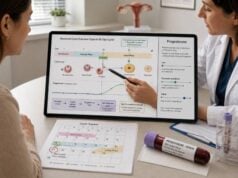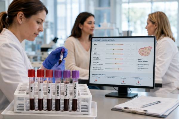
A female infertility blood test panel measures hormones involved in follicle development, ovulation, ovarian reserve, and pituitary signaling. It commonly includes follicle-stimulating hormone (FSH), luteinizing hormone (LH), anti-Müllerian hormone (AMH), estradiol, and prolactin. These tests can help identify reduced ovarian response, irregular or absent ovulation, polycystic ovary syndrome, primary ovarian insufficiency, or a pituitary-related problem.
The panel cannot measure egg quality, prove that ovulation has occurred, show whether the fallopian tubes are open, or predict natural pregnancy with certainty. Age, menstrual history, ultrasound findings, semen analysis, and tubal and uterine assessment often matter more than any single hormone value. Timing is also important: FSH and estradiol are usually assessed early in the menstrual cycle, while AMH can generally be measured on any day. Results should therefore be interpreted as a coordinated pattern within a complete fertility evaluation.
- FSH and estradiol are usually checked on cycle days 2–4 to assess early-follicular ovarian signaling.
- AMH estimates the remaining pool of recruitable follicles and expected response to ovarian stimulation, not egg quality or the chance of natural conception by itself.
- High FSH with low estradiol may suggest reduced ovarian function, while high estradiol can mask an otherwise elevated FSH result.
- LH is most useful when interpreted with cycle timing and symptoms; the LH/FSH ratio alone does not diagnose PCOS.
- Prolactin is most helpful with irregular periods, absent periods, nipple discharge, or suspected pituitary disease and is not required routinely for every infertility evaluation.
- Normal blood tests do not rule out endometriosis, tubal blockage, uterine conditions, male-factor infertility, or age-related decline in egg quality.
Table of Contents
- What the Female Infertility Panel Evaluates
- Who May Need Hormone Testing
- Timing and Preparation
- What Each Hormone Result Means
- Common Result Patterns
- Ranges and Important Limitations
- Other Tests That Complete the Evaluation
- Next Steps After the Results
What the Female Infertility Panel Evaluates
The ovaries, hypothalamus, and pituitary gland communicate through the hypothalamic-pituitary-ovarian axis. At the start of a menstrual cycle, the pituitary releases FSH to recruit a group of follicles. Growing follicles produce estradiol and inhibin, which feed back to the brain and help regulate FSH. A dominant follicle eventually develops, rising estradiol helps trigger the LH surge, and ovulation follows. After ovulation, the corpus luteum produces progesterone.
The standard infertility panel samples different parts of this system:
| Test | Main role | What an abnormal result may suggest |
|---|---|---|
| FSH | Stimulates follicle recruitment and estrogen production | Reduced ovarian feedback when high; central suppression when low with low estradiol |
| LH | Supports follicle maturation, ovulation, and corpus-luteum function | Anovulation, PCOS-related patterns, pituitary suppression, or ovarian failure depending on context |
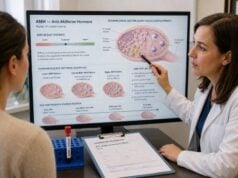
| AMH | Produced by small growing ovarian follicles | Lower or higher recruitable follicle pool; altered expected response to stimulation |
| Estradiol | Main estrogen during reproductive years | Follicle activity, ovarian feedback, cycle stage, medication effect, or persistent estrogen production |
| Prolactin | Pituitary hormone involved in lactation | Reproductive-axis suppression when persistently elevated |
These blood tests answer different questions. FSH and estradiol provide a cycle-specific snapshot. AMH is a more cycle-stable marker of follicle quantity. LH may help explain irregular ovulation, but it fluctuates substantially. Prolactin looks for a potentially reversible pituitary or medication-related cause of menstrual disruption.
An AMH test and antral follicle count by ultrasound are often the most useful predictors of how the ovaries may respond to fertility medicines. They do not count every remaining egg and do not establish whether an individual egg can produce a healthy embryo.
The panel should not be confused with a complete infertility diagnosis. Fertility also depends on ovulation, the fallopian tubes, uterine anatomy, sperm, sexual timing, and the genetic competence of eggs and embryos. Female age remains the strongest general predictor of reproductive potential because egg quality declines even when AMH remains within a laboratory range.
Who May Need Hormone Testing
An infertility evaluation is commonly started after 12 months of regular unprotected intercourse without pregnancy when the woman is younger than 35, after 6 months at age 35 or older, and more promptly after age 40. Evaluation should begin earlier when there are irregular or absent periods, known endometriosis, prior pelvic infection, previous ovarian surgery, chemotherapy or pelvic radiation, suspected male-factor infertility, or another condition likely to reduce fertility.
Hormone testing is particularly useful when there is:
- Amenorrhea, oligomenorrhea, or markedly irregular cycles
- A history of early menopause or primary ovarian insufficiency in the family
- Symptoms of androgen excess, such as new facial hair growth or persistent acne
- Hot flashes, vaginal dryness, or other symptoms of low estrogen before the expected age of menopause
- Galactorrhea, headaches, visual changes, or medicines known to raise prolactin
- Previous low response to ovarian stimulation
- Planned in vitro fertilization, egg freezing, or fertility preservation before gonadotoxic treatment
Testing does not need to be identical for every patient. In women with regular cycles of 21–35 days, additional testing to prove ovulation is often unnecessary unless the history raises concern. Prolactin is not routinely needed in every woman with infertility when periods are regular and there are no related symptoms. Likewise, broad hormone panels ordered without reference to cycle day can create misleading results.
The evaluation should include the partner or sperm source at the same time. Delaying semen analysis while repeatedly testing ovarian hormones can lose valuable time. A fertility hormone test panel may organize the endocrine portion of care, but it does not replace the rest of the couple-based or individual family-building assessment.
Timing and Preparation
Correct timing makes FSH, LH, and estradiol much easier to interpret. “Cycle day 1” is the first day of full menstrual flow, not light spotting. Baseline testing is usually performed on cycle day 2, 3, or 4, when ovarian hormone production should be relatively quiet.
| Test | Common timing | Why timing matters |
|---|---|---|
| FSH | Cycle day 2–4 | Provides a baseline before dominant follicle selection |
| Estradiol | Cycle day 2–4 with FSH | Detects early estrogen elevation that may suppress FSH |
| LH | Often cycle day 2–4 when part of a baseline panel | LH varies through the cycle and surges before ovulation |
| AMH | Usually any cycle day | Has less within-cycle variation than FSH or estradiol |
| Prolactin | Morning or a few hours after waking, after quiet rest | Sleep, stress, exercise, and venipuncture can temporarily raise it |
Hormonal contraception and fertility medicines can alter results. Combined birth-control pills may suppress AMH and antral follicle count modestly in some users; GnRH medicines, gonadotropins, clomiphene, letrozole, and estrogen or progesterone therapy also change cycle hormones. The clinician may test while treatment continues, pause it when medically appropriate, or interpret the value in that context.
Before the draw:
- Confirm which cycle day the clinic wants and whether irregular bleeding changes the plan.
- Ask whether fasting is required. It is usually not essential for these hormones alone, but fasting may be requested if glucose, insulin, or lipids are included.
- Avoid unusually strenuous exercise immediately before prolactin testing.
- Sit quietly for 15–30 minutes if a previous prolactin result was mildly elevated.
- Provide a full list of prescriptions, supplements, biotin, fertility drugs, and hormonal products.
- Report pregnancy possibility because pregnancy changes several hormones and must be considered in amenorrhea.
A result collected on the wrong day is not necessarily useless, but it may answer a different question. For example, a high estradiol value near ovulation is expected and should not be compared with an early-follicular reference interval.
What Each Hormone Result Means
FSH
FSH rises when the ovaries provide less negative feedback to the pituitary. A persistently high early-follicular FSH level may indicate diminished ovarian reserve or primary ovarian insufficiency, particularly when estradiol is low and periods are irregular or absent. FSH can vary considerably between cycles, so one normal value does not erase a previously high result, and one borderline high result may need confirmation.
Low or normal FSH is not always reassuring. When both FSH and estradiol are low in someone with amenorrhea, the pattern may reflect hypothalamic suppression from low energy availability, intensive exercise, major stress, weight loss, chronic illness, or pituitary disease. The FSH blood test must therefore be interpreted with estradiol and menstrual history.
LH
LH triggers ovulation after a midcycle surge. A random LH measurement cannot reliably confirm that a surge occurred because the peak may be brief and pulsatile. High LH can occur near ovulation, after menopause, with primary ovarian insufficiency, and in some people with PCOS. Low LH with low estradiol may point toward central suppression.
The LH/FSH ratio was historically emphasized in PCOS, but it is neither required nor sufficiently accurate to diagnose the condition. Many women with PCOS have no elevated ratio, and healthy women can have a ratio above 1 depending on cycle timing and assay. Diagnosis instead uses ovulatory dysfunction, clinical or biochemical hyperandrogenism, and ovarian morphology or AMH within an age-appropriate diagnostic pathway after excluding other causes.
AMH
AMH is produced by granulosa cells in preantral and small antral follicles. It correlates with the number of follicles available to respond to stimulation and can help estimate whether a patient may have a low, average, or high ovarian response during IVF. AMH generally declines with age, but values overlap widely among people of the same age.
Low AMH does not mean pregnancy is impossible, and high AMH does not guarantee pregnancy. AMH is a quantity marker, not a direct egg-quality test. It is also a poor stand-alone screening test for how quickly a person who is not infertile will conceive naturally. Very high AMH may occur with PCOS and may signal increased risk of an excessive response to ovarian stimulation.
Estradiol
Estradiol reflects follicle activity. During baseline testing, an unexpectedly elevated estradiol level may arise from an early-developing follicle or functional ovarian cyst. Because estradiol suppresses FSH, a high baseline estradiol can make FSH appear normal even when ovarian reserve is reduced.
Low estradiol with high FSH supports reduced ovarian function. Low estradiol with low or normal FSH suggests insufficient central stimulation. Estradiol later in the cycle rises before ovulation and is monitored during ovarian stimulation, but no single estradiol target predicts a healthy pregnancy.
Prolactin
Persistently high prolactin can reduce GnRH signaling, disrupt LH and FSH release, interfere with ovulation, and cause irregular periods or amenorrhea. Causes include pregnancy, breastfeeding, stress, pituitary adenoma, hypothyroidism, kidney disease, and medicines such as antipsychotics, some antidepressants, metoclopramide, and opioids.
Mild elevations are often repeated. The laboratory may test for macroprolactin when the value is elevated but symptoms are absent. A dedicated prolactin blood test evaluation is important when levels remain high, especially with headaches, vision changes, or nipple discharge.
Common Result Patterns
Hormone combinations narrow the possibilities more effectively than individual results.
| Result pattern | Possible meaning | Typical follow-up |
|---|---|---|
| High FSH, low estradiol, irregular or absent periods | Primary ovarian insufficiency or advanced ovarian aging | Repeat testing, pregnancy test, AMH, ultrasound, thyroid and genetic/autoimmune evaluation when indicated |
| High or borderline FSH with high baseline estradiol | Estradiol may be masking FSH; possible reduced ovarian reserve or active cyst | Repeat early-cycle testing and antral follicle count |
| Low AMH with otherwise normal cycle hormones | Lower follicle quantity or expected stimulation response | Age-based counseling, antral follicle count, treatment planning |
| High AMH, irregular cycles, androgen excess | PCOS is possible | Testosterone or free androgen assessment, pelvic ultrasound or AMH-based pathway, metabolic screening, exclusion of mimics |
| Low FSH and LH with low estradiol | Hypothalamic or pituitary hypogonadism | Nutrition, exercise, weight and illness review; prolactin, thyroid, iron studies, and pituitary assessment as indicated |
| High prolactin with low/normal FSH and LH | Prolactin-related ovulatory suppression | Repeat prolactin, medication review, macroprolactin, thyroid testing, possible pituitary imaging |
| Normal panel with infertility | Hormones may not be the cause | Tubal, uterine, semen, endometriosis, timing, and unexplained-infertility evaluation |
Primary ovarian insufficiency is not diagnosed from AMH alone. In a woman younger than 40 with menstrual disturbance, diagnosis generally relies on clinical history and elevated FSH on appropriately interpreted testing. AMH can support the assessment in some settings but should not replace established diagnostic criteria.
PCOS also cannot be diagnosed from high AMH or a high LH/FSH ratio alone. The current diagnostic approach accounts for age, cycle pattern, hyperandrogenism, ultrasound or AMH findings, and exclusion of thyroid disease, high prolactin, nonclassic congenital adrenal hyperplasia, and other androgen excess disorders. The PCOS hormone panel is therefore broader than this infertility panel.
Ranges and Important Limitations
Laboratory ranges differ by assay, age, cycle phase, menopausal status, and medication use. The report’s own interval should be used. The following broad examples explain scale only.
| Test | Common units | Broad early-cycle example |
|---|---|---|
| FSH | IU/L or mIU/mL | Often about 3–10 IU/L before menopause, with lab-specific cutoffs |
| LH | IU/L or mIU/mL | Often about 2–12 IU/L in the follicular phase; much higher during the surge |
| Estradiol | pg/mL or pmol/L | Often about 20–80 pg/mL in the early follicular phase |
| AMH | ng/mL or pmol/L | Strongly age- and assay-dependent; no universal “fertile” range |
| Prolactin | ng/mL or mIU/L | Often below about 20–25 ng/mL in nonpregnant women, depending on the assay |
Many clinics use FSH cutoffs near 10 IU/L to flag possible reduced response, but thresholds vary and become more concerning as values rise. Estradiol above roughly 60–80 pg/mL at baseline may complicate FSH interpretation, but the meaning depends on ultrasound and the clinic’s assay. AMH cutoffs used to define low or high response vary among laboratories and IVF protocols.
Five limitations prevent simple scorekeeping:
- Age changes meaning. An AMH value that is expected at 40 may be unusually low at 25, yet age still contributes more to egg quality.
- Assays differ. Results from different laboratories are not always directly interchangeable.
- Cycle hormones fluctuate. FSH, LH, and estradiol can change within hours or between cycles.
- Hormonal contraception and fertility treatment alter values. Suppression may be temporary.
- Pregnancy is an outcome of several systems. A favorable panel cannot show that tubes are open or sperm are adequate.
Direct-to-consumer “fertility scores” often overstate what a blood test can predict. A low AMH result may be emotionally alarming, but it should lead to context-specific counseling rather than a conclusion that conception cannot occur.
Other Tests That Complete the Evaluation
Hormone testing is usually combined with structural and partner assessment. Depending on the history, clinicians may recommend:
- Transvaginal ultrasound for antral follicle count, ovarian cysts, fibroids, adenomyosis, or other pelvic findings
- Hysterosalpingography or contrast sonography to assess tubal patency and the uterine cavity
- Semen analysis at the beginning of the evaluation
- Midluteal progesterone when ovulation needs confirmation; the sample is usually timed about seven days before the expected next period rather than automatically on day 21
- Thyroid-stimulating hormone when thyroid disease or ovulatory dysfunction is possible
- Androgen testing for hirsutism, acne, or suspected PCOS
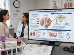
- 17-hydroxyprogesterone when nonclassic congenital adrenal hyperplasia is a concern
- Genetic testing or specialist evaluation for primary ovarian insufficiency
An ovulation blood test panel answers a different question from ovarian reserve testing. It focuses on whether and when ovulation occurred, while AMH and baseline FSH estimate follicle quantity and stimulation response. Neither replaces tubal testing.
Laparoscopy is not a routine first test for every patient, but it may be considered when symptoms or imaging suggest endometriosis or pelvic disease. Endometrial biopsy, postcoital testing, routine immunologic panels, and many add-on tests are generally not part of the standard initial infertility workup without a specific indication.
Next Steps After the Results
Results should lead to a defined plan rather than repeated testing without action. A practical review includes the exact cycle day, medications, age, menstrual pattern, pregnancy history, ultrasound findings, and semen results.
Possible next steps include:
- Repeat a questionable result under correct conditions. This is common for borderline FSH, unexpected estradiol, or mildly high prolactin.
- Complete missing parts of the evaluation. Hormones cannot substitute for semen analysis or tubal and uterine assessment.
- Address a reversible cause. Treatment may involve managing hypothyroidism or hyperprolactinemia, restoring adequate energy intake, changing a medication, or treating ovulatory PCOS.
- Plan treatment according to age and duration of infertility. Options may include timed intercourse, ovulation induction, intrauterine insemination, IVF, donor eggs, or fertility preservation.
- Refer promptly when time is important. Markedly diminished reserve, suspected primary ovarian insufficiency, age over 40, severe male-factor findings, or bilateral tubal disease often warrants early reproductive-endocrinology consultation.
Seek urgent medical care for severe pelvic pain with a positive pregnancy test, heavy bleeding with fainting, or sudden one-sided pelvic pain with vomiting. Persistent severe headache or new visual changes with high prolactin also deserves prompt assessment.
The most useful interpretation is individualized. A low AMH result at age 32, a high FSH result at age 42, and a mildly elevated prolactin result during acute stress have different implications. The panel becomes clinically meaningful only when the numbers are connected to the person’s cycle, symptoms, examination, and family-building goals.
References
- Guideline for the prevention, diagnosis and treatment of infertility 2025 (Guideline)
- Fertility evaluation of infertile women: a committee opinion 2021 (Guideline)
- Definition of infertility: a committee opinion 2023 (Guideline)
- ESHRE evidence-based guideline on Unexplained Infertility 2023 (Guideline)
- International Evidence-based Guideline for the assessment and management of polycystic ovary syndrome 2023 2023 (Guideline)
- Testing and interpreting measures of ovarian reserve: a committee opinion 2020 (Guideline)
Disclaimer
This article is for general education and does not diagnose infertility or determine an individual chance of pregnancy. Hormone values must be interpreted with age, cycle timing, medications, symptoms, ultrasound, semen analysis, and other fertility tests by a qualified clinician. Seek urgent care for severe pelvic pain, heavy bleeding with fainting, or pregnancy symptoms that could indicate an ectopic pregnancy.