
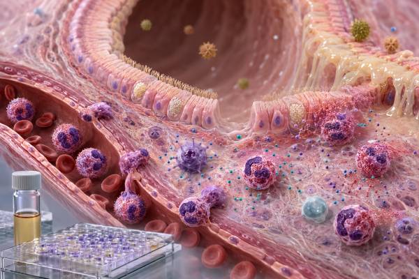
An interleukin-5 (IL-5) test measures a cytokine that promotes eosinophil development, survival, activation, and movement from bone marrow into blood and tissues. IL-5 is central to many eosinophilic diseases, including some forms of severe asthma, eosinophilic granulomatosis with polyangiitis, chronic rhinosinusitis with nasal polyps, and hypereosinophilic syndromes. Yet a direct serum or plasma IL-5 level is not the standard way to diagnose these conditions or decide whether an anti-IL-5 biologic is appropriate. Circulating concentrations are often very low, may not match tissue activity, and differ substantially by assay. Laboratories may measure unstimulated IL-5, cytokine released after immune-cell stimulation, IL-5-producing cells, or a multiplex type 2 inflammation panel. High values can support evidence of immune activation but are nonspecific. Low or undetectable values are common and do not exclude eosinophilic inflammation. Interpretation should begin with the exact test method and then incorporate symptoms, absolute eosinophil counts, medicine exposure, organ involvement, lung testing, imaging, pathology, and other disease-specific evidence.
- IL-5 is the most eosinophil-focused cytokine in type 2 immunity, but it is not the only signal controlling eosinophils.
- Blood eosinophil counts are more established clinically than direct IL-5 concentrations.
- Tissue eosinophilia can be active even when blood eosinophils or serum IL-5 are low.
- Anti-IL-5 drugs neutralize the cytokine, while anti-IL-5 receptor drugs target eosinophils through IL-5Rα.
- A single IL-5 result cannot identify an allergen, diagnose asthma, or determine disease severity.
Table of Contents
- How IL-5 Controls Eosinophils
- What an IL-5 Test Actually Measures
- When IL-5 Testing Is Considered
- Sample Handling and Result Interpretation
- What a High IL-5 Result Can Mean
- What a Low or Undetectable Result Can Mean
- Follow-Up Tests and IL-5-Targeted Biologics
How IL-5 Controls Eosinophils
IL-5 is produced mainly by activated Th2 cells and group 2 innate lymphoid cells, with contributions from mast cells, eosinophils, and other immune populations in selected settings. Epithelial alarm signals such as IL-25, IL-33, and thymic stromal lymphopoietin can activate type 2 networks after allergens, parasites, viruses, pollutants, or tissue injury. IL-4 and IL-13 help establish and amplify the broader type 2 program, while IL-5 is particularly important for eosinophil biology.
The IL-5 receptor has a cytokine-specific alpha chain, IL-5Rα, paired with a common beta chain also used by receptors for IL-3 and granulocyte-macrophage colony-stimulating factor. IL-5Rα is expressed strongly on eosinophils and their precursors and is also present on basophils. Receptor signaling activates JAK-STAT, MAP kinase, and PI3K-related pathways that support cell differentiation, survival, priming, adhesion, and effector function.
In bone marrow, IL-5 encourages committed progenitors to mature into eosinophils. In blood and tissue, it prolongs eosinophil survival and makes the cells more responsive to chemokines such as eotaxins. IL-5 alone does not determine where eosinophils travel. Adhesion molecules, CCR3 ligands, epithelial signals, and tissue-specific inflammatory cues guide migration into airways, skin, gastrointestinal tissue, blood vessels, nerves, and other organs.
Eosinophils contain granules with major basic protein, eosinophil peroxidase, eosinophil-derived neurotoxin, and other molecules. These substances can damage parasites and participate in immune defense, but uncontrolled release can injure epithelium, blood vessels, myocardium, nerves, and other tissues. Eosinophils also release cytokines, lipid mediators, growth factors, and extracellular traps, so their role extends beyond granule toxicity.
Normal eosinophils contribute to tissue homeostasis, immune regulation, and host defense. The problem is not the existence of IL-5 or eosinophils but excessive, persistent, or misdirected activity. Some eosinophilic diseases are strongly IL-5-dependent, while others use multiple survival pathways. This is why anti-IL-5 treatment can dramatically reduce blood eosinophils yet not eliminate every symptom or tissue finding.
An IL-5 level is therefore a narrow snapshot of one signal. It cannot be treated as an “eosinophil activity score.” The number of eosinophils, their activation state, the organ they occupy, and the damage they cause may be more clinically important than the amount of circulating cytokine.
What an IL-5 Test Actually Measures
A direct IL-5 assay measures protein in serum, plasma, bronchoalveolar lavage fluid, sputum supernatant, tissue fluid, or cell-culture media. Enzyme-linked immunosorbent assays, chemiluminescent methods, bead-based multiplex platforms, and ultrasensitive digital immunoassays may be used. Results are commonly reported in picograms per milliliter, but identical units do not make results interchangeable.
Serum and plasma can produce different concentrations because clotting activates cells and changes the specimen matrix. Collection tube, processing delay, temperature, storage, and freeze-thaw cycles also influence recovery. IL-5 is frequently close to an assay’s detection limit, where small analytical differences can look like large percentage changes.
A stimulated cytokine assay measures production capacity rather than resting circulation. Peripheral blood mononuclear cells may be exposed to an allergen, parasite antigen, mitogen, drug antigen, or receptor-directed reagent. The laboratory measures IL-5 released after a defined incubation. A high response means the sampled cells produced IL-5 under those conditions; it does not prove that the same amount was present in the body.
ELISpot and FluoroSpot assays count individual IL-5-secreting cells. Intracellular cytokine staining can identify whether CD4 T cells, innate lymphoid cells, or another population produced IL-5 and can examine co-production of IL-4, IL-13, or other signals. These methods are valuable in research, vaccine studies, drug-allergy investigation, and mechanistic immunology but are not routine stand-alone tests for common asthma or allergy.
Tissue techniques may measure IL5 messenger RNA, immunostaining, or a broader type 2 gene signature. A biopsy can demonstrate eosinophils and local pathology even when blood cytokines are undetectable. Protein concentration, transcript abundance, and cell counts are different biological measures and require separate interpretation.
An IL-5 result may be part of a cytokine panel. Multiplex panels can reveal a pattern, but each marker has different kinetics and technical limitations. One mildly abnormal analyte among dozens can be incidental. Clinical utility depends on whether the panel has been validated for the particular disease and decision.
The most common practical confusion is between IL-5 and eosinophils. An eosinophil count measures cells in blood; an IL-5 test measures a signaling protein. High IL-5 can occur without marked eosinophilia, and eosinophilia can persist when IL-5 is low because other signals or tissue compartments are involved.
When IL-5 Testing Is Considered
Direct IL-5 testing is used mainly in research, clinical trials, and selected specialist evaluations. It is generally not part of the initial workup for asthma, rhinitis, eczema, eosinophilic esophagitis, or a high eosinophil count. The result is most defensible when the clinician can state the question the assay is intended to answer.
Asthma researchers may measure IL-5 to characterize type 2 inflammation, compare phenotypes, or evaluate pharmacodynamic effects. In routine care, severe eosinophilic asthma is identified through symptoms, exacerbation history, inhaler use, spirometry, adherence, exposure assessment, blood eosinophils, and sometimes sputum eosinophils or exhaled nitric oxide. A serum IL-5 cutoff is not required to diagnose asthma or select most biologics.
Eosinophilic granulomatosis with polyangiitis can involve asthma, sinus disease, eosinophilia, neuropathy, pulmonary infiltrates, skin findings, cardiac disease, kidney disease, and vasculitis. IL-5 contributes to eosinophil survival, but diagnosis relies on the clinical pattern, blood counts, antineutrophil cytoplasmic antibodies when relevant, imaging, organ studies, and sometimes biopsy. A high cytokine result cannot establish vasculitis.
Hypereosinophilic syndromes are defined by persistent or significant eosinophilia with organ involvement after careful evaluation. Causes include clonal blood disorders, reactive cytokine-driven disease, infections, medicines, autoimmune conditions, and idiopathic syndromes. IL-5 measurement may contribute to research or a specialized assessment of reactive disease, but molecular testing, bone marrow evaluation, flow cytometry, and organ studies are often more decisive.
Eosinophilic gastrointestinal diseases are tissue diagnoses. Eosinophilic esophagitis requires symptoms of esophageal dysfunction and adequate biopsy findings. Blood eosinophils and IL-5 can be normal. Emerging blood biomarkers are under study, but endoscopy and histology remain central.
Parasitic infections can produce eosinophilia and type 2 cytokines. Travel, residence, raw or undercooked foods, soil exposure, animal contact, symptoms, and targeted microbiology or serology guide testing. A high IL-5 result does not identify the organism and should not trigger unsupervised antiparasitic treatment.
An IL-5 assay may also be included in a drug-development protocol for mepolizumab, reslizumab, benralizumab, depemokimab, or another pathway agent. Protocol-defined results are interpreted with drug concentration, eosinophils, receptor occupancy, exacerbations, and organ outcomes. They should not be converted into a general diagnostic range outside the study.
Drug reactions are another setting in which eosinophils and type 2 cytokines may rise. A simple itchy eruption, drug reaction with eosinophilia and systemic symptoms, acute eosinophilic pneumonia, and medication-triggered organ inflammation are clinically different problems. Timing after a new medicine, fever, facial swelling, lymph-node enlargement, liver or kidney injury, lung findings, and the complete blood count carry more diagnostic weight than IL-5. The suspected medicine should be reviewed promptly with a clinician, but essential treatment should not be stopped without a safe plan unless emergency care is required. Cytokine testing cannot determine whether future exposure will be safe.
The absence of an obvious allergy does not exclude an IL-5-related disorder. Many adults with severe eosinophilic asthma have no clear allergen sensitization, and clonal or idiopathic eosinophilic syndromes may present with cardiovascular, neurologic, skin, or gastrointestinal findings. Conversely, common seasonal allergy can produce mild eosinophilia without dangerous organ disease. The scale, persistence, symptoms, and organ involvement distinguish these situations far better than one cytokine value.
Sample Handling and Result Interpretation
Fasting is usually unnecessary unless other ordered tests require it. Patients should report corticosteroids, biologics, immune suppressants, recent infection, vaccination, allergic exposure, parasite treatment, and the timing of asthma attacks or infusions. Oral and systemic corticosteroids can lower eosinophils and cytokine production. A result obtained soon after treatment may describe drug effect rather than untreated disease.
Blood is collected into the laboratory’s specified tube. Venipuncture risks are limited to brief pain, bruising, lightheadedness, and rarely infection or prolonged bleeding. For specialized cytokine work, preanalytical control is more important than the physical risk: cells can release or degrade cytokines while a sample waits to be processed.
There is no universal normal IL-5 range. A laboratory may provide a reference interval, a detection limit, or no clinical range if the assay is research use only. Use only the performing laboratory’s method-specific information. An online range from another platform can be misleading.
“Below detection” means the analyte was not measurable above the assay threshold. It does not mean no IL-5 was produced. Cytokine may remain in airway or tissue, bind receptors, be neutralized by a drug, or fluctuate outside the sampling window. A normal result cannot exclude active eosinophilic disease.
For stimulated assays, verify the negative and positive controls. High spontaneous background can mask a specific response. A failed positive control can make a low allergen response uninterpretable. Cell number, viability, incubation, and background subtraction should be documented.
Trends are most useful when the same specimen type, assay, processing conditions, and timing relative to treatment are maintained. Even then, a change in IL-5 is not automatically a change in disease severity. Blood eosinophils may fall rapidly with treatment while tissue symptoms improve more slowly, or a cytokine level may change without a clinically meaningful outcome.
Unexpected extreme values should prompt review of units, dilution, calibration range, heterophile interference, and whether the assay measures free or drug-bound IL-5. Anti-IL-5 therapy can complicate measurement because different assays recognize cytokine-antibody complexes differently.
What a High IL-5 Result Can Mean
A high IL-5 result supports increased detectable type 2 cytokine activity in the tested specimen, but it is not disease-specific. Allergic inflammation, eosinophilic disorders, parasitic infection, immune-cell activation, selected malignancies, and technical factors can all contribute.
In eosinophilic asthma, increased IL-5 may support the biological pattern, especially when blood or sputum eosinophils are elevated and exacerbations persist despite appropriate inhaled treatment. However, asthma must still be confirmed objectively, and other causes of poor control—incorrect inhaler technique, nonadherence, smoking, occupational exposure, reflux, vocal-cord dysfunction, bronchiectasis, or infection—must be addressed.
Chronic rhinosinusitis with nasal polyps can involve strong local IL-5 production and tissue eosinophilia. Serum levels may or may not be high. Nasal endoscopy, symptom burden, smell loss, imaging, surgery history, and coexisting asthma guide care.
EGPA and hypereosinophilic syndromes may produce marked eosinophil-driven inflammation. A high IL-5 result can fit the picture but cannot distinguish reactive disease from a clonal eosinophil disorder. Cardiac symptoms, neuropathy, breathing difficulty, abdominal pain, thrombosis, or organ dysfunction require urgent assessment because eosinophil-mediated injury can progress even before a complete diagnosis is established.
Allergic sensitization can increase IL-5-producing cells, but a high result cannot identify the responsible allergen or predict anaphylaxis. Specific IgE and skin testing must be matched to exposure-related symptoms. A total IgE test is also nonspecific and should not be used as a universal allergy screen.
Helminth infection is another consideration, especially with travel or exposure risk. Some parasites cause tissue migration and pronounced eosinophilia, while others do not. Diagnosis uses organism-specific tests and clinical context.
A high stimulated IL-5 response means cells reacted to the laboratory stimulus. It may support allergen-specific Th2 memory, but cross-reactivity, nonspecific activation, and high background are possible. Such testing should be interpreted by a specialist familiar with the protocol.
The appropriate response to a high value is to identify the underlying disease and organ risk. Trying to lower IL-5 without a diagnosis can delay treatment for infection, malignancy, vasculitis, or another cause.
Absolute eosinophil count and percentage should be read together. A high percentage can occur because other white-cell populations are low, while a normal percentage can hide an elevated absolute count when the total white-cell count is high. Counts also vary during the day and can fall rapidly after systemic corticosteroids. Reviewing older results often shows whether eosinophilia is new, intermittent, or persistent. That history can help separate a transient allergic or infectious response from a chronic hematologic or inflammatory process.
What a Low or Undetectable Result Can Mean
Low circulating IL-5 is common because the cytokine acts locally and is rapidly consumed. A person can have eosinophilic inflammation in lungs, sinuses, skin, or gastrointestinal tissue while serum IL-5 remains below detection. Therefore, a low result is not a rule-out test.
Treatment can reduce or alter measurable IL-5. Corticosteroids suppress type 2 cytokine transcription. Mepolizumab, reslizumab, and other IL-5-neutralizing antibodies bind the cytokine, while benralizumab targets IL-5Rα and depletes eosinophils through antibody-dependent mechanisms. Depending on assay design, drug-bound cytokine may be undetected, detected differently, or occasionally accumulate as complexes.
A low stimulated response can result from poor cell viability, too few responsive lymphocytes, recent systemic steroids, an inappropriate antigen, or technical failure. It may also mean that the tested immune response is not IL-5-dominant. Positive controls and other cytokine readouts determine whether the finding is meaningful.
Low IL-5 does not diagnose immune deficiency. Most primary immune disorders are evaluated with infection history, lymphocyte subsets, immunoglobulins, vaccine responses, proliferation assays, and genetics. Rare pathway defects require targeted specialist testing, not a stand-alone serum cytokine level.
There is no evidence-based reason to raise IL-5 in a person with a low laboratory value. Increasing eosinophil survival could worsen allergic or eosinophilic disease. Supplements marketed to “balance” cytokines cannot reliably target this pathway and may interact with treatment.
When low blood eosinophils and low IL-5 conflict with strong symptoms, clinicians look for other inflammatory patterns and noninflammatory mimics. Neutrophilic asthma, infection, fixed airway disease, dysfunctional breathing, reflux, cardiac disease, anemia, and medication effects can resemble eosinophilic symptoms. The goal is not to force every case into an IL-5 pathway but to identify the mechanism that best explains the person’s current illness and responds to appropriate treatment safely, consistently, and over meaningful follow-up in everyday care.
Follow-Up Tests and IL-5-Targeted Biologics
The first follow-up test for suspected eosinophilic inflammation is often a complete blood count with differential and calculation of the absolute eosinophil count. Repeated counts matter because corticosteroids, time of day, acute illness, and natural fluctuation affect the number. Marked or persistent eosinophilia requires evaluation even if the person feels well.
Organ-directed testing may include spirometry, bronchodilator response, exhaled nitric oxide, sputum analysis, chest or sinus imaging, cardiac tests, nerve studies, stool or parasite testing, endoscopy with biopsy, skin biopsy, bone marrow examination, and molecular studies. The selection depends on symptoms and the degree of eosinophilia.
Anti-IL-5 biologics neutralize IL-5, while anti-IL-5Rα therapy targets the receptor-bearing eosinophil lineage. These medicines are used for specific approved indications and patient groups, including severe eosinophilic asthma and selected eosinophilic disorders. Eligibility commonly depends on diagnosis, exacerbation history, prior therapy, eosinophil counts, and local prescribing criteria—not on a measured serum IL-5 threshold.
Treatment response is assessed through fewer asthma exacerbations, improved control, reduced oral corticosteroid exposure, better lung function or symptoms, lower eosinophil counts, and improvement in the affected organ. Some patients improve substantially despite a modest change in lung function; others have biologic suppression of eosinophils but persistent symptoms from non-eosinophilic causes.
A very low eosinophil count during receptor-targeted therapy can be expected. It should not be interpreted as bone marrow failure without considering the medicine. Conversely, a low blood count does not guarantee that tissue disease is absent, especially early in treatment or in organ-restricted conditions.
Patients receiving biologics should follow product-specific guidance for infection assessment, vaccination, hypersensitivity monitoring, pregnancy discussions, and dosing. Abrupt discontinuation of systemic corticosteroids can be dangerous; tapering requires supervision. Acute asthma attacks still need rescue treatment because biologics are maintenance therapies, not immediate bronchodilators.
The central lesson is that IL-5 biology is clinically important even though direct IL-5 testing is rarely decisive. Eosinophil counts, organ findings, validated disease criteria, and treatment response usually provide the actionable information.
References
- Biological functions and clinical efficacy of IL-5/IL-5Rα-targeted therapies in eosinophil-associated diseases 2026 (Review)
- Effectiveness of anti-IL-5/5Rα biologics in severe asthma in real-world settings: a systematic review and meta-analysis 2025 (Systematic Review and Meta-Analysis)
- Proteomic Identification of Plasma Biomarkers of Response to Interleukin-5 Inhibitors in Severe Asthma 2025 (Research Article)
- Anti-IL-5 Pathway Agents in Eosinophilic-Associated Disorders Across the Lifespan 2024 (Review)
- Targeting the IL-5 pathway in eosinophilic asthma: A review of the evidence to date 2024 (Review)
- Effectiveness and safety of anti-IL-5/Rα biologics in eosinophilic granulomatosis with polyangiitis: a two-year multicenter observational study 2023 (Observational Study)
Disclaimer
This article is for general education and does not diagnose asthma, allergy, vasculitis, parasitic infection, or an eosinophilic disorder. IL-5 assays are specialized and method-dependent; results should be interpreted with eosinophil counts, symptoms, organ findings, and treatment history by a qualified clinician. Seek urgent care for severe breathing difficulty, chest pain, fainting, new weakness, or signs of organ injury.





