
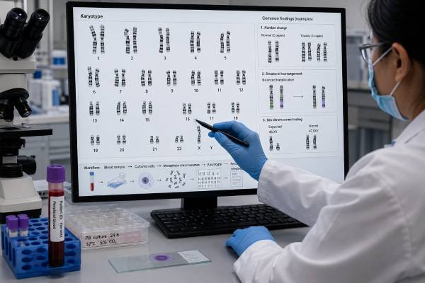
A karyotype test for infertility examines the number and large-scale structure of chromosomes in a blood sample. It can identify findings such as Klinefelter syndrome, Turner syndrome or mosaicism, balanced translocations, inversions, and other chromosome changes that may interfere with egg or sperm production, increase miscarriage risk, or create unbalanced embryos. Karyotyping is not needed for every person with infertility. It is most useful when the history, hormone pattern, semen analysis, ovarian function, recurrent pregnancy loss, or family history suggests a higher chance of a chromosome condition. A normal karyotype does not rule out smaller DNA changes, Y-chromosome microdeletions, CFTR variants, or most single-gene causes of infertility. An abnormal result also requires careful interpretation: a balanced rearrangement may not affect general health but can have major reproductive consequences. The complete chromosome notation, not a shortened portal summary, guides genetic counseling, partner testing, reproductive planning, and decisions about prenatal or embryo testing.
- Sample: Usually peripheral blood with cultured white blood cells
- What it detects: Extra, missing, rearranged, or mosaic chromosomes
- Common infertility findings: 47,XXY, 45,X/46,XX mosaicism, translocations, and inversions
- What it misses: Small gene variants, most microdeletions, and many molecular causes
- Why results matter: Diagnosis, treatment planning, miscarriage risk, and offspring risk
- Next step: Review the exact notation with reproductive genetics professionals
Table of Contents
- When an Infertility Karyotype Is Useful
- How the Test Is Performed
- Chromosome Findings in Male Infertility
- Chromosome Findings in Female Infertility and Pregnancy Loss
- How to Read Karyotype Notation
- What a Normal or Uncertain Result Does Not Exclude
- Reproductive Options After an Abnormal Result
- Preparing for Testing and Counseling
When an Infertility Karyotype Is Useful
A karyotype is a broad chromosome test, but its diagnostic yield is highest in selected groups. Ordering it routinely for every person with infertility may add cost without answering the most likely question. The clinical indication should be clear.
For males, current AUA/ASRM guidance recommends karyotype testing in primary infertility when azoospermia or severe oligozoospermia is accompanied by evidence of impaired sperm production, such as elevated follicle-stimulating hormone, testicular atrophy, or another suspected cause of abnormal spermatogenesis. The guideline threshold includes azoospermia or a sperm concentration below 5 million per milliliter in the appropriate clinical context.
Reasons a clinician may consider a male karyotype include:
- Nonobstructive azoospermia
- Severe oligozoospermia
- Small, firm testes or hypergonadotropic hypogonadism
- Delayed or incomplete puberty
- Gynecomastia with infertility
- A suspected sex chromosome condition
- Recurrent pregnancy loss in the couple
- A family history of chromosome rearrangement or infertility
For females, karyotyping may be considered with primary amenorrhea, premature or primary ovarian insufficiency, short stature or other features suggesting Turner syndrome, recurrent pregnancy loss, or a family history of a structural chromosome change. Some people with mosaic sex chromosome findings have few physical signs and first present because of irregular periods, low ovarian reserve, or infertility.
Parental karyotyping after recurrent pregnancy loss is not automatically recommended for every couple in all guidelines. Its usefulness depends on the pregnancy history, products-of-conception results, age, family history, and local practice. A balanced translocation in one partner is found in a small minority of couples with repeated losses, and identifying it can clarify recurrence and reproductive options.
A karyotype may also be ordered when testing of a miscarriage, fetus, or child shows an unbalanced translocation. The parents are then tested to determine whether the rearrangement arose de novo or came from a balanced carrier.
The test is not a substitute for a complete infertility evaluation. Semen analysis, ovulation assessment, uterine and tubal evaluation, hormone testing, and medical history remain essential. A chromosome finding may be the main cause, a contributing factor, or an unrelated finding.
How the Test Is Performed
Most constitutional karyotypes use a blood sample. The laboratory cultures lymphocytes, encourages them to divide, stops cell division at metaphase, stains the chromosomes to create a banding pattern, and photographs or digitally arranges them into pairs. A cytogenetics professional examines chromosome number and structure.
The process takes longer than many DNA tests because living cells must grow. Turnaround commonly ranges from about one to several weeks, depending on the laboratory, cell growth, urgency, and whether additional analysis is needed.
A standard report includes:
- Total chromosome count
- Sex chromosome complement
- Any extra or missing chromosome
- Structural rearrangements and breakpoints
- Whether more than one cell line was observed
- The number of cells counted or analyzed
- An interpretation and recommendations
Resolution is described by the number of visible chromosome bands. Higher-resolution banding can identify smaller structural changes than lower-resolution analysis, but karyotype still cannot detect most small deletions, duplications, or gene variants.
The usual sample is blood, but other tissues may be relevant in special situations. If low-level mosaicism is strongly suspected and blood is normal, a genetics specialist may discuss a second tissue such as skin fibroblasts or buccal cells. This is not routine, because mosaicism can be tissue-specific and additional testing may still not capture gonadal cells.
Before testing, tell the laboratory or clinician about:
- Previous stem-cell or bone-marrow transplant
- Recent blood transfusion
- Known cancer or chemotherapy
- A prior chromosome report
- A pregnancy or child with a chromosome finding
- Fertility treatments and planned procedures
A bone-marrow transplant can cause blood cells to carry the donor’s chromosomes, making peripheral blood unsuitable for a constitutional karyotype. Recent transfusion is less likely to alter a standard lymphocyte culture but should still be disclosed.
The test itself requires no fasting. Risks are limited to ordinary blood-draw effects such as bruising, discomfort, fainting, or rarely infection.
A karyotype is different from chromosomal microarray. Microarray detects much smaller gains and losses but generally does not identify balanced translocations or inversions. For infertility and recurrent loss, the ability to see balanced structure is often the reason karyotype is chosen.
Chromosome Findings in Male Infertility
Chromosome abnormalities become more common as sperm concentration decreases, especially in nonobstructive azoospermia. Findings may involve sex chromosomes or autosomes.
Klinefelter syndrome is the best-known sex chromosome cause of male infertility. The classic karyotype is 47,XXY, meaning there is an additional X chromosome. Testicular development and testosterone production may be affected, and most adults have azoospermia or severe oligozoospermia. Mosaic forms, such as 46,XY/47,XXY, can have a broader range of sperm production. The Klinefelter syndrome genetic test article explains diagnosis and health follow-up.
Some males with Klinefelter syndrome may have small areas of sperm production. Microdissection testicular sperm extraction, followed by IVF with intracytoplasmic sperm injection, may retrieve sperm in selected patients. Success is not guaranteed, and counseling should address hormonal health, procedural risks, embryo options, and prenatal testing.
46,XX testicular difference of sex development can occur when sex-determining genetic material, often SRY, is moved to an X chromosome. A person may have male external anatomy but no Y chromosome and usually has azoospermia because Y-linked sperm-production regions are absent. Karyotype starts the diagnosis, while FISH or molecular testing may clarify the rearrangement.
Structural autosomal rearrangements include Robertsonian translocations, reciprocal translocations, and inversions. A balanced carrier may have all necessary genetic material and no general health effects, yet meiosis can produce sperm with an unbalanced chromosome complement. Consequences may include infertility, failed implantation, miscarriage, or a child with a chromosome condition.
A Robertsonian translocation joins the long arms of two acrocentric chromosomes, commonly chromosomes 13, 14, 15, 21, or 22. A balanced carrier often has 45 chromosomes because two chromosomes are fused, but no important genetic material is missing. Reproductive risk depends on the chromosomes involved.
A reciprocal translocation exchanges segments between two chromosomes. The carrier usually has 46 chromosomes. The report’s breakpoints determine which embryo imbalances are possible.
An inversion reverses a chromosome segment. Many inversions are benign variants, while others can interfere with pairing and recombination, producing unbalanced gametes. Risk depends on inversion size and location.
A normal karyotype does not complete the genetic evaluation of severe male infertility. Y-chromosome microdeletion testing may be indicated for azoospermia or very low sperm concentration, and CFTR testing is relevant to congenital bilateral absence of the vas deferens. The CFTR test for male infertility addresses obstructive causes that a karyotype cannot detect.
Chromosome Findings in Female Infertility and Pregnancy Loss
Chromosome findings in female infertility often involve the X chromosome or a balanced structural rearrangement.
Turner syndrome classically has a 45,X karyotype. Many affected individuals have ovarian insufficiency because ovarian follicles are lost rapidly. Some are diagnosed in childhood, while others present with absent periods, infertility, or early menopause. Mosaic results such as 45,X/46,XX can be associated with spontaneous puberty and occasional fertility, but ovarian reserve may still decline early.
A Turner chromosome finding has health implications beyond reproduction, including possible heart, aortic, kidney, thyroid, hearing, and metabolic concerns. Pregnancy may carry serious cardiovascular risk. Anyone with a Turner-related karyotype needs specialist medical assessment before fertility treatment or pregnancy; donor eggs do not remove the maternal heart risk.
X chromosome structural abnormalities can disrupt ovarian function, particularly when breakpoints or missing material involve regions important for ovarian maintenance. Examples include X deletions, ring X chromosomes, and X-autosome translocations. The phenotype depends on which genes are affected and how X inactivation occurs.
47,XXX may be found incidentally. Many individuals have typical fertility, while some have premature ovarian insufficiency. The karyotype alone cannot predict ovarian lifespan.
Balanced translocations and inversions can be present in any sex. In a person producing eggs, the rearrangement can lead to embryos with duplicated or deleted chromosome segments. The carrier may conceive naturally but experience repeated miscarriage, infertility, or an affected pregnancy.
When miscarriage tissue undergoes chromosome testing, most abnormal results are sporadic embryo aneuploidies related to egg age rather than an inherited parental rearrangement. If the tissue shows an unbalanced structural finding, parental karyotypes become particularly important.
A balanced parental result does not mean every pregnancy will miscarry. Reproductive outcomes depend on the specific rearrangement, age, fertility factors, and chance. Some carriers have healthy children without intervention. Others have repeated losses or pregnancies with imbalance.
Karyotyping may be part of a primary ovarian insufficiency evaluation, but gene-level tests can also be relevant. FMR1 premutation testing, for example, is not detected by karyotype. Autoimmune, treatment-related, and unexplained causes must also be considered.
How to Read Karyotype Notation
Karyotype notation follows the International System for Human Cytogenomic Nomenclature. The formula can be decoded in steps.
The first number is the total chromosome count. Next come the sex chromosomes. Then any change is listed.
| Example | Meaning |
|---|---|
46,XX | 46 chromosomes with two X chromosomes; no visible abnormality |
46,XY | 46 chromosomes with X and Y chromosomes; no visible abnormality |
47,XXY | Extra X chromosome, consistent with Klinefelter syndrome |
45,X | One X chromosome, consistent with Turner syndrome |
46,XX,t(2;8)(q21;p23) | Balanced reciprocal translocation between chromosomes 2 and 8 at stated breakpoints |
45,XY,rob(13;14)(q10;q10) | Balanced Robertsonian translocation involving chromosomes 13 and 14 |
46,XY,inv(9)(p11q13) | Inversion involving chromosome 9; interpretation depends on the exact finding |
A slash separates cell lines in mosaicism. For example, 45,X[10]/46,XX[20] indicates that 10 counted cells had monosomy X and 20 had a 46,XX result. The proportion in blood may not equal the proportion in ovaries or other tissues.
The word balanced means there is no large visible net gain or loss of chromosome material. It does not mean “no reproductive effect.” The word unbalanced means material is missing or duplicated and may affect health or pregnancy viability.
Breakpoints are described by chromosome arm and band. The short arm is p; the long arm is q. More digits identify a more specific location. For a translocation, these breakpoints help a laboratory design targeted prenatal or embryo testing.
Reports may mention a variant, heteromorphism, or normal variant involving repetitive chromosome regions. Some visible differences have no established reproductive effect. They should not automatically be blamed for infertility.
A finding may be labeled apparently balanced because standard banding cannot exclude a small deletion or duplication at the breakpoint. If the person has congenital anomalies or developmental differences, microarray or sequencing may be recommended. In an otherwise healthy infertility patient, additional testing depends on the exact rearrangement and counseling question.
Ask for the full signed report. A clinic note stating “abnormal chromosomes” is not enough to estimate reproductive risk.
What a Normal or Uncertain Result Does Not Exclude
A normal karyotype means no chromosome-number or large structural abnormality was visible at the test’s resolution in the cells examined. It does not prove that infertility has no genetic cause.
Conditions commonly missed include:
- Y-chromosome AZF microdeletions
- CFTR gene variants causing congenital absence of the vas deferens
- FMR1 premutations associated with ovarian insufficiency
- Small copy-number variants below karyotype resolution
- Single-gene causes of spermatogenic failure or ovarian dysfunction
- Mitochondrial DNA conditions
- Epigenetic changes
- Low-level or tissue-limited mosaicism
The next genetic test should be driven by phenotype. A man with nonobstructive azoospermia may need Y microdeletion testing. A man with absent vas deferens may need CFTR analysis. A woman with primary ovarian insufficiency may need FMR1 testing. Broad panels or exome sequencing may be considered in selected cases but can produce uncertain findings.
A variant of uncertain significance is uncommon as a standard karyotype conclusion but may arise when a structural change cannot be clearly classified. Parental karyotyping can show whether it was inherited. Inheritance from a healthy parent may be reassuring, though it does not always eliminate reproductive significance.
A normal blood result also does not exclude gonadal mosaicism. Eggs or sperm may contain chromosome changes not found in blood. Age-related embryo aneuploidy occurs through errors in individual gametes and is not detected by a parental karyotype.
If infertility remains unexplained, repeating a normal karyotype usually adds little unless the first test had technical limitations or new clinical information suggests mosaicism. The evaluation should return to reproductive anatomy, hormones, gamete quality, and other genetic mechanisms.
Chromosomal microarray is sometimes suggested after a normal karyotype, but it is not an automatic next step for isolated infertility. It is more likely to help when the person also has congenital anomalies, developmental differences, or a family history suggesting a small deletion or duplication syndrome. Genome or exome sequencing may identify rare gene causes in selected patients, yet results can be uncertain and may not alter treatment. Testing should therefore be matched to the phenotype rather than ordered as an unrestricted search.
Reproductive Options After an Abnormal Result
An abnormal karyotype does not create one required treatment plan. Options depend on whether the finding affects gamete production, embryo chromosome balance, maternal health, or all three.
For a balanced translocation or inversion, possibilities include:
- Natural conception with no prenatal genetic testing
- Natural conception with chorionic villus sampling or amniocentesis
- IVF with preimplantation genetic testing for structural rearrangements, or PGT-SR
- Use of donor eggs or sperm
- Donor embryos
- Adoption or choosing not to pursue pregnancy
PGT-SR tests embryo biopsy cells for chromosome imbalance related to the rearrangement. It may reduce transfer of clearly unbalanced embryos, but it requires IVF and may produce no embryo suitable for transfer. Some platforms cannot distinguish a chromosome-normal embryo from a balanced carrier embryo. The embryo genetic testing guide explains PGT-SR limitations.
Prenatal diagnosis remains available after PGT-SR because embryo biopsy is not perfectly accurate and does not assess every fetal cell. CVS offers earlier testing but samples placenta; amniocentesis samples amniotic-fluid cells.
For Klinefelter syndrome or severe spermatogenic failure, treatment may include hormonal care, testicular sperm retrieval, IVF with ICSI, donor sperm, or other family-building options. Exogenous testosterone should not be started casually when fertility is desired because it can suppress sperm production.
For Turner syndrome or related mosaicism, the first priority is medical safety. Cardiac and aortic evaluation may determine that pregnancy is high risk or contraindicated. Fertility preservation, donor eggs, gestational carrier arrangements where legal, or other options require coordinated reproductive and cardiovascular care.
Risk estimates for a balanced rearrangement are case-specific. They depend on the chromosomes, breakpoints, carrier sex, segregation patterns, and observed family history. A generic percentage from another translocation should not be applied.
Emotional impact also matters. A carrier may feel guilt even though the chromosome arrangement was inherited or occurred by chance and was not caused by behavior. Genetic counseling can separate biological recurrence from personal responsibility.
Preparing for Testing and Counseling
A well-planned karyotype evaluation begins with the question the result is meant to answer.
Before the blood draw, gather:
- Semen-analysis results and hormone values
- Menstrual, ovarian reserve, and amenorrhea history
- Records from pregnancy losses or prior prenatal testing
- Family history of infertility, miscarriage, stillbirth, congenital anomalies, or intellectual disability
- Any known chromosome report in a relative
- Previous chemotherapy, radiation, surgery, or gonadotoxic exposure
- Information about stem-cell transplant or blood disorders
Ask the clinician:
- Why is karyotyping indicated in my case?
- Is the test being ordered for one partner or both?
- Which other genetic tests should be ordered at the same time?
- How many cells and what banding resolution will the laboratory use?
- What happens if mosaicism or an apparently balanced rearrangement is found?
- Will parental or relative samples be needed?
- How would the result change fertility treatment?
- Would PGT-SR, prenatal diagnosis, or donor gametes be relevant?
- Does this finding require medical care outside fertility treatment?
After testing, request a genetics appointment for any numerical abnormality, structural rearrangement, mosaic result, or unclear variant. The counselor can create a pedigree, explain notation, estimate reproductive outcomes, and coordinate testing of relatives when useful.
Keep the report permanently. A balanced rearrangement can matter to siblings, adult children, pregnancy care, and embryo-testing laboratories years later. Exact breakpoints and laboratory wording are difficult to reconstruct from memory.
A karyotype is most valuable when it is used neither too broadly nor too narrowly. In the right clinical setting, it can reveal the cause of impaired fertility, identify risks that semen or hormone tests cannot show, and turn an unexplained reproductive history into a specific plan.
References
- Diagnosis and Treatment of Infertility in Men: AUA/ASRM Guideline 2024 (Clinical Guideline)
- Male Infertility: Guideline Amendment Summary 2024 (Guideline Summary)
- Recurrent Pregnancy Loss 2023 (Clinical Guideline)
- ACGS Best Practice Guidelines for Constitutional Karyotype Analysis and Targeted Chromosome Analysis 2024 (Laboratory Guideline)
- Karyotype Genetic Test 2025 (Medical Test Guide)
- Genetic tests for reproduction Explained 2025 (Professional Patient Resource)
Disclaimer
This article provides general education about constitutional karyotyping in infertility and recurrent pregnancy loss. Testing indications, laboratory resolution, reproductive risks, and treatment choices depend on the complete clinical history and exact chromosome notation. Review individual results with a fertility specialist, clinical geneticist, urologist, gynecologist, or genetic counselor before making reproductive or medical decisions.





