
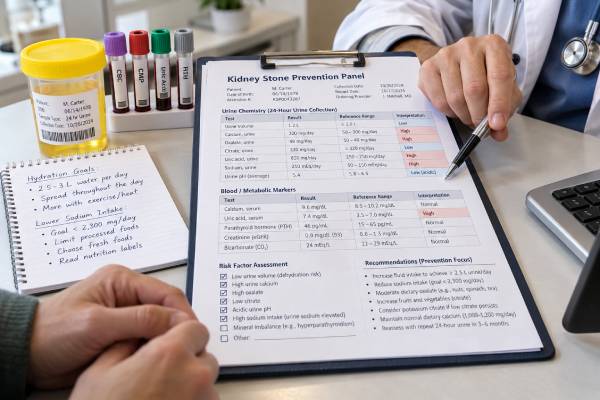
A kidney stone prevention test panel combines urine testing, blood work, stone composition, and clinical risk factors to identify why stones are forming and what can be changed to reduce recurrence. The most useful prevention evaluation is not one universal laboratory bundle. It is a structured workup that usually starts with urinalysis, kidney function, electrolytes, blood calcium, and stone analysis when available, then adds one or two 24-hour urine collections for people at higher risk or those who want individualized prevention. The 24-hour urine profile typically measures volume, calcium, oxalate, citrate, uric acid, sodium, pH, and creatinine, with other analytes added by the laboratory. Results guide practical targets such as increasing urine volume, lowering excess sodium, correcting hypocitraturia, evaluating hypercalciuria or hyperoxaluria, and identifying systemic causes such as primary hyperparathyroidism or cystinuria.
- A prevention panel usually combines blood tests, urinalysis, stone analysis, and 24-hour urine chemistry rather than relying on one marker.
- High-risk or recurrent stone formers generally benefit most from detailed metabolic testing, often with two 24-hour urine collections.
- Core urine targets include volume, calcium, oxalate, citrate, uric acid, sodium, pH, and creatinine, with supersaturation calculations often included.
- Blood calcium, creatinine, electrolytes, bicarbonate, and uric acid can reveal systemic causes that urine testing alone may miss.
- Repeat testing after treatment is important because prevention should be confirmed by measurable improvement in urine chemistry and stone activity.
Table of Contents
- What a Kidney Stone Prevention Panel Includes
- Who Needs a Detailed Prevention Workup
- How to Interpret the 24-Hour Urine Chemistry
- Blood Minerals and Kidney Function Tests
- Stone Type, Medical History, and Risk Factors
- Turning Test Results Into a Prevention Plan
- Repeat Testing, Follow-Up, and Common Pitfalls
What a Kidney Stone Prevention Panel Includes
A prevention panel is best understood as a metabolic stone evaluation, not a single fixed test. The exact components depend on whether the stone is a first event or a recurrence, the patient’s age, stone type, medical history, and risk of future stones.
A basic evaluation commonly includes:
- urinalysis, with urine culture when infection is suspected;
- serum creatinine or estimated glomerular filtration rate;
- sodium, potassium, bicarbonate, and other electrolytes;
- serum calcium and albumin;
- serum uric acid;
- stone composition analysis when a stone is available.
A more complete evaluation adds one or two 24-hour urine collections. Current guideline summaries commonly include urine volume, calcium, oxalate, citrate, uric acid, sodium, potassium, pH, and creatinine, with magnesium and phosphate often included as well.
Many commercial stone panels also report calculated supersaturation for calcium oxalate, calcium phosphate, and uric acid. Supersaturation integrates several measured values and estimates how strongly the urine favors crystal formation.
A 24-hour urine kidney stone risk test is therefore one central part of the prevention panel, but it should not be mistaken for the whole evaluation. Blood tests can reveal endocrine or kidney disorders, while stone analysis can identify the crystal type that actually formed.
Who Needs a Detailed Prevention Workup
Every stone former benefits from at least a basic evaluation, but detailed metabolic testing is most strongly indicated when recurrence risk is high or the result is likely to change treatment.
Common high-risk features include:
- recurrent stones;
- bilateral or multiple stones;
- stones in childhood or adolescence;
- a single functioning kidney;
- nephrocalcinosis;
- cystine, uric acid, infection, or other unusual stone types;
- family history of recurrent stones;
- bowel disease, chronic diarrhea, short bowel, or bariatric surgery;
- osteoporosis or recurrent fractures with calcium stones;
- gout, diabetes, metabolic syndrome, or primary hyperparathyroidism;
- reduced kidney function;
- stones that continue despite general prevention measures.
A first-time stone former without high-risk features may still choose detailed testing, especially when they want specific prevention targets rather than broad advice. The value of testing rises when the person is willing to use the results to change diet, fluid intake, or medication.
The distinction between “low risk” and “high risk” is not simply the number of previous stones. One first stone can still justify a detailed workup if it occurs in childhood, is made of cystine or uric acid, appears with nephrocalcinosis, accompanies bowel disease, or occurs in a person with a solitary kidney. Conversely, some adults with one uncomplicated calcium oxalate stone and no concerning history may reasonably start with general prevention and basic screening.
Patient preference matters because metabolic testing is most valuable when it leads to action. A person who has already had repeated procedures may want a full evaluation even if a guideline leaves room for discretion. The clinician can use the stone burden, recurrence pattern, occupation, travel, climate exposure, and family history to decide how comprehensive the initial panel should be.
Timing matters. A baseline 24-hour urine collection is best obtained after the acute stone episode has settled, infection has resolved, and the patient is back on their usual diet and fluid routine. European guidance recommends performing specific metabolic evaluation after the acute phase rather than immediately during renal colic or temporary overhydration.
If a stone fragment is available, a kidney stone analysis should be incorporated early because composition determines which metabolic findings deserve the most attention.
How to Interpret the 24-Hour Urine Chemistry
The urine panel is interpreted as a pattern. One borderline abnormality rarely explains recurrent stones by itself.
| Urine marker | Why it matters | Common prevention focus |
|---|---|---|
| Volume | Low volume concentrates all stone-forming solutes | Increase fluid intake; many adults aim for >2.5 L urine/day |
| Calcium | High excretion promotes calcium oxalate and calcium phosphate stones | Review sodium, blood calcium, diet, and possible thiazide therapy |
| Oxalate | Higher values strongly raise calcium oxalate supersaturation | Normal calcium with meals, moderate high-oxalate foods, investigate malabsorption |
| Citrate | Low citrate removes a natural inhibitor of calcium crystallization | Dietary alkali or prescribed citrate when appropriate |
| Uric acid | High excretion can contribute to uric acid or calcium oxalate stones | Review purine intake and systemic urate disorders |
| pH | Low pH favors uric acid; higher pH can favor calcium phosphate | Match alkalinization strategy to stone type |
| Sodium | High sodium can increase urinary calcium | Reduce excess salt intake |
| Creatinine | Helps assess collection completeness | Repeat questionable collections |
Low urine volume is one of the most universal modifiable risks. Many guidelines advise enough fluid to produce more than 2.5 L of urine per day in adult stone formers. Cystinuria generally requires a higher urine volume target.
Urine calcium should not be interpreted as a reason to eliminate calcium from the diet. Normal dietary calcium is usually recommended because calcium in the intestine binds oxalate. Excess sodium often has a more direct relationship with hypercalciuria than normal food calcium does.
A urine oxalate test is particularly important after bowel disease or bariatric surgery because enteric hyperoxaluria can be a major driver of calcium oxalate stones. Markedly elevated oxalate deserves evaluation for secondary or rare genetic causes rather than simple food avoidance.
Urine citrate and pH must be interpreted together. Potassium citrate can raise citrate and pH, which is useful for hypocitraturia and uric acid stones, but excessive alkalinization may increase calcium phosphate risk in susceptible patients.
Blood Minerals and Kidney Function Tests
Blood tests identify systemic conditions that can produce abnormal urine chemistry. They also show whether medications used for prevention can be prescribed safely.
Serum calcium is one of the most important screening tests. Persistent hypercalcemia in a stone former raises concern for primary hyperparathyroidism. If calcium is high or inappropriately high-normal in the right clinical setting, parathyroid hormone testing may be indicated. A blood calcium test for kidney stones is therefore a key part of basic screening.
Creatinine and estimated GFR assess kidney function. Reduced function can affect interpretation of urine excretion, medication choice, potassium safety, and the urgency of preventing recurrent obstruction.
Bicarbonate helps identify acid-base disorders. Low serum bicarbonate with high urine pH and low citrate can suggest distal renal tubular acidosis, a condition associated with calcium phosphate stones and nephrocalcinosis.
Serum uric acid can support evaluation for gout, high urate production, or reduced urate clearance. It does not diagnose uric acid stone composition. For uric acid stones, urine pH and urine volume are usually more important than serum urate alone.
Phosphate, magnesium, vitamin D, and PTH may be added when the history or initial results point toward a mineral or endocrine disorder. A prevention panel should be targeted rather than expanded indiscriminately; each test should answer a clinical question.
Blood testing may also be repeated after starting thiazide-type diuretics, potassium citrate, allopurinol, or cystine-binding drugs because prevention medications can affect electrolytes, kidney function, glucose, uric acid, liver tests, blood counts, or urine protein depending on the drug.
Serum and urine results should also be checked for internal consistency. A very high blood calcium with a completely unremarkable PTH history deserves confirmation and endocrine review; a very low serum bicarbonate with persistently alkaline urine may warrant investigation beyond ordinary diet counseling. Similarly, severe hyperoxaluria or unusually low urine citrate can point toward a disorder that generic hydration advice will not correct. The purpose of the panel is to recognize these patterns early enough to investigate them.
Stone Type, Medical History, and Risk Factors
The same urine number can mean something different depending on stone type. That is why prevention starts with the clinical context rather than a spreadsheet of laboratory values.
For calcium oxalate stones, common targets are urine volume, calcium, oxalate, citrate, and sodium. For calcium phosphate stones, urine pH, calcium, citrate, bicarbonate, and PTH become especially important. For uric acid stones, persistently low urine pH is usually central. For cystine stones, very high urine volume, alkalinization, and cystine-specific monitoring are required. For struvite, infection and complete stone clearance dominate the plan.
Medical history can uncover causes that the panel alone cannot. Important questions include bowel surgery, inflammatory bowel disease, chronic diarrhea, gout, diabetes, recurrent urinary infection, osteoporosis, endocrine disease, eating patterns, occupation, climate exposure, and family history.
Medication review is equally important. Topiramate and other carbonic anhydrase inhibitors can promote alkaline urine and low citrate. Loop diuretics can increase urinary calcium. High-dose vitamin C can raise oxalate. Some medications can crystallize directly and form drug stones.
Supplements also deserve review. Calcium supplements are not equivalent to calcium-rich foods, and their timing can matter. Large vitamin D doses may affect calcium handling in susceptible patients. Sodium-containing products can increase total sodium intake without being obvious from food labels alone.
A useful prevention panel therefore combines measured chemistry with why that chemistry is abnormal. The laboratory result identifies the target; the history identifies the likely cause.
Turning Test Results Into a Prevention Plan
The strongest prevention plans are specific and measurable. “Drink more water” is less useful than “adjust fluids so your next 24-hour urine volume is above the agreed target.” “Eat less salt” becomes more actionable when a high urine sodium result confirms that sodium intake is contributing.
Common result-based strategies include:
- Low urine volume: distribute fluids across the day and increase intake during heat, exercise, or sweating.
- High urine calcium: reduce excess sodium, maintain normal food calcium, and consider thiazide-type therapy when clinically appropriate.
- High urine oxalate: pair normal dietary calcium with meals, reduce frequently consumed high-oxalate foods, avoid excessive vitamin C, and investigate malabsorption when indicated.
- Low urine citrate: increase fruits and vegetables and consider potassium citrate when appropriate.
- Low urine pH with uric acid stones: use supervised urine alkalinization and improve urine volume.
- High urine uric acid: review purine intake and determine whether urate-lowering treatment is indicated.
- Cystinuria: pursue much higher urine volume, controlled alkalinization, lower sodium intake, and cystine-specific medication if needed.
The goal is not to “normalize every number.” Treatment should reduce supersaturation and future stone activity without creating new problems. For example, pushing urine pH too high to raise citrate can increase calcium phosphate supersaturation.
Priorities are often chosen by impact and feasibility. Correcting a urine volume of 1.1 L may have a larger immediate effect than trying to fine-tune a mildly high uric acid value. Reducing a very high sodium intake can lower urinary calcium without restricting healthy food calcium. In enteric hyperoxaluria, timing calcium with meals may matter more than broadly avoiding every food that appears on an oxalate list. A good prevention plan therefore translates numbers into a small set of high-value changes rather than a long list of restrictions.
The panel can also show whether a treatment is doing what it was intended to do. A thiazide-type drug should generally lower urinary calcium; potassium citrate should raise citrate and usually pH; more fluid should increase urine volume. If the expected marker does not move, the clinician can check adherence, dose, diet, collection quality, or whether the original assumption about the cause was wrong.
Dietary advice should also remain nutritionally balanced. Very low calcium diets, extreme low-oxalate diets, or excessive fluid intake can create problems of their own. A dietitian familiar with stone disease can help when several restrictions seem to conflict.
Repeat Testing, Follow-Up, and Common Pitfalls
Prevention should be verified. European guidance suggests repeating 24-hour urine testing about 8–12 weeks after starting pharmacologic prevention, then adjusting the plan if the targeted abnormalities have not improved. Once urine risk factors are stable, annual testing is often sufficient for many high-risk patients, although follow-up is individualized.
Imaging is also part of prevention follow-up. Better urine chemistry is encouraging, but the real clinical outcomes are fewer new stones, less growth of existing stones, fewer emergency visits, and fewer procedures.
Common testing pitfalls include incomplete 24-hour urine collection, collecting longer than 24 hours, changing diet dramatically for the test day, intentional overhydration, testing during acute illness, and interpreting a single mildly abnormal value without context.
Two baseline collections can be useful because stone-risk chemistry varies day to day. A surprising result should be checked against collection quality, creatinine, recent diet, weather, exercise, and medication adherence before major treatment changes are made.
Collection completeness is a frequent source of error. Missing one large void can make calcium, oxalate, citrate, sodium, and uric acid all appear lower than they really are. Collecting for longer than 24 hours can push the same values upward. Creatinine excretion is commonly used as a plausibility check, but it is influenced by age, sex, muscle mass, body size, diet, and kidney function, so it should not be used as a rigid pass-fail rule without context.
A normal panel does not eliminate recurrence risk. Anatomy, intermittent dietary exposures, infection, genetic factors, and day-to-day variation can still contribute. Conversely, one abnormal result does not guarantee another stone will form.
Seek urgent care for flank pain with fever or chills, inability to urinate, persistent vomiting, severe uncontrolled pain, pregnancy, or symptoms in a person with one functioning kidney. A prevention panel is for long-term risk reduction; it does not replace emergency evaluation of obstruction or infection.
A well-designed kidney stone prevention panel turns recurrence prevention into a feedback loop: identify the stone type, measure the blood and urine environment, target the dominant risks, repeat the measurements, and confirm that both chemistry and stone activity improve.
References
- Metabolic Evaluation and Recurrence Prevention – EAU Guidelines on Urolithiasis – Uroweb 2026 (Guideline)
- Summary of the clinical practice guideline for the management of urinary stones, third edition 2025 (Guideline)
- Metabolic Evaluation and Recurrence Prevention for Urinary Stone Patients: An EAU Guidelines Update 2024 (Review)
- Kidney Stone Pathophysiology, Evaluation and Management: Core Curriculum 2023 2023 (Review)
- Kidney Stone Prevention 2023 (Review)
- UPDATE – Canadian Urological Association guideline: Evaluation and medical management of kidney stones 2022 (Guideline)
Disclaimer
This article is for general education and does not replace individualized medical care. Kidney stone reference ranges, prevention targets, medication choices, and follow-up intervals vary with stone type, kidney function, other illnesses, and laboratory methods. Review abnormal results and treatment changes with a qualified clinician.