
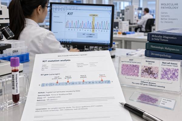
A KIT mutation test looks for DNA changes that activate the KIT receptor tyrosine kinase. The result can be highly important, but its meaning depends on the disease. In gastrointestinal stromal tumor (GIST), KIT mutations are common and often guide selection and dosing of tyrosine kinase inhibitors. In melanoma, KIT mutations occur mainly in selected subtypes such as mucosal, acral, and chronically sun-damaged melanoma and may create a targeted-treatment option in advanced disease. In systemic mastocytosis, the KIT D816V mutation is a major molecular finding that supports diagnosis and can influence treatment selection. A KIT mutation test is different from CD117 immunohistochemistry: a tumor can stain positive for KIT protein without having a KIT gene mutation. For that reason, the report should always be interpreted with the exact variant, exon, specimen, tumor type, and other pathology findings rather than as a simple positive-or-negative cancer marker.
- A positive KIT mutation result means a reportable KIT gene variant was detected; the clinical meaning depends on the exact exon and disease.
- In GIST, KIT exon 11 mutations are most common, while exon 9 and secondary exon 13, 14, 17, or 18 mutations can affect drug response and resistance.
- KIT D816V is found in most patients with systemic mastocytosis and requires a sensitive assay because the mutant allele can be present at very low levels.
- CD117-positive staining is not the same as a KIT mutation; immunohistochemistry measures protein expression, while molecular testing detects DNA changes.
- A negative KIT result may lead to testing of other drivers such as PDGFRA in GIST or broader molecular profiling in melanoma and mast cell disease.
Table of Contents
- What KIT Testing Measures
- How the Test Is Performed
- Understanding KIT Results
- KIT Mutations in GIST
- KIT Mutations in Melanoma
- KIT Mutations in Mast Cell Disease
- Limitations and Next Steps
What KIT Testing Measures
KIT is a gene that encodes a receptor tyrosine kinase, a protein on the cell surface that helps regulate growth and survival after binding its ligand, stem cell factor. Certain activating mutations keep KIT signaling switched on even when the normal growth signal is absent. Persistent signaling can drive uncontrolled growth in several cancers and clonal disorders.
A KIT mutation test examines DNA for these activating changes. Depending on the laboratory, the assay may use targeted polymerase chain reaction, Sanger sequencing, droplet digital PCR, or next-generation sequencing (NGS). Broader NGS panels are common because they can identify KIT together with other clinically relevant genes.
The exact location of a KIT mutation matters. KIT contains several functional domains encoded by different exons. In GIST, primary mutations most often occur in exon 11, followed by exon 9, with less common primary variants in exons 13 and 17. During treatment, secondary mutations can emerge in exons 13, 14, 17, and 18 and create resistance to specific tyrosine kinase inhibitors.
In systemic mastocytosis, the classic finding is KIT p.D816V, located in exon 17. This mutation stabilizes KIT in an active state and is present in the great majority of systemic mastocytosis cases. Because the mutant mast cells may represent only a small fraction of all cells in blood or marrow, a highly sensitive assay is often needed.
In melanoma, KIT alterations include point mutations and, less consistently, amplification. The most clinically meaningful activating variants commonly involve exons 11 and 13, although the spectrum is broader. The presence of a KIT alteration is most relevant in mucosal, acral, and chronically sun-damaged melanomas rather than in the more common cutaneous melanomas driven by BRAF or NRAS.
KIT mutation versus CD117 staining
KIT is also known as CD117, but these terms describe different kinds of testing. CD117 immunohistochemistry detects KIT protein in tissue. Molecular testing identifies a change in the KIT gene itself. Most GISTs stain strongly for CD117 regardless of whether the tumor carries a KIT mutation, and some non-GIST tumors can also express CD117. Therefore, positive staining does not prove that a targetable KIT mutation is present.
This distinction prevents a common interpretation error. A pathology report that says “CD117 positive” should not be read as “KIT mutation positive.” If molecular status matters for treatment or classification, DNA-based testing is needed.
How the Test Is Performed
The specimen depends on the condition being evaluated. GIST and melanoma are usually tested on formalin-fixed, paraffin-embedded tumor tissue from a biopsy or surgical resection. A pathologist selects an area with adequate viable tumor and may mark the tissue for microdissection before DNA extraction.
For suspected systemic mastocytosis, KIT testing may be performed on bone marrow aspirate, peripheral blood, or another involved tissue. High-sensitivity testing is especially important for KIT D816V. Conventional sequencing can miss the mutation when the variant allele frequency is low, so allele-specific quantitative PCR or droplet digital PCR is often preferred when mastocytosis is strongly suspected.
No special fasting is required for the genetic analysis itself. Any preparation relates to the biopsy or bone marrow procedure used to obtain the sample.
What appears on the report
A molecular report may include:
- the gene and transcript tested;
- the protein change, such as KIT p.D816V;
- the DNA-level variant notation;
- the exon involved;
- variant classification, such as pathogenic or likely pathogenic;
- variant allele frequency when measured;
- assay coverage and limit of detection; and
- an interpretation describing diagnostic or treatment significance.
The variant allele frequency is not a direct measurement of the percentage of tumor cells. Normal-cell contamination, copy-number changes, tumor heterogeneity, and clonality all affect it. In mastocytosis, a very low VAF can still be clinically meaningful if the assay is validated and the variant is the characteristic D816V driver.
Understanding KIT Results
A KIT result should be interpreted at the variant level.
Positive for a pathogenic or likely pathogenic mutation
A positive result means the laboratory found a KIT alteration supported as disease-causing or oncogenic. The next question is which alteration. KIT exon 11 GIST, KIT exon 9 GIST, KIT D816V mastocytosis, and KIT L576P melanoma are not interchangeable findings. They may differ in disease association, drug sensitivity, prognosis, and resistance patterns.
Negative or not detected
A negative KIT test means no reportable mutation was found within the regions and sensitivity of the assay. It does not mean the tumor is benign, and it does not exclude the disease being considered.
For example, a GIST can be KIT-wild-type and instead harbor a PDGFRA mutation, SDH deficiency, NF1-related biology, BRAF alteration, or rare fusion. A melanoma can be KIT-negative and have other drivers. A patient with suspected systemic mastocytosis can have a false-negative result if a low-sensitivity method was used or if the tested specimen contains very few clonal mast cells.
Variant of uncertain significance
A KIT variant of uncertain significance has insufficient evidence for a clear benign or pathogenic classification. A VUS should not automatically be used to select a kinase inhibitor. Clinical laboratories may later reclassify a VUS as more evidence becomes available, and consultation with molecular pathology can help when an unusual variant sits in a known functional domain.
Amplification is not the same as mutation
Some reports identify increased KIT copy number rather than a sequence mutation. Amplification can increase the amount of gene material but does not necessarily create the same biology or drug sensitivity as an activating mutation. This distinction is particularly important in melanoma, where clinical responses to KIT inhibitors have been more closely associated with specific activating mutations than with amplification alone.
KIT Mutations in GIST
GIST is the disease in which KIT genotyping has the most established day-to-day treatment role. Most GISTs are driven by mutually exclusive activating mutations in KIT or PDGFRA, and approximately three-quarters have a KIT mutation.
Exon 11
KIT exon 11 encodes the juxtamembrane region and is the most commonly mutated part of the gene in GIST. Deletions, substitutions, and insertions can occur. Exon 11-mutant GISTs are generally sensitive to imatinib, although individual risk also depends on tumor size, mitotic rate, site, rupture, and the specific mutation.
Certain exon 11 deletions involving codons 557 and 558 have been associated with more aggressive behavior in some settings. This is one reason a report that merely says “KIT positive” provides less useful information than a report that gives the exact variant.
Exon 9
KIT exon 9 mutations occur less often and are particularly associated with small-bowel GIST. The classic alteration is a duplication involving codons 502 and 503. Exon 9 status can influence the dose-response relationship to imatinib in advanced GIST, so the genotype can affect treatment planning rather than simply confirming the diagnosis.
Secondary mutations and acquired resistance
A GIST that initially responds to a tyrosine kinase inhibitor can later progress because additional KIT mutations emerge under treatment pressure. Secondary variants cluster in the ATP-binding pocket and activation loop, especially exons 13, 14, 17, and 18. Different secondary mutations have different sensitivities to later-line agents.
This creates tumor heterogeneity: one patient can have several resistant subclones in different metastases. Repeat tissue or circulating-tumor-DNA testing may therefore be considered when advanced disease progresses and the molecular information could influence trial selection or treatment strategy.
Why molecular testing is recommended even when CD117 is positive
Most GISTs express CD117, but the genotype adds predictive information that immunostaining cannot provide. Molecular testing can also help confirm GIST in tumors with unusual morphology or weak KIT expression. If KIT and PDGFRA are both negative, broader testing can identify alternative molecular classes with different hereditary implications and treatment sensitivity.
KIT Mutations in Melanoma
KIT mutations are uncommon across melanoma as a whole, but they are enriched in mucosal melanoma, acral melanoma, and melanoma arising in chronically sun-damaged skin. Testing is therefore most useful when the clinical and pathologic subtype makes KIT alteration plausible or when broad molecular profiling is being performed for advanced disease.
A positive KIT result may create an option for a KIT inhibitor, particularly when the mutation is a known activating and drug-sensitive variant. Responses have been reported with agents such as imatinib in selected patients, but the benefit is less predictable than in GIST. Not every KIT mutation is sensitive, and amplification alone has not shown the same consistency.
The report should therefore specify whether the alteration is a known activating mutation and whether clinical evidence supports a particular drug. Other melanoma biomarkers still matter. BRAF, NRAS, NF1, tumor mutational features, disease site, prior immunotherapy, and overall disease burden can all affect treatment planning.
A KIT-positive melanoma result also does not replace immune-based treatment considerations. Checkpoint inhibitors remain central in advanced melanoma, and KIT-directed therapy is usually considered in the context of subtype, prior treatment, symptoms, disease tempo, and available evidence.
KIT Mutations in Mast Cell Disease
In systemic mastocytosis, KIT testing has a different purpose. The key alteration is KIT D816V, which is present in more than 90% of adults with systemic mastocytosis when sensitive methods and appropriate specimens are used.
Finding an activating KIT mutation in bone marrow, blood, or another extracutaneous organ can satisfy a minor diagnostic criterion for systemic mastocytosis when interpreted with the rest of the diagnostic framework. Diagnosis does not rest on KIT alone. Pathologists and hematologists also assess mast-cell aggregates, cell morphology, aberrant expression of markers such as CD25, CD2, or CD30, and serum tryptase in the appropriate context.
Why assay sensitivity is crucial
A patient can have systemic mastocytosis even when only a very small fraction of blood or marrow cells carry D816V. Standard NGS panels may not detect such low-level disease. High-sensitivity PCR or droplet digital PCR can detect much lower allele burdens and is preferred when clinical suspicion remains high.
If D816V is negative with a sensitive method, broader KIT sequencing may be appropriate in selected cases because other activating KIT variants can occur. A negative blood test may also lead to bone marrow testing if the clinical picture strongly suggests systemic mastocytosis.
Treatment implications
KIT genotype can influence selection of kinase inhibitors. The D816V mutation is resistant to some older KIT inhibitors that work well in KIT-mutant GIST, which illustrates why drug sensitivity cannot be generalized across diseases. Newer agents that inhibit D816V have changed treatment of advanced systemic mastocytosis, and avapritinib is also used in appropriate patients with indolent disease. Treatment choice depends on disease subtype, symptoms, organ damage, mutation profile, blood counts, and toxicity considerations.
Limitations and Next Steps
The main limitation of KIT testing is that “KIT positive” can refer to several different things: protein expression, sequence mutation, amplification, or low-level mutation detected by a high-sensitivity assay. These findings should never be assumed to have identical meaning.
Pre-analytic factors can also cause false-negative or uninterpretable results. Small biopsies may contain little tumor, formalin can damage DNA, and decalcification can reduce molecular-test quality. In mastocytosis, the mutant cells may be rare enough that a conventional sequencing assay misses D816V. In GIST, an older low-sensitivity method can occasionally label a tumor wild-type when a more sensitive NGS assay detects a low-frequency KIT or PDGFRA mutation.
If the result is unexpected, useful follow-up questions include:
- Was this molecular KIT testing or CD117 immunohistochemistry?
- Which exon and exact variant were tested and detected?
- Was the specimen adequate, and what was the assay’s limit of detection?
- Are there other relevant biomarkers that need testing for this disease?
- Does the specific variant have evidence for sensitivity or resistance to the proposed drug?
For GIST, review KIT and PDGFRA together and consider broader classification if both are wild-type. For melanoma, interpret KIT in the context of melanoma subtype and the rest of the molecular profile. For suspected systemic mastocytosis, make sure D816V testing used a sufficiently sensitive method and that the molecular result is integrated with bone marrow pathology and clinical criteria.
Practical interpretation examples
Consider three reports that all contain the word “KIT.” A gastric GIST has a KIT exon 11 deletion, a mucosal melanoma has KIT K642E, and a patient with unexplained anaphylaxis has KIT D816V detected at a very low allele fraction in peripheral blood. These are three different clinical problems. The GIST result can help predict sensitivity to specific tyrosine kinase inhibitors and may affect adjuvant or metastatic treatment planning. The melanoma result may support a targeted option in advanced disease, but response is less predictable and depends on the specific activating variant. The D816V result supports a mast-cell neoplasm workup and must be integrated with systemic mastocytosis criteria.
Another useful example is a GIST that is strongly CD117-positive but reports “KIT mutation not detected.” That is not a contradiction. Protein expression and gene mutation are different measurements. The next question is whether PDGFRA was tested and whether the tumor belongs to a KIT/PDGFRA-wild-type molecular subgroup. Conversely, weak or negative CD117 staining does not completely exclude GIST, particularly when DOG1, morphology, and molecular results support the diagnosis.
For treated GIST, timing also changes interpretation. A KIT exon 11 mutation found in the original tumor is usually a primary driver. A new exon 17 mutation found after years of kinase-inhibitor therapy may represent acquired resistance. If several progressing lesions are present, different secondary variants can coexist, so a single biopsy may not capture every resistant clone. This is one reason treatment decisions in advanced GIST combine molecular data with imaging pattern, prior drug exposure, and the known resistance profile of each agent.
References
- 2023 GEIS Guidelines for gastrointestinal stromal tumors 2023 (Guideline)
- KIT Mutations and Other Genetic Defects in Mastocytosis: Implications for Disease Pathology and Targeted Therapies 2023 (Review)
- Recent advances in diagnosis and therapy in systemic mastocytosis 2023 (Review)
- Standardized indolent systemic mastocytosis evaluations across a health care system: implications for screening accuracy 2024
- Tyrosine Kinase Inhibitors in Non-advanced Systemic Mastocytosis 2023 (Review)
- KIT and Melanoma: Biological Insights and Clinical Implications 2020 (Review)
Disclaimer
KIT results must be interpreted with the disease type, pathology, assay method, exact variant, and other molecular findings. This article is educational and does not replace individualized diagnosis or treatment advice from an oncologist, hematologist, pathologist, dermatologist, surgeon, or genetics professional.





