
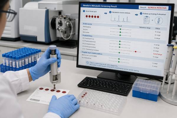
Newborn blood-spot screening looks for signs of serious conditions before a baby becomes visibly ill. Many of the target conditions are inherited metabolic disorders, in which the body cannot properly break down, use, or store nutrients and other chemicals. The screen may measure amino acids, acylcarnitines, enzymes, hormones, proteins, or DNA from a few drops of heel blood. It is often called a genetic test because most screened disorders have a genetic cause, but the first test is usually biochemical rather than a direct reading of the baby’s genes. An out-of-range result means more testing is needed; it does not prove that the baby has the named condition. Some metabolic disorders can cause rapid illness after feeding or fasting, so “time-critical” results require same-day action even when the newborn appears well. Accurate interpretation depends on collection time, prematurity, nutrition, transfusion, medications, and the specific state or national screening program.
- Newborn screening is a public-health screen, not a complete genetic diagnosis.
- Most metabolic markers are measured from a dried blood spot, often with tandem mass spectrometry or enzyme assays.
- Panels differ by state and country, and newly recommended conditions may not yet be implemented everywhere.
- Out-of-range results are commoner than confirmed disease because screening favors sensitivity.
- Some results are time-critical, requiring immediate metabolic consultation and treatment before confirmation is complete.
- A normal screen cannot exclude every metabolic disorder, especially rare conditions outside the panel or disorders not detectable at the collection time.
Table of Contents
- What Newborn Screening Tests—and What It Does Not
- How Metabolic Disorders Create Screening Markers
- Blood-Spot Collection, Timing, and Special Circumstances
- Understanding In-Range, Borderline, and Out-of-Range Results
- What Happens With a Time-Critical Result
- Confirmatory Biochemical and Genetic Testing
- Major Metabolic Disorder Groups and Early Treatment
- Records, Family Testing, and Future Pregnancies
What Newborn Screening Tests—and What It Does Not
Newborn screening is a coordinated system, not one laboratory test. It includes specimen collection, laboratory analysis, rapid reporting, diagnostic follow-up, treatment, and long-term care. In the United States, every state runs its own program. The Recommended Uniform Screening Panel, or RUSP, is a federal recommendation, but each state decides which conditions to include and when to implement additions.
The dried-blood-spot panel covers many inherited metabolic disorders along with endocrine, blood, immune, neuromuscular, and other conditions. Hearing screening and pulse oximetry for critical congenital heart disease are separate parts of newborn screening and do not use the blood spot.
Why screen before symptoms
A newborn with a metabolic disorder may look healthy while maternal support and regular feeding temporarily keep harmful chemicals low. Illness, longer fasting, or increasing protein intake can trigger a crisis. Depending on the disorder, toxic metabolites may injure the brain, liver, heart, or other organs within hours or days.
Screening identifies a biochemical pattern early enough to start protective measures. For phenylketonuria, early dietary treatment prevents severe neurologic injury. For medium-chain acyl-CoA dehydrogenase deficiency, avoiding prolonged fasting and providing glucose during illness can prevent hypoglycemia and sudden death. The purpose is not merely to name a condition; it is to change the outcome.
Why “genetic test” is only partly accurate
Most inherited metabolic disorders result from variants in genes that encode enzymes, transporters, or helper proteins. However, the first screen commonly measures the consequence of altered metabolism rather than the DNA change itself. For example, it may find high phenylalanine, an unusual acylcarnitine pattern, or low enzyme activity.
Some programs add DNA as a second-tier test to improve specificity, distinguish related disorders, or identify a common variant. A molecular finding on the screening card can still require confirmation from a new specimen. Newborn screening is designed for population detection, while diagnostic genetic testing is designed to establish the cause in one child.
What a normal result cannot promise
An in-range screen does not mean the baby has been tested for every genetic disease. It cannot exclude:
- Metabolic disorders not included on the local panel
- A screened disorder whose marker was not abnormal at collection
- Mild or later-onset forms below the cutoff
- Rare variants that escape a DNA tier
- Specimen, timing, transfusion, or treatment effects
- Disorders that require urine, cerebrospinal fluid, tissue, or specialized functional testing
A symptomatic baby needs medical evaluation even if the newborn screen was reported as normal. Screening should never delay testing for hypoglycemia, hyperammonemia, sepsis-like illness, or another urgent problem.
How Metabolic Disorders Create Screening Markers
Metabolism is a network of chemical reactions. Enzymes convert one molecule into another, transporters move substances across membranes, and cofactors help reactions occur. When a step is blocked, material before the block may accumulate, material after it may become deficient, or an alternate toxic pathway may become active.
The newborn screen looks for one or more signals produced by that disruption.
Tandem mass spectrometry
Tandem mass spectrometry can measure many amino acids and acylcarnitines from one small blood-spot punch. Amino acids are building blocks of proteins. Acylcarnitines are molecules formed as fats and some amino acids are moved through metabolic pathways.
A pattern may be more informative than one value. Elevated C8 acylcarnitine suggests MCAD deficiency, while a combination of C3 and related ratios can raise concern for propionic acidemia, methylmalonic acidemia, or vitamin B12-related conditions. High leucine-related signals can indicate maple syrup urine disease, but the initial method may not distinguish leucine from similar molecules without second-tier testing.
Laboratories use ratios because they can separate disease patterns from normal variation. The report may therefore list an abnormal marker, related markers, and ratios rather than one simple “level.”
Enzyme and protein assays
Some disorders are screened by measuring enzyme activity. Examples include biotinidase deficiency, Pompe disease, mucopolysaccharidosis type I or II, and other lysosomal storage disorders, depending on the panel. Low activity can reflect disease, carrier status in some assays, prematurity, specimen handling, or a pseudodeficiency variant that lowers laboratory activity without causing the clinical disorder.
Other screens measure proteins or hormones. Galactosemia programs may measure total galactose, galactose-1-phosphate, GALT enzyme activity, or a combination. Congenital hypothyroidism screening measures thyroid-related hormones, which is endocrine rather than an inherited metabolic disorder but uses the same card.
DNA-based tiers
DNA tests can target a common gene change, sequence selected genes, or check for a deletion. They may reduce false positives or provide fast etiologic information. Yet DNA alone has limitations: variants may be uncertain, only common changes may be covered, and finding one recessive variant may indicate carrier status rather than disease.
The relationship between marker and gene is not always one-to-one. The same metabolite can rise in several genetic disorders, maternal vitamin deficiency, liver disease, or treatment. Confirmatory testing must start from the biochemical pattern and the baby’s clinical status, not from a screening label alone.
Blood-Spot Collection, Timing, and Special Circumstances
A healthcare professional warms and cleans the baby’s heel, makes a small puncture, and allows blood to soak through printed circles on a filter-paper card. The card is dried and transported to the newborn screening laboratory. In many U.S. programs the routine specimen is collected around 24 to 48 hours after birth.
Collection too early can make some markers harder to interpret because the baby has had little protein feeding or because hormone and enzyme values are still changing. Delaying collection can miss the window for preventing illness. Programs use age-specific rules and may request another specimen after an early discharge.
Some states use a one-screen model, while others routinely collect a second specimen at about 1 to 2 weeks. A second screen does not mean the first was abnormal; it is part of that jurisdiction’s protocol.
Information on the card matters
The laboratory may need:
- Birth date and time
- Collection date and time
- Gestational age and birth weight
- Feeding type and timing
- Total parenteral nutrition, or TPN
- Transfusion history
- Medications and oxygen support
- NICU status
- Whether the specimen was collected before 24 hours
- Contact information for the responsible clinician
Missing or incorrect data can change the cutoff or delay follow-up. The hospital should identify who will receive results after discharge, especially when the family changes pediatricians or the baby remains in another facility.
Prematurity and intensive care
Premature and critically ill infants have more out-of-range results. Immature liver and kidney function, catabolism, respiratory distress, medications, and nutrition alter metabolite concentrations. This does not make screening unnecessary. It means the program may use serial specimens and interpret findings with neonatal specialists.
TPN contains amino acids and lipids that can mimic amino acid or fatty-acid oxidation disorders. Whenever medically safe and allowed by protocol, a specimen may be timed before TPN begins, but urgent nutrition should never be withheld just to improve a screen. The laboratory should be told when TPN was running.
Transfusion and treatment
Red-blood-cell transfusion can mask or distort disorders measured in red cells, including some galactosemia and hemoglobin screening methods. Exchange transfusion has an even larger effect. Programs specify when a pretransfusion specimen and later repeat are needed.
Antibiotics, carnitine, vitamin supplements, dialysis, and metabolic treatment can alter markers. A baby treated because a sibling has a known disorder may have a normal-looking screen despite being affected. Diagnostic testing should be arranged from the family history rather than relying on the routine result.
Specimen quality
Too little blood, blood applied from both sides, compressed spots, contamination, heat, humidity, or failure to dry the card can cause an unsatisfactory result. “Repeat needed” may therefore mean the laboratory could not perform a valid screen, not that a metabolic marker was abnormal. The replacement specimen should be obtained promptly.
Understanding In-Range, Borderline, and Out-of-Range Results
The exact wording differs by program, but reports generally fall into four categories.
| Result category | Meaning | Next step |
|---|---|---|
| In range or screen negative | No marker crossed the program’s action threshold | Routine care; investigate symptoms or family history separately |
| Borderline | A marker is near a cutoff or needs a repeat measurement | Repeat blood spot or specified diagnostic test promptly |
| Out of range or presumptive positive | The pattern could represent a screened disorder | Immediate contact and disorder-specific confirmatory testing |
| Unsatisfactory | The specimen or control was not valid | Repeat collection as directed |
Why screen positives often do not become diagnoses
Newborn screening prioritizes sensitivity because missing a treatable disorder can be devastating. Cutoffs are set to identify nearly all affected babies, which also captures unaffected infants. Prematurity, diet, illness, maternal factors, carrier status, pseudodeficiency, and ordinary biological variation contribute to false positives.
The positive predictive value varies widely by disorder and program. A rare condition can have many more false positives than true positives even when the laboratory method is excellent. Parents should hear both parts of the message: most abnormal screens require confirmation, and some require action before confirmation.
Marker name versus disorder name
A report may list “elevated C3—possible propionic or methylmalonic acidemia” or “elevated phenylalanine—possible PKU.” The named disorder is the condition the pattern is designed to detect, not a diagnosis.
Secondary conditions can be found because one marker pattern overlaps several diseases. For example, screening intended for one fatty-acid oxidation defect may identify another. The RUSP separates core and secondary conditions, but clinically both can require evaluation.
Borderline results
A borderline result is not always low urgency. The program’s follow-up instructions determine the timeline. Some repeat specimens can be collected in the outpatient clinic; others need plasma or urine testing the same day. If the baby is ill, diagnostic evaluation should replace routine repeat screening.
Result communication
Parents may receive a phone call before the full report appears in the patient portal. Ask for the exact marker, value, cutoff, collection age, and action category. Confirm which specialist is involved and where testing will occur. Do not assume the absence of a phone call proves that every result was normal; verify the screen at an early well-baby visit.
What Happens With a Time-Critical Result
Some metabolic conditions can deteriorate rapidly after feeding, fasting, or minor illness. ACMG newborn screening ACT sheets identify time-critical findings and give clinicians immediate steps. Examples include maple syrup urine disease, urea-cycle disorders, several organic acidemias, classic galactosemia, and some fatty-acid oxidation disorders.
A time-critical call usually triggers:
- Immediate contact with the family and assessment of the baby’s condition
- Consultation with a metabolic specialist
- Same-day confirmatory blood and urine collection
- Instructions about feeding, fasting, or emergency evaluation
- Treatment based on the suspected disorder and laboratory pattern
The baby should not wait for a routine clinic appointment. If the newborn has poor feeding, repeated vomiting, unusual sleepiness, low temperature, rapid breathing, seizures, limpness, jaundice, or an abnormal odor, emergency evaluation is needed.
Do not improvise dietary treatment
Metabolic treatment can involve stopping a specific sugar, limiting a particular amino acid, avoiding fasting, or giving specialized formula. The wrong restriction can cause malnutrition or worsen another disorder. Parents should not stop breastfeeding, dilute formula, give sugar water, or purchase a metabolic formula unless the specialist provides exact instructions.
For suspected fatty-acid oxidation disease, the immediate danger may be fasting, so frequent feeding or intravenous glucose is central. For suspected galactosemia, lactose and galactose intake may need to stop. For a urea-cycle defect, excess protein can worsen ammonia, but calories are needed to prevent catabolism. These differences explain why one generic “metabolic diet” is unsafe.
Emergency laboratory evaluation
Depending on the screen, urgent tests may include glucose, electrolytes, blood gas, ammonia, lactate, liver tests, ketones, plasma amino acids, acylcarnitines, urine organic acids, and disease-specific metabolites. An ammonia result requires proper collection and fast handling; a falsely high value from poor technique should be repeated quickly, but a concerning baby is treated while clarification occurs.
Early treatment may begin before the diagnosis is final. This is not overreaction. It is a reversible safety step when delay carries greater risk than short-term therapy.
Confirmatory Biochemical and Genetic Testing
Confirmation uses a fresh specimen and methods designed for diagnosis. The exact tests depend on the marker pattern.
Biochemical confirmation
Common tests include:
- Quantitative plasma amino acids
- Plasma acylcarnitine profile
- Urine organic acids
- Urine acylglycines
- Total and free carnitine
- Enzyme activity in blood cells, plasma, or cultured cells
- Disease-specific metabolites such as succinylacetone or very-long-chain fatty acids
- Vitamin and cofactor measurements when maternal or infant deficiency can mimic disease
A normal repeat marker does not always close the case. Some disorders fluctuate with feeding and illness. The specialist considers whether the confirmatory sample was collected after treatment, during good health, or at an age when the marker may have fallen.
Molecular confirmation
Genetic testing can confirm the responsible gene, distinguish related disorders, and identify carrier relatives. A panel may sequence several genes associated with the biochemical pattern. Deletion/duplication analysis, mitochondrial DNA testing, or genome sequencing may be needed when routine sequencing is negative.
Two pathogenic variants in an autosomal recessive gene often confirm the cause, but phase matters: the variants usually need to be on opposite gene copies. One variant may indicate carrier status, incomplete testing, or a different diagnosis. A variant of uncertain significance should not be treated as proof without biochemical and clinical support.
Maternal findings
Newborn screening occasionally detects a maternal condition. Low infant carnitine can reflect primary carnitine deficiency in the mother. Abnormal methylmalonic or propionylcarnitine markers can reflect maternal vitamin B12 deficiency. The baby still needs evaluation, but testing the mother may explain the pattern and identify a treatable health issue.
This is not the same as a false laboratory result. The screen has detected a real biochemical signal whose source is maternal rather than an inherited disorder in the infant.
Final classifications
After follow-up, the outcome may be:
- Confirmed disorder
- Carrier status
- Benign variant or pseudodeficiency
- Maternal condition affecting the newborn marker
- Transient abnormality related to prematurity, nutrition, or illness
- False positive with normal diagnostic testing
- Unresolved finding needing repeat monitoring
Families should receive the final classification in writing. A chart that only says “abnormal newborn screen” can cause confusion years later.
Major Metabolic Disorder Groups and Early Treatment
The following groups show why one screening panel uses different markers and follow-up plans.
Amino acid disorders
Phenylketonuria causes high phenylalanine and is treated with a phenylalanine-restricted diet and condition-specific therapies. Maple syrup urine disease impairs breakdown of branched-chain amino acids and can cause encephalopathy; urgent dietary management and removal of toxic metabolites may be needed. Homocystinuria and tyrosinemia require different markers and treatments despite also involving amino acids.
Organic acidemias
Propionic acidemia, methylmalonic acidemia, isovaleric acidemia, and related conditions can cause metabolic acidosis, hyperammonemia, low blood counts, cardiomyopathy, or neurologic injury. Treatment may include stopping catabolism with glucose, restricting precursor nutrients, carnitine, vitamins or cofactors, and emergency toxin removal. The first acylcarnitine pattern often overlaps, so urine organic acids and genetics refine the diagnosis.
Fatty-acid oxidation and carnitine disorders
These conditions impair the use of fat for energy during fasting. Babies may develop low blood sugar with few ketones, liver dysfunction, cardiomyopathy, muscle breakdown, or sudden death. Core management includes avoiding prolonged fasting and using an emergency illness plan. Some disorders also require fat modification or carnitine under specialist supervision.
Urea-cycle disorders
The urea cycle removes nitrogen by converting ammonia to urea. A block can cause rapidly rising ammonia, often with respiratory alkalosis and few early signs. Treatment may include high-calorie support, temporary protein interruption, nitrogen-scavenging medicines, arginine or citrulline for selected defects, and dialysis when ammonia is severe. Not every urea-cycle disorder is detected reliably by newborn screening.
Carbohydrate, cofactor, and storage disorders
Classic galactosemia requires removal of galactose and lactose after a concerning result. Biotinidase deficiency can be treated with biotin. Pompe disease and other lysosomal storage conditions are identified by low enzyme activity, followed by biomarker and genetic testing; enzyme replacement may be time-sensitive in severe infantile forms.
The treatment examples illustrate why a disorder-specific team matters. A result that appears similar on the initial card can lead to very different nutrition, medication, and emergency plans.
Records, Family Testing, and Future Pregnancies
Most inherited metabolic disorders on newborn screening are autosomal recessive. When both parents are carriers of pathogenic variants in the same gene, each pregnancy usually has a 25% chance of an affected child, a 50% chance of a carrier child, and a 25% chance of a child who inherited neither familial variant. Some disorders are X-linked, mitochondrial, or follow other patterns.
A confirmed molecular diagnosis allows targeted testing of parents and siblings. An older sibling who seems healthy may still need testing because some disorders have mild or intermittent forms. Testing relatives should be planned with genetics or metabolic specialists rather than inferred from the newborn’s carrier report.
Future options may include targeted prenatal testing by CVS or amniocentesis, donor gametes, adoption, or IVF with preimplantation genetic testing. Screening a future newborn remains important even when prenatal testing was reassuring because newborn panels cover many unrelated conditions.
Keep a durable result packet
Families should retain:
- The original newborn screening report
- Confirmatory biochemical results
- Exact gene and variant names
- The final diagnosis or resolved classification
- Emergency letters and feeding plans
- Medication and formula instructions
- Specialist contacts
- Guidance for siblings and future pregnancies
For a confirmed disorder, an emergency letter should explain what to do during vomiting, poor intake, fever, surgery, or fasting. Copies belong with caregivers, the primary clinician, and the nearest emergency department.
Questions after an abnormal metabolic screen
- Which marker or ratio was abnormal, and is the result time-critical?
- What conditions can produce this pattern?
- Could prematurity, TPN, transfusion, or early collection affect it?
- Should the baby feed normally while testing is arranged?
- Which symptoms require emergency care today?
- What blood and urine tests must be collected before treatment?
- Has a metabolic specialist reviewed the result?
- Is DNA testing diagnostic or only a second-tier screen?
- Could a maternal vitamin or metabolic condition explain the marker?
- When and how will the family receive the final classification?
Newborn screening works best as a chain: timely collection, reliable transport, sensitive laboratory detection, immediate follow-up, accurate diagnosis, and treatment. No link should be mistaken for the entire process. An abnormal card is an alert, while the confirmatory evaluation determines whether the baby has an inherited metabolic disorder and what care will protect the child.
References
- About Newborn Dried Blood Spot Screening — 2026, Centers for Disease Control and Prevention.
- Newborn Screening Process — 2024, HRSA Newborn Screening Information Center.
- Recommended Uniform Screening Panel — 2024, Health Resources and Services Administration.
- Recommended Uniform Screening Panel: Core Conditions and Secondary Conditions — 2025, federal screening panel.
- ACT Sheets and Algorithms — 2024, American College of Medical Genetics and Genomics.
- Newborn Screening Quality Assurance Program — 2024, Centers for Disease Control and Prevention.
Disclaimer
This article provides general education about newborn metabolic screening and cannot interpret a baby’s individual result. An out-of-range or time-critical screen may require immediate specialist-directed testing and treatment even if the baby appears healthy. Do not change feeding, formula, supplements, or medication without instructions from the newborn screening, metabolic, or emergency care team.





