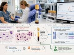
A plasma histamine test measures histamine circulating in the liquid portion of blood. Histamine rises quickly when mast cells or basophils release their stored mediators during an allergic or other activation event, but it also disappears from blood within minutes. That short window makes the test difficult to use well. A high result may support recent mediator release when the sample was collected promptly during typical symptoms and handled exactly as required. A normal result, especially from a delayed or poorly processed sample, does not rule out anaphylaxis, mast cell activation syndrome, or another mast-cell disorder. Plasma histamine is therefore an adjunct rather than a stand-alone diagnosis. Doctors usually interpret it with the event history, acute and baseline serum tryptase, urinary histamine metabolites, prostaglandin or leukotriene markers, and tests directed at possible triggers. During a suspected severe allergic reaction, treatment comes first; blood testing must never delay epinephrine or emergency care.
- A high plasma histamine level can support very recent mast-cell or basophil activation, but it is not specific to one disease.
- The most informative sample is usually collected during symptoms, ideally within about 15–30 minutes of onset.
- Histamine falls rapidly, so a normal delayed sample cannot exclude anaphylaxis or mast cell activation.
- The tube must be chilled, separated quickly, and frozen; handling errors can change the result.
- Laboratory reference ranges and units vary, so use the range printed on the report.
- Breathing difficulty, throat swelling, faintness, or rapidly worsening multisystem symptoms require immediate treatment, not laboratory confirmation.
Table of Contents
- What the Plasma Histamine Test Measures
- Collection Timing and Sample Handling
- What a High Plasma Histamine Result Means
- Normal, Low, and Borderline Results
- Use in Anaphylaxis and Mast Cell Activation
- Other Causes and Confounding Factors
- Follow-Up Testing and Next Steps
What the Plasma Histamine Test Measures
Histamine is a small signaling molecule made from the amino acid histidine. Mast cells store large amounts in tissue, especially in the skin, airway, and gastrointestinal tract. Basophils, a type of circulating white blood cell, also contain histamine. When these cells activate, histamine can enter nearby tissue and the bloodstream.
Histamine acts through several receptors. Its effects help explain familiar allergic symptoms:
- widening of blood vessels, which can cause warmth, flushing, and low blood pressure;
- increased leakage from small blood vessels, contributing to hives and swelling;
- stimulation of sensory nerves, causing itching;
- contraction and mucus production in the airway, contributing to wheeze or congestion;
- effects on the stomach and intestine, sometimes causing cramps, nausea, vomiting, or diarrhea.
The plasma test measures free histamine in blood after blood cells have been removed. It is different from a whole-blood histamine test, a basophil activation or histamine-release assay, and a urine test for N-methylhistamine. These tests answer related but different questions.
Plasma histamine is an event marker. It is most likely to rise when mediator release is occurring at or shortly before the blood draw. It does not measure how many mast cells a person has, identify the trigger, or prove that symptoms came from histamine. A person with systemic mastocytosis may have a normal plasma histamine level between episodes. Conversely, a temporary high result can occur without a chronic mast-cell disease.
Laboratories commonly report histamine in nanograms per milliliter, nanomoles per liter, or another assay-specific unit. The upper reference limit varies with the method, specimen preparation, and population used to establish the range. Some laboratories use an upper limit near 1.0 ng/mL, while others report a different interval. Converting a result or applying a cutoff from another laboratory can be misleading. The report’s own reference interval and specimen type should guide interpretation.
The assay also cannot show which receptor caused a symptom or whether an antihistamine would prevent a future reaction. Antihistamines block histamine receptors; they generally do not remove histamine from the sample or erase every other mast-cell mediator. A clinical response to an antihistamine may be informative, but it is not proof that a high plasma value came from a specific disorder.
Collection Timing and Sample Handling
Timing and handling determine whether plasma histamine testing has a reasonable chance of detecting an event. Histamine has a very short life in circulation. Enzymes rapidly metabolize it, and concentrations may return toward baseline within 30–60 minutes. For this reason, plasma histamine is less forgiving than serum tryptase or urine metabolite testing.
Timing the blood draw
When the purpose is to document an acute reaction, the sample should be collected as soon as practical after symptoms begin. A draw within roughly 15–30 minutes offers the best opportunity. A sample obtained later may still be requested, but its sensitivity falls quickly. In the 2023 anaphylaxis practice parameter, plasma histamine was not recommended routinely because of rapid degradation; it may have value in the unusual situation where blood can be obtained within 30 minutes.
A baseline sample taken when the person feels well can help in selected evaluations, although plasma histamine has more biological and preanalytical variability than serum tryptase. The clinician may instead prioritize paired acute and baseline tryptase or paired urine mediator measurements.
Record the following with the sample whenever possible:
- exact symptom-onset time;
- blood-draw time;
- foods, medicines, stings, exercise, heat, alcohol, infection, or other exposures;
- symptoms by organ system;
- treatment given before collection and the response;
- whether the sample remained cold and when it was processed.
A value without this timeline is hard to interpret. “Drawn the same day” is not precise enough for a marker that can change within minutes.
How the specimen is handled
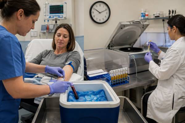
Plasma histamine usually requires EDTA plasma. Collection protocols commonly instruct staff to place the tube on ice immediately, centrifuge it under refrigerated conditions within about 20 minutes, transfer the upper portion of plasma without disturbing cells, and freeze it promptly. The testing laboratory’s instructions take priority because method requirements differ.
Poor handling can cause both false elevation and false reassurance. Delayed separation may allow blood cells to release histamine after collection. Warm storage can speed degradation. Hemolysis, marked lipemia, the wrong tube, repeated thawing, or contamination with the cellular layer may make a sample unacceptable or unreliable. A result from a routine tube left at room temperature should not be interpreted as though it came from a correctly handled acute sample.
Before going to a collection site for an outpatient event kit, confirm that the site can meet the full protocol. Many ordinary phlebotomy locations cannot centrifuge cold or freeze a sample immediately. Written orders should specify the test, tube, processing steps, and destination laboratory. Patients should not place themselves at risk or delay emergency treatment to reach a particular laboratory.
What a High Plasma Histamine Result Means
A value above the laboratory range means more histamine was present in plasma at the time represented by the sample. In the right setting, it supports recent activation of mast cells, basophils, or both. It does not by itself diagnose anaphylaxis, mast cell activation syndrome, systemic mastocytosis, food allergy, histamine intolerance, or any single cause.
Interpretation becomes stronger when four pieces align:
- Symptoms begin suddenly and resemble mediator release, such as hives, flushing, swelling, wheeze, vomiting, or a blood-pressure drop.
- Two or more organ systems are involved in a systemic episode.
- The sample is collected very early and handled correctly.
- Other findings, such as an acute tryptase rise or elevated urine mediators, support the same event.
The size of the elevation does not provide a dependable severity scale. A person can have severe anaphylaxis without a measurable plasma histamine rise if collection was late. Another person may have a noticeable laboratory elevation with mainly skin symptoms. The concentration reflects release, distribution, metabolism, timing, and specimen quality—not just clinical danger.
A high result also does not establish an IgE-mediated trigger. Mast cells can activate through IgE after exposure to a food, venom, latex, or medicine, but they can also respond through other receptors and pathways. Certain drugs can activate mast cells directly. Physical factors, complement pathways, and clonal mast-cell disorders may produce similar mediator patterns.
When the result was unexpected, review preanalytical details before launching a broad diagnostic workup. Questions include whether the tube was separated on time, whether the plasma was visibly hemolyzed, whether the test was truly plasma histamine rather than whole-blood histamine, and whether the reference range matched the method. Repeating a poorly collected random level is often less useful than planning a properly timed event sample and baseline measurements.
A persistently elevated random plasma level is unusual and deserves clinical review, but persistence still does not point automatically to mastocytosis. The evaluation should consider recurrent symptoms, baseline serum tryptase, complete blood count findings, kidney function, medications, and laboratory quality. Bone marrow testing or genetic testing is not justified solely by plasma histamine.
Normal, Low, and Borderline Results
A normal plasma histamine level is common outside the brief release window. It means the assay did not find an elevation in that specimen; it does not prove that no histamine was released earlier or in tissue.
Common reasons for a normal result despite a convincing episode include:
- the sample was collected more than 30–60 minutes after onset;
- symptoms began gradually, making the peak time unclear;
- histamine release was mainly local rather than systemic;
- another mediator, such as prostaglandin D2 or leukotrienes, dominated the episode;
- the specimen warmed, remained unseparated, or was otherwise mishandled;
- the assay’s detection limit or biological variation obscured a small rise.
A low plasma histamine result generally has no recognized disease meaning. Histamine is active mainly in tissue and is normally cleared rapidly from blood. There is no accepted “optimal” plasma histamine target for wellness, and treatment is not aimed at raising a low value.
Borderline values require restraint. A result just above the cutoff may reflect genuine mild release, normal variation near the reference boundary, or sample handling. A result just below the cutoff may still be compatible with an event if the draw was delayed. The clinical timeline often carries more weight than the decimal place.
Do not compare unlike samples as though they were a trend. Plasma histamine and whole-blood histamine have different reference ranges. A value reported in ng/mL cannot be compared directly with one in nmol/L without correct conversion, and even converted values from different assay platforms may not be equivalent. The same laboratory and protocol provide the cleanest event-to-baseline comparison.
Antihistamine use does not make a normal result definitive. H1 and H2 blockers occupy receptors rather than directly stopping every mast cell from releasing histamine. Some other medicines may affect activation pathways, but medication effects are not standardized enough to justify stopping needed treatment simply to “improve” the test. Any medication hold should come from the ordering clinician and must not compromise safety.
Use in Anaphylaxis and Mast Cell Activation
Plasma histamine can fit into an acute evaluation, but anaphylaxis remains a clinical diagnosis. Sudden skin or mucosal symptoms plus breathing difficulty or low blood pressure, or rapid involvement of multiple systems after a likely trigger, can justify treatment without laboratory confirmation. Epinephrine is first-line treatment. Antihistamines may help itching and hives after stabilization, but they do not treat airway obstruction or shock.
Anaphylaxis testing
For suspected anaphylaxis, clinicians more often obtain serum tryptase because its useful collection window is longer. Tryptase typically rises within minutes, peaks around one to two hours, and can remain elevated for several hours. An acute sample is compared with a baseline collected at least 24 hours after recovery or during a symptom-free period. A rise above the individual’s baseline can support systemic mast-cell activation even when both numbers lie within the laboratory’s population range.
Plasma histamine may add evidence when blood is drawn extremely early. It can also be elevated in some reactions where tryptase remains normal, including certain food reactions. Neither test has perfect sensitivity. A normal marker should not be used to reverse a well-supported clinical diagnosis or withhold an epinephrine prescription after a qualifying event.
Mast cell activation syndrome
Mast cell activation syndrome, or MCAS, requires more than chronic symptoms and one high histamine result. Consensus approaches generally look for recurrent, severe, systemic episodes typical of mast-cell mediator release; objective evidence of mediator elevation during an event; and improvement with treatment directed at mast-cell mediators or activation. Other disorders must be considered.
Plasma histamine is biologically relevant but is not the preferred sole laboratory criterion because it is unstable and can come from basophils as well as mast cells. More practical options may include:
- paired acute and baseline serum tryptase;
- urinary N-methylhistamine;
- urinary 2,3-dinor-11β-prostaglandin F2α;
- urinary leukotriene E4.
Urine mediator measurements are often collected during a flare and compared with baseline. They have their own dietary, medication, kidney, and collection limitations. Multiple abnormal mediators can strengthen evidence, but broad testing without a compatible symptom pattern increases the chance of incidental results.
Systemic mastocytosis is a clonal disorder involving an abnormal accumulation of mast cells. Its evaluation centers on baseline tryptase, KIT mutation testing, tissue findings, and other established criteria—not plasma histamine alone. A high plasma histamine event value may document activation in a person with mastocytosis, but it does not measure mast-cell burden.
Other Causes and Confounding Factors
A high level can arise in several clinical and technical settings. The result must be interpreted with the whole picture rather than assigned automatically to MCAS.
Allergic and immune triggers
IgE-mediated reactions to foods, insect venom, medicines, or latex can release histamine. Severe seasonal allergy, asthma, or urticaria may involve histamine, but localized symptoms do not always produce a measurable plasma rise. Complement-mediated reactions and some immune complexes can also activate mast cells or basophils.
Non-IgE mast-cell activation
Some medicines activate mast cells through non-IgE pathways. Examples may include certain opioids, radiocontrast exposures, and drugs that interact with mast-cell receptors. Exercise, temperature change, pressure, or alcohol can act as triggers in susceptible people. A non-IgE mechanism can look clinically identical to allergy during the event, so specialist assessment may be needed before future exposure.
Other biological sources
Basophils are a legitimate source of circulating histamine. Platelets and other cellular interactions can influence measured concentrations indirectly. Histamine is also involved in gastric acid regulation and nervous-system signaling, although plasma testing does not localize the source.
Food naturally contains histamine, and intestinal metabolism influences symptoms after high-histamine meals. However, plasma histamine is not a validated stand-alone test for “histamine intolerance.” Symptoms attributed to dietary histamine overlap with migraine, flushing disorders, medication effects, gastrointestinal disease, and allergy. Diamine oxidase testing and low-histamine diets also have limitations. Prolonged restrictive diets should not be started from one plasma result without a structured clinical assessment.
Collection-related elevation
The most avoidable confounder is release after the blood has left the body. Rough handling, prolonged tourniquet time, failure to keep the tube cold, delayed centrifugation, or contamination of plasma with cells can increase measured histamine. These problems can create a number that looks biologically important but reflects the specimen rather than the patient.
Stress, exercise, acute illness, and recent procedures may change mediator release, but their effects are variable. Instead of trying to control every possible factor, the clinician should define the testing purpose. For an event sample, capturing the episode quickly is more important than fasting. For a baseline sample, collecting during a stable period with the same protocol improves comparability.
Follow-Up Testing and Next Steps
The next step depends on why the test was ordered and how well the result matches the episode.
After a suspected anaphylactic event, the clinical priorities are documenting the trigger and symptoms, arranging baseline tryptase, reviewing emergency treatment, and deciding whether allergy testing is needed. Trigger evaluation may include targeted skin testing, specific IgE blood testing, supervised challenge, or medication-allergy assessment. Broad panels without a history can detect sensitization that is unrelated to the event.
For recurrent unexplained episodes, a specialist may create a written collection plan. It can specify:
- which symptoms count as an event;
- when to obtain acute tryptase and any plasma histamine sample;
- whether to collect spot or timed urine mediators;
- when to obtain baseline samples;
- how the laboratory must process each specimen;
- which medicines should be continued or held;
- when emergency treatment takes priority over collection.
Paired results are often more useful than isolated values. For tryptase, the acute value is interpreted against the person’s own baseline using an event-related increase formula. For urine mediators, an event-to-baseline ratio may provide context. Plasma histamine lacks a universally accepted paired-rise formula, but a clearly elevated acute value with a normal properly handled baseline can still support the narrative.
Further evaluation may include a complete blood count, metabolic panel, kidney function, baseline tryptase, allergy testing, and tests for conditions that mimic mediator episodes. Depending on symptoms, clinicians may consider arrhythmia, vasovagal syncope, panic episodes, endocrine causes of flushing, carcinoid syndrome, medication effects, hereditary angioedema, asthma, or gastrointestinal disease. Testing should follow the symptom pattern rather than expand indefinitely after a borderline laboratory value.
Seek emergency help for trouble breathing, throat tightness, tongue swelling, collapse, severe dizziness, or rapidly progressing symptoms involving more than one body system. Use epinephrine immediately when an emergency plan indicates it. Do not wait for hives, and do not wait for a laboratory result.
Useful questions for the ordering clinician include:
- Was the sample collected early enough to be meaningful?
- Did the collection site follow the required cold-processing protocol?
- Is the elevation large enough to exceed analytical and biological variation?
- How does it compare with acute and baseline tryptase or urine mediators?
- Does the symptom pattern meet criteria for anaphylaxis or systemic mast-cell activation?
- Which trigger-specific tests are appropriate?
- What should be collected during the next episode?
A plasma histamine test is most valuable when it is part of a planned, time-stamped evaluation. Its main strength is capturing very recent mediator release. Its main weakness is the same: the signal can be gone before the patient reaches care. Good interpretation respects both facts.
References
- Biomarkers in the diagnosis of mast cell activation 2024 (Review)
- Mast cell activation syndrome: Current understanding and research needs 2024 (Review)
- Mast cell activation syndrome: An up-to-date review of literature 2024 (Review)
- Anaphylaxis: A 2023 practice parameter update 2024 (Practice Parameter)
- Secretory and Membrane-Associated Biomarkers of Mast Cell Activation and Proliferation 2023 (Review)
- Histamine, Plasma | ARUP Laboratories Test Directory 2026 (Official Laboratory Guide)
Disclaimer
This article provides general information and cannot diagnose anaphylaxis, mast cell activation syndrome, mastocytosis, or histamine intolerance. Laboratory results must be interpreted by a qualified clinician using the symptom timeline and specimen-quality details. Suspected anaphylaxis requires immediate treatment; testing should never delay epinephrine or emergency care.