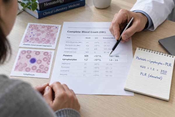
The platelet-to-lymphocyte ratio (PLR) is calculated from two components of a complete blood count: the platelet count and the absolute lymphocyte count. Platelets help form clots, but they also participate in inflammatory signaling, blood-vessel responses, and interactions with immune cells. Lymphocytes coordinate adaptive immunity through B cells, T cells, and natural killer cells. When platelets rise, lymphocytes fall, or both changes occur together, the PLR increases. Researchers have linked higher PLR values with worse outcomes in several inflammatory, cardiovascular, infectious, and cancer settings. Those associations do not make PLR a diagnostic test. There is no universal healthy interval or disease cutoff, and the same ratio can result from very different blood-count patterns. Iron deficiency, recent surgery, corticosteroids, smoking, pregnancy, infection, immune suppression, and bone marrow disorders can all alter the value. Proper interpretation begins with the individual platelet and lymphocyte counts and then considers symptoms, medications, other CBC findings, and the reason the ratio was calculated.
- PLR equals the platelet count divided by the absolute lymphocyte count.
- A high ratio may reflect high platelets, low lymphocytes, or both.
- PLR is an adjunctive inflammatory and prognostic index, not a stand-alone diagnosis.
- Published cutoffs are condition-specific and cannot be applied universally.
- The underlying counts matter more than the ratio when deciding what to investigate.
Table of Contents
- How PLR Is Calculated and What the Two Cell Types Do
- How to Read a PLR Number Without a Universal Range
- High PLR: Platelet, Lymphocyte, and Combined Patterns
- Where PLR Is Used in Clinical Research and Risk Assessment
- When PLR Can Be Misleading
- Evaluation, Follow-Up, and Treatment
How PLR Is Calculated and What the Two Cell Types Do
PLR is usually derived from a CBC with differential rather than ordered as a separate laboratory assay. The formula is:
PLR = platelet count ÷ absolute lymphocyte count
If the platelet count is 250 × 109/L and the absolute lymphocyte count is 2.0 × 10⁹/L, the PLR is 125. The units cancel, so the ratio is reported as a plain number. A laboratory information system, research database, or clinician may calculate it automatically.
Platelets are small cell fragments produced by megakaryocytes in the bone marrow. Their best-known job is to adhere and aggregate at damaged blood vessels, helping stop bleeding. Platelets also release signaling molecules, interact with white cells and the vessel lining, and contribute to inflammatory and immune processes. Their number can rise reactively during infection, inflammation, bleeding, iron deficiency, tissue injury, or recovery from low counts.
Lymphocytes are white blood cells that include B cells, T cells, and natural killer cells. They produce antibodies, direct cellular immune responses, regulate inflammation, create immune memory, and remove infected or abnormal cells. The absolute lymphocyte count may decline during acute physiological stress, severe infection, corticosteroid exposure, immune suppression, malnutrition, and some chronic diseases.
The ratio is intended to capture a relationship between platelet-related inflammatory activity and lymphocyte-mediated immune capacity. That interpretation is a simplification. Platelet number does not measure platelet activation or function, and lymphocyte count does not describe the proportions or performance of lymphocyte subtypes. A normal count can coexist with abnormal function, while an abnormal count may be temporary and clinically harmless.
The mathematical result also hides the mechanism. Consider three examples:
| Platelets | Absolute lymphocytes | PLR | What drives the ratio |
|---|---|---|---|
| 400 × 109/L | 2.0 × 10⁹/L | 200 | Higher platelet count |
| 200 × 109/L | 1.0 × 10⁹/L | 200 | Lower lymphocyte count |
| 400 × 109/L | 1.0 × 10⁹/L | 400 | Both changes |
The first pattern may suggest reactive thrombocytosis; the second may focus attention on lymphopenia; the third may occur during a strong inflammatory or stress response. Identical PLR values therefore do not guarantee identical causes or risks.
How to Read a PLR Number Without a Universal Range
Most routine laboratory reports provide reference intervals for platelets and lymphocytes but not for PLR. Population studies often find typical adult values somewhere around 100 to 200, yet ranges vary substantially by age, sex, ethnicity, pregnancy status, smoking, health conditions, and study design. Numbers in that broad area should not be used as universal boundaries.
Research studies commonly select a cutoff by analyzing which value best separates groups with and without a particular outcome. A cancer study might use 150, 180, or 200; a cardiovascular study might choose another threshold; and an infection study may divide patients into quartiles rather than use a fixed value. A cutoff optimized for one dataset may perform poorly elsewhere.
Differences arise because studies vary in:
- the disease, stage, and severity being examined;
- whether PLR is measured before treatment, during hospitalization, or after surgery;
- the outcome, such as mortality, recurrence, complications, or treatment response;
- the age and health of the comparison population;
- the handling of medications, infection, anemia, and other confounders;
- the statistical method used to choose the threshold.
A high PLR label on a wellness report or online calculator may therefore create more certainty than the science supports. PLR has not been standardized as a general-population screening test for “immune balance,” hidden inflammation, cardiovascular disease, or cancer. A single value cannot quantify how inflamed a person is.
Ratios are especially sensitive to a small denominator. When lymphocytes are already low, a modest further decrease can produce a large jump in PLR even if platelets barely change. For example, platelets of 240 with lymphocytes falling from 1.2 to 0.6 raise PLR from 200 to 400. That mathematical doubling does not necessarily mean the biological process doubled in severity.
The most defensible comparison is often with the person’s own previous CBCs, obtained in similar circumstances. Even then, trends should be interpreted alongside clinical change. A rising ratio after surgery may be expected; the same trend in a stable outpatient with persistent thrombocytosis may prompt a different evaluation.
A low PLR also lacks a universal cutoff. It can result from low platelets, high lymphocytes, or both. The urgency depends on the component count. Significant thrombocytopenia can create bleeding risk even if the ratio itself is not emphasized, while lymphocytosis may accompany infection or a hematologic condition. The ratio should never distract from a dangerously abnormal numerator or denominator.
High PLR: Platelet, Lymphocyte, and Combined Patterns
A high PLR is best approached by asking which component is abnormal.
High platelets with lymphocytes in range
Reactive thrombocytosis is more common than a primary bone marrow disorder. Potential causes include:
- acute or chronic infection;
- autoimmune or inflammatory disease;
- iron deficiency, with or without obvious anemia;
- recent bleeding, surgery, trauma, or tissue injury;
- recovery after infection, chemotherapy, or a period of thrombocytopenia;
- removal or reduced function of the spleen;
- some cancers.
Persistent unexplained thrombocytosis can also occur in myeloproliferative neoplasms such as essential thrombocythemia. Those disorders require evaluation of the platelet trend, blood smear, symptoms, iron status, inflammatory markers, and sometimes molecular tests. PLR cannot distinguish reactive from clonal thrombocytosis.
Low lymphocytes with platelets in range
Lymphopenia may be transient during acute illness or a stress response. Corticosteroids can lower circulating lymphocytes. Other possibilities include severe infection, autoimmune disease, protein-calorie malnutrition, HIV or other immune disorders, chemotherapy, radiation, immunosuppressive drugs, bone marrow disease, and loss of lymphocytes through certain gastrointestinal or kidney conditions.
The depth and duration of lymphopenia matter. A mildly low count during an acute illness often resolves. Persistent or severe lymphopenia, recurrent unusual infections, weight loss, enlarged lymph nodes, or abnormalities in other cell lines may require more detailed immune or hematologic testing.
High platelets plus low lymphocytes
This combination can produce a striking PLR. It may occur with substantial inflammation, major surgery, trauma, severe infection, active malignancy, or concurrent iron deficiency and physiological stress. The pattern can support concern that the body is under stress, but it cannot identify the cause or prove the condition is severe.
Other influences include smoking, obesity, pregnancy, aging, chronic kidney disease, liver disease, and medications. These factors can shift baseline platelet or lymphocyte counts and may partly explain why population-specific reference values differ.
A high ratio is not the same as a blood clot. Platelet count and clot risk have a complicated relationship, and PLR does not measure platelet function, coagulation factors, blood flow, or whether thrombosis is present. Suspected deep-vein thrombosis, pulmonary embolism, stroke, or heart attack must be evaluated with symptoms, examination, imaging, electrocardiography, and appropriate laboratory tests—not PLR.
Where PLR Is Used in Clinical Research and Risk Assessment
PLR has attracted attention because it is inexpensive and available from routine blood work. Studies have evaluated it in cancer, cardiovascular disease, infection, critical illness, autoimmune conditions, kidney disease, pregnancy complications, and perioperative care. In many analyses, groups with higher PLR have experienced more complications or poorer survival.
The most established role remains research-supported prognosis rather than diagnosis. A prognostic association means that, within a defined patient population, higher values correlate statistically with an outcome. It does not mean PLR causes the outcome, identifies the disease, or predicts an individual future with certainty.
In oncology, pretreatment PLR has been studied across many solid tumors. Elevated values sometimes correlate with advanced disease, lower treatment response, recurrence, or reduced survival. However, cancer type, stage, therapy, infection, anemia, steroid use, and the chosen cutoff differ across studies. PLR cannot screen for cancer or replace biopsy, imaging, molecular testing, and established staging systems.
In cardiovascular research, higher PLR has been associated with adverse outcomes in acute coronary syndromes and other vascular conditions. Platelet activation and inflammation are biologically relevant to atherosclerosis, but a simple cell-count ratio does not measure coronary blockage or diagnose a heart attack. Troponin, electrocardiography, symptoms, and imaging remain central.
In infectious and critical care settings, PLR may be combined with other CBC-derived indices, including the neutrophil-to-lymphocyte ratio or systemic immune-inflammation index. Reviews find that these measures can add prognostic information, yet their stand-alone accuracy for a specific condition is limited. They generally perform better as pieces of a multivariable assessment than as isolated thresholds.
Possible uses include:
- Describing baseline inflammatory status before surgery or treatment.
- Adding a low-cost variable to a validated risk model.
- Following a trend when the same disease and treatment context are maintained.
- Generating research hypotheses about platelet-immune interactions.
PLR should not be used alone to start antibiotics, diagnose autoimmune disease, change chemotherapy, prescribe antiplatelet medication, or determine whether a person needs hospitalization. Those decisions demand condition-specific evidence and direct clinical assessment.
When PLR Can Be Misleading
Because PLR is built from routine counts, every source of error or biological variation affecting those counts can alter the ratio.
Temporary physiological changes: Exercise, acute anxiety, pain, sleep disruption, and recent illness can redistribute blood cells. Pregnancy produces expected hematologic changes. A single sample may not represent a stable baseline.
Medication effects: Corticosteroids can lower lymphocytes. Chemotherapy, immunotherapy, radiation, antiplatelet drugs, immune suppressants, and growth factors may alter counts directly or through marrow recovery. Medication timing should be recorded when trends are compared.
Iron deficiency: Iron deficiency can increase platelets and therefore PLR. In that setting, the ratio may reflect iron status more than an independent inflammatory process. Ferritin and iron studies may be relevant, but ferritin itself can rise with inflammation.
Splenic function: Platelet counts may remain higher after splenectomy, creating a different baseline. The spleen also helps regulate immune-cell populations.
Laboratory artifacts: Platelets sometimes clump in an EDTA blood tube, causing a falsely low automated platelet count. Very small red cell fragments or other particles can occasionally be miscounted as platelets. Analyzer flags or unexpected changes may lead to repeat testing, a different anticoagulant tube, or a peripheral blood smear.
Hematologic disease: Leukemia, lymphoma, marrow failure, myeloproliferative neoplasms, and immune thrombocytopenia can produce ratios that do not fit a simple inflammation narrative. Abnormal cells, extreme counts, anemia, or persistent multi-line abnormalities deserve direct evaluation.
Timing: Platelets and lymphocytes do not change at the same speed. A ratio during the onset of illness, after fluid resuscitation, after surgery, or during marrow recovery may differ substantially from a later value. A fall in PLR can reflect lymphocyte recovery, platelet decline, or both; it is not automatically proof that treatment worked.
PLR can also disagree with other inflammatory markers. Such discordance is not necessarily an error. CRP reflects a liver-produced acute-phase protein, ESR reflects red-cell sedimentation, and PLR reflects cell counts. Each responds to different biological inputs.
Evaluation, Follow-Up, and Treatment
When PLR is unexpectedly high, the first step is not to lower the ratio. It is to identify why the platelet or lymphocyte count changed. A clinician may review the full CBC, prior results, recent infections, medications, menstrual or other blood loss, diet, surgery, smoking, inflammatory symptoms, cancer history, and immune risk.
Depending on the pattern, follow-up may include:
- a repeat CBC with differential to confirm persistence;
- a peripheral blood smear if counts are extreme or analyzer flags appear;
- ferritin, transferrin saturation, and other iron studies;
- CRP or ESR when inflammatory disease is suspected;
- kidney and liver testing;
- infection testing guided by symptoms and exposure;
- immune evaluation for persistent lymphopenia or recurrent infection;
- hematology referral for sustained unexplained thrombocytosis, abnormal cells, or multiple blood-count abnormalities.
Treatment is directed at the cause. Iron replacement may normalize reactive thrombocytosis caused by iron deficiency. Platelets may fall as an infection or inflammatory flare resolves. Medication-related lymphopenia may improve after a clinically supervised change. A myeloproliferative neoplasm or immune disorder requires specialist management.
There is no validated supplement, food, detox, or exercise program specifically designed to normalize PLR. Healthy habits can support overall cardiovascular and immune health, but attempting to manipulate a ratio may delay investigation of anemia, infection, medication effects, or a blood disorder. Do not start aspirin or another blood thinner because PLR is high; those medicines have real bleeding risks and require a specific indication.
Seek urgent care for chest pain, sudden shortness of breath, coughing blood, one-sided leg swelling, new weakness or speech difficulty, severe headache, uncontrolled bleeding, confusion, very high fever, or rapidly worsening illness. These symptoms require evaluation whether PLR is high, normal, or unknown.
For a stable outpatient result, useful questions include:
- Which component is driving my ratio?
- Are my platelet and lymphocyte counts actually outside the laboratory ranges?
- Is the PLR being used for a validated purpose in my condition?
- Could iron deficiency, medication, smoking, surgery, or recent illness explain it?
- Would repeating the CBC change the plan?
PLR is best understood as a compact description of two blood counts, not as a direct measure of “immune balance.” It can add context in selected diseases and research models, but safe interpretation returns to the platelet count, absolute lymphocyte count, the rest of the CBC, and the person’s clinical story.
When following PLR over time, record whether the blood samples were taken during illness, after treatment, or under otherwise comparable circumstances. A ratio that returns toward a personal baseline can be reassuring when symptoms and the component counts improve as well. A ratio that remains high for months deserves attention to the platelet and lymphocyte trends, even when the person feels well, because persistence changes the range of possible explanations.
References
- Platelet Tests 2024
- Blood Differential 2024
- Complete Blood Count (CBC) 2024
- Unraveling the Clinical Significance and Prognostic Value of Neutrophil-to-Lymphocyte Ratio, Platelet-to-Lymphocyte Ratio, Systemic Immune-Inflammation Index, Systemic Inflammation Response Index, and Delta Neutrophil Index: An Extensive Literature Review 2024
- The Prognostic Role of Platelet-to-Lymphocyte Ratio in Patients with Acute Coronary Syndrome: A Systematic Review and Meta-Analysis 2023
Disclaimer
This article is for general education and does not diagnose inflammation, infection, cancer, or a clotting disorder. PLR has no universal cutoff, and results must be interpreted from the individual platelet and lymphocyte counts, clinical context, and laboratory findings. Seek urgent care for symptoms of a blood clot, stroke, severe infection, or significant bleeding.