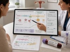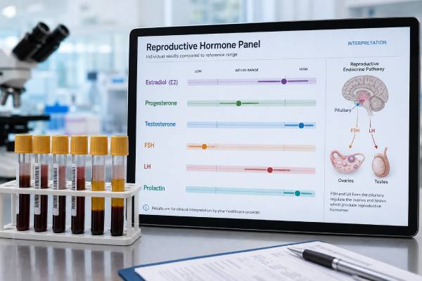
A reproductive hormone panel measures estrogen—usually estradiol—progesterone, testosterone, follicle-stimulating hormone (FSH), luteinizing hormone (LH), and prolactin. The panel can help evaluate irregular periods, infertility, ovulation, menopause, low testosterone, androgen excess, and pituitary or gonadal disorders. It is not one universal test with one normal pattern. Expected results differ by sex, age, menstrual-cycle phase, pregnancy status, menopause, time of day, medications, and laboratory method. In people with menstrual cycles, FSH, LH, and estradiol may be checked early in the cycle, while progesterone is usually measured about seven days after ovulation. Testosterone is generally collected in the morning when evaluating men, and unexpected results often need confirmation. Prolactin can rise temporarily with stress, sleep, exercise, or nipple stimulation. Clinicians interpret the hormones as a feedback system: pituitary FSH and LH signal the ovaries or testes, sex hormones provide feedback, and prolactin can suppress the entire reproductive axis when elevated.
- The panel’s meaning depends on the clinical question and timing.
- Estradiol, FSH, LH, and progesterone change across the menstrual cycle.
- Morning repeat testing is important for low testosterone in men.
- High prolactin can suppress ovulation or testosterone production.
- High gonadotropins with low sex hormones suggest gonadal failure.
- Low gonadotropins with low sex hormones suggest hypothalamic or pituitary suppression.
Table of Contents
- What a Reproductive Hormone Panel Measures
- Why the Panel Is Ordered
- Timing and Preparation
- How to Interpret Each Hormone
- Common Result Patterns in Women
- Common Result Patterns in Men
- Limitations, Follow-Up, and Next Steps
What a Reproductive Hormone Panel Measures
The reproductive system is controlled by the hypothalamic-pituitary-gonadal axis. The hypothalamus releases gonadotropin-releasing hormone in pulses. The pituitary responds by releasing FSH and LH. These hormones act on the ovaries or testes, which produce estradiol, progesterone, testosterone, inhibins, and other signals. Sex hormones then feed back to the brain and pituitary.
A typical panel includes:
| Hormone | Main source | Main information |
|---|---|---|
| Estradiol | Ovaries; smaller amounts from testes and peripheral conversion | Follicle activity, ovarian estrogen status, or estrogen exposure |
| Progesterone | Corpus luteum and placenta | Evidence of recent ovulation or pregnancy-related support |
| Testosterone | Testes; ovaries and adrenal pathways | Androgen status, gonadal function, or androgen excess |
| FSH | Pituitary | Ovarian follicle recruitment or testicular Sertoli-cell stimulation |
| LH | Pituitary | Ovulation signaling or testicular testosterone stimulation |
| Prolactin | Pituitary | Lactation signal and possible suppression of reproductive function |
The hormones are linked. A low sex-hormone level should be interpreted with FSH and LH. If the gonads are failing, feedback falls and FSH/LH generally rise. If the hypothalamus or pituitary is not signaling adequately, sex hormones and gonadotropins may all be low or inappropriately normal.
Prolactin sits alongside this axis. Persistent elevation can reduce GnRH pulses, lowering FSH, LH, estradiol, progesterone, or testosterone. This can create a secondary hypogonadism pattern even though the ovaries or testes are structurally capable of functioning.
The panel does not directly measure egg quality, sperm count, fallopian tube openness, uterine anatomy, sexual function, or pregnancy potential. It provides endocrine clues that must be combined with symptoms and other tests. A reproductive hormone panel is best viewed as a map of signaling, not a fertility score.
Why the Panel Is Ordered
The same panel may be ordered for very different clinical questions.
In people with ovaries, reasons include:
- Irregular, infrequent, or absent periods
- Infertility or suspected lack of ovulation
- Symptoms of PCOS or androgen excess
- Possible primary ovarian insufficiency
- Perimenopause or menopause when diagnosis is uncertain
- Galactorrhea or suspected high prolactin
- Delayed or early puberty
- Monitoring fertility treatment or hormone therapy
In people with testes, reasons include:
- Low libido or erectile dysfunction with other signs of low testosterone
- Infertility or abnormal semen analysis
- Reduced facial or body hair, low muscle mass, or gynecomastia
- Delayed or early puberty
- Testicular injury, chemotherapy, radiation, or surgery
- Pituitary symptoms or high prolactin
- Monitoring testosterone or gonadotropin treatment
Clinicians may order all six hormones, but a targeted panel is often better. Progesterone is useful only when timed to ovulation or a treatment protocol. Estradiol is important with FSH in early-cycle or amenorrhea evaluation. SHBG and free testosterone may be needed when total testosterone does not fit symptoms. TSH is commonly added because thyroid disease can alter cycles, prolactin, sexual function, and fertility.
For infertility, hormone tests are only one component. Women may need assessment of ovulation, ovarian reserve, the uterus, and fallopian tubes. Men generally need at least one semen analysis, because normal testosterone, FSH, and LH do not guarantee normal sperm concentration or movement. An infertility panel for men supports but does not replace semen testing.
Testing is also ordered to monitor treatment. The expected pattern differs during ovarian stimulation, testosterone therapy, gender-affirming hormone therapy, ovulation induction, menopause therapy, or prolactinoma treatment. The prescribing clinician’s targets take priority over general reference intervals.
Timing and Preparation
Timing is one of the most important parts of reproductive hormone testing.
Menstrual-Cycle Testing
FSH, LH, and estradiol are commonly checked on cycle days 2–4 when evaluating ovarian reserve or baseline ovarian function. Cycle day 1 is the first day of full bleeding. These hormones can be measured on other days for different questions, but the reference range must match the phase.
Progesterone should be measured about six to eight days after ovulation or approximately seven days before the next expected period. The common “day 21” test applies only to a roughly 28-day cycle. A positive urine LH test or ultrasound can guide timing.
Testosterone in women can be measured in the morning, preferably during the early follicular phase when practical, especially when an unexpected elevation needs confirmation. Hormonal contraception suppresses ovarian androgen production and changes SHBG, making interpretation difficult.
Testosterone Testing in Men
Total testosterone is usually drawn early in the morning, often between 7 and 10 a.m., after a normal night’s sleep. A low result should be repeated on another morning before diagnosing testosterone deficiency. Acute illness, sleep deprivation, calorie restriction, and some medications can temporarily lower it.
Prolactin Testing
For a mild elevation, repeat prolactin in the morning at least a few hours after waking, after resting quietly. Avoid strenuous exercise, sex, and nipple stimulation beforehand when possible. A stressful blood draw can raise the value.
Fasting is not universally required, but follow the laboratory’s instructions, especially when glucose or lipid tests are included. Tell the clinician about:
- Pregnancy or breastfeeding
- Hormonal contraception or menopause therapy
- Fertility medication and progesterone support
- Testosterone, anabolic steroids, DHEA, or bodybuilding products
- Antipsychotics, anti-nausea drugs, opioids, and other prolactin-raising medicines
- Biotin supplements
- Recent illness, intense exercise, major weight change, or poor sleep
Do not stop prescribed hormones or psychiatric medications without medical guidance. A planned medication washout is sometimes useful but must be safe and paired with alternative contraception or treatment when needed.
How to Interpret Each Hormone
Estradiol
Estradiol is the main estrogen during the reproductive years. In cycling women, it is relatively low early in the follicular phase, rises as a dominant follicle develops, peaks before ovulation, and has a smaller luteal rise. Low estradiol with high FSH suggests reduced ovarian function; low estradiol with low or normal FSH/LH suggests hypothalamic or pituitary suppression. High estradiol can occur near ovulation, in pregnancy, with ovarian cysts, multiple stimulated follicles, or estrogen medication.
In men, estradiol comes mainly from conversion of testosterone in fat and other tissues. It supports bone and sexual function. Testing may be helpful with gynecomastia, very low testosterone, liver disease, or therapy monitoring, but it is not required in every evaluation.
Progesterone
Progesterone below about 1 ng/mL is expected before ovulation. A luteal value above 3 ng/mL generally supports recent ovulation. Because secretion is pulsatile, one result cannot diagnose a defective luteal phase or predict pregnancy. The progesterone test must be matched to the actual ovulation date.
Testosterone
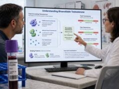
Total testosterone includes free hormone and hormone bound to SHBG or albumin. Free testosterone becomes important when SHBG is abnormal or symptoms conflict with total testosterone. In men, diagnosis of deficiency requires compatible symptoms plus consistently low morning values. In women, mild elevation is common in PCOS; marked elevation or rapid virilization requires prompt evaluation.
FSH
FSH stimulates ovarian follicles and Sertoli cells in the testes. High FSH with low estradiol in women suggests ovarian insufficiency or menopause. High FSH in men, especially with low testosterone or abnormal semen results, suggests impaired testicular function. Low FSH can result from hypothalamic or pituitary suppression.
LH
LH triggers ovulation and corpus luteum formation in women and stimulates Leydig-cell testosterone production in men. A midcycle surge is normal. High LH with low sex hormones suggests gonadal failure. Low LH with low sex hormones suggests central suppression. An elevated LH/FSH ratio can occur in PCOS but is not diagnostic.
Prolactin
Persistent high prolactin can suppress the axis. Mild elevations should be repeated and evaluated for pregnancy, medication effects, hypothyroidism, kidney disease, stress, and macroprolactin. A very high or persistent unexplained level may lead to pituitary MRI. The prolactin blood test should never be interpreted without medication review.
Common Result Patterns in Women
Patterns are more informative than isolated flags.
| Pattern | Possible interpretation | Next considerations |
|---|---|---|
| High FSH, low estradiol, irregular or absent periods | Primary ovarian insufficiency or menopause | Repeat testing, pregnancy exclusion, age and symptom review |
| Low/normal FSH and LH, low estradiol | Hypothalamic or pituitary suppression | Nutrition, exercise, stress, chronic illness, pituitary assessment |
| Mildly high testosterone, low SHBG, irregular cycles | Common PCOS pattern | Exclude thyroid disease, high prolactin, and nonclassic CAH |
| Markedly high testosterone with rapid virilization | Possible ovarian/adrenal tumor or other severe androgen disorder | Urgent repeat accurate assay and specialist evaluation |
| High prolactin, low estradiol, low/normal gonadotropins | Prolactin-related reproductive suppression | Repeat, medication/thyroid review, macroprolactin, possible MRI |
| Progesterone above 3 ng/mL in the luteal phase | Recent ovulation likely | Does not prove egg quality or implantation |
| High FSH with elevated early-cycle estradiol | Possible diminished ovarian reserve | AMH and antral follicle count may help treatment planning |
Pregnancy changes the entire pattern: estradiol and progesterone rise, prolactin increases, and pituitary FSH and LH are suppressed. Nonpregnant ranges should not be used. Hormonal contraception likewise creates a medication-driven pattern that may not reflect natural ovarian function.
During perimenopause, FSH and estradiol fluctuate greatly. In people over 45 with typical symptoms and cycle changes, menopause is usually a clinical diagnosis rather than a panel diagnosis. Testing is more useful when symptoms occur early, the presentation is atypical, or another disorder is possible.
Common Result Patterns in Men
In men, testosterone must be interpreted with LH, FSH, symptoms, and sometimes SHBG.
| Pattern | Possible interpretation | Next considerations |
|---|---|---|
| Low testosterone, high LH and FSH | Primary testicular hypogonadism | Testicular history, karyotype when indicated, fertility counseling |
| Low testosterone, low or normal LH/FSH | Secondary hypogonadism | Obesity, illness, medication, prolactin, iron, pituitary assessment |
| Low testosterone, high prolactin | Prolactin-related suppression | Repeat prolactin, medication review, pituitary evaluation |
| Normal testosterone, high FSH, abnormal semen analysis | Possible impaired spermatogenesis | Andrology evaluation and genetic testing when severe |
| Low total testosterone, low SHBG, normal free testosterone | Binding-protein effect rather than clear androgen deficiency | Assess metabolic and thyroid factors |
| High testosterone with low LH/FSH | Exogenous testosterone or androgen exposure | Review therapy; sperm production may be suppressed |
Exogenous testosterone suppresses LH and FSH and can sharply reduce intratesticular testosterone and sperm production. Men who want current or future fertility should discuss alternatives before starting therapy. A normal serum testosterone level during treatment does not mean fertility is preserved.
High FSH is more directly associated with impaired sperm-producing tissue than low testosterone is. Even so, semen analysis remains the key test. Hormones cannot show sperm concentration, movement, shape, or obstruction.
Limitations, Follow-Up, and Next Steps
Reference ranges vary widely. Menstrual-phase ranges overlap, testosterone assays differ, and pediatric or pregnancy values require specialized intervals. Results from two laboratories may not be directly comparable. Unexpected testosterone or estradiol results are best confirmed with high-quality mass-spectrometry methods when available.
The panel has several limitations:
- It cannot diagnose infertility by itself.
- It does not measure egg quality or sperm quality.
- A single sample can miss an LH surge or progesterone pulse.
- Prolactin and testosterone are affected by collection conditions.
- Hormonal medications can mask the natural axis.
- Normal values do not exclude structural disease.
- Abnormal values do not always identify the cause without follow-up.
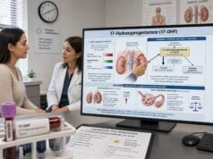
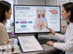
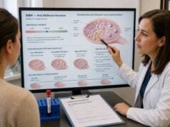
Additional testing may include pregnancy testing, TSH and free T4, SHBG and calculated free testosterone, AMH, inhibin B, DHEA-S, 17-hydroxyprogesterone, iron studies, hCG, semen analysis, pelvic or testicular ultrasound, pituitary MRI, bone age, or genetic tests. The choice should be driven by the pattern rather than ordering every possible hormone.
Seek urgent care for severe pelvic pain or bleeding in pregnancy, sudden severe headache or vision loss, a new testicular mass, rapidly progressive virilization, or symptoms of adrenal or pituitary crisis. Otherwise, schedule review with the ordering clinician and bring cycle dates, medication lists, and prior results.
Useful questions include: What was the panel intended to diagnose? Was each sample timed correctly? Which result is truly abnormal for my age and phase? Does the pattern point to the gonads or the pituitary? What test will confirm the cause? The value of a reproductive panel comes from answering those questions, not from counting how many entries are marked high or low.
Age changes the interpretation even when the laboratory range does not. In adolescence, pubertal stage and progression are central. During reproductive years, cycle day and pregnancy possibility dominate. In perimenopause, large fluctuations are expected, and in older adults the same concentration may carry different bone, sexual, or treatment implications. Results should therefore be compared with an appropriate population, not simply labeled “optimal” against a single adult chart.
Discordant findings are common and should trigger a check of timing and assay quality. A normal FSH with unexpectedly high early-cycle estradiol may be falsely reassuring because estradiol suppresses FSH. Low total testosterone with low SHBG may coexist with normal free testosterone. High prolactin during a stressful collection may normalize after rest. Repeating the right test under standardized conditions is often better than expanding immediately to a large panel.
The panel is also affected by exogenous hormones. Combined contraceptives suppress LH and FSH, alter estradiol measurements, and raise SHBG. Testosterone therapy suppresses gonadotropins. Progesterone support makes endogenous luteal assessment difficult. After an hCG trigger or ovarian stimulation, expected ranges differ from a natural cycle. Every report should be reviewed with the exact product, route, dose, and time of last administration.
When multiple values are abnormal, clinicians prioritize safety and reversible causes. Pregnancy is excluded where relevant; severe pituitary, adrenal, ovarian, or testicular symptoms receive prompt attention; and medication or acute-illness effects are checked. Only then are chronic syndromes assigned. This order reduces both missed urgent disease and overdiagnosis from temporary hormone changes.
For people with irregular cycles, the laboratory requisition should record the last menstrual period and whether bleeding was spontaneous or medication-induced. A withdrawal bleed after a progestogen does not prove that ovulation occurred, and “cycle day 3” after such bleeding may answer a different question from a natural early-follicular sample. The clinician can decide whether testing should proceed or be interpreted cautiously.
Home tests can complement but not duplicate serum testing. Urine LH kits estimate a surge, while urine pregnancy tests detect hCG. Consumer devices that estimate estrogen or progesterone metabolites use different analytes and thresholds from blood laboratories. Their trends may help with timing, but abnormal consumer readings should be confirmed with standard clinical evaluation before medication or fertility decisions are made.
A normal panel can still coexist with significant symptoms. Endometriosis, fibroids, structural pituitary lesions, medication adverse effects, sexual pain, and many infertility factors may not change these six hormones. Clinicians should continue the evaluation when the history or examination remains concerning instead of using normal values as a reason to stop.
References
- Guideline for the prevention, diagnosis and treatment of infertility 2025 (Guideline)
- Diagnosis and Treatment of Infertility in Men: AUA/ASRM Guideline 2024 (Guideline Amendment)
- International Evidence-based Guideline for the assessment and management of polycystic ovary syndrome 2023 2023 (Guideline)
- Diagnosis and management of prolactin-secreting pituitary adenomas: a Pituitary Society international Consensus Statement 2023 (International Consensus Statement)
- Fertility evaluation of infertile women: a committee opinion 2021 (Committee Opinion)
- Testosterone Deficiency Guideline 2024 (Clinical Guideline)
Disclaimer
This article is for general education and does not replace medical evaluation or treatment. Reproductive hormone results vary with age, sex, cycle phase, time, medication, pregnancy, and assay; review the complete pattern with a qualified clinician.