
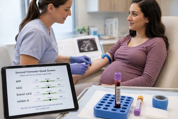
The second-trimester quad screen is a maternal blood test that combines four pregnancy-related markers—alpha-fetoprotein (AFP), human chorionic gonadotropin (hCG), unconjugated estriol (uE3), and inhibin A—with clinical information to estimate the chance of certain fetal conditions. It can screen for trisomy 21, trisomy 18, and open neural tube defects, but it cannot diagnose any of them. The result is calculated rather than read from one “high” or “low” hormone level, so accurate gestational age and complete maternal information are essential. Quad screening remains useful when first-trimester or cell-free DNA screening was not performed, is unavailable, or is not the chosen approach. It also provides AFP-based information about open fetal defects that cell-free DNA screening does not provide. An increased-chance result is a reason to review dates, perform targeted ultrasound, and discuss additional screening or diagnostic testing—not evidence that the fetus definitely has a condition.
- The quad screen is a probability test, not a diagnosis. It estimates chance from four markers, age, gestational dating, and other factors.
- AFP has a second job. In addition to contributing to chromosome-condition calculations, elevated maternal serum AFP can flag an open neural tube or abdominal wall defect.
- Gestational age can change the answer substantially. A dating error may make normal marker levels appear abnormal for the stated week.
- A screen-positive result has several possible explanations. These include a fetal condition, incorrect dates, more than one fetus, placental factors, or normal biologic variation.
- Do not compare the four raw numbers with another patient’s results. Laboratories convert them to gestation-specific multiples of the median and apply a validated risk algorithm.
Table of Contents
- What the quad screen measures and when
- The four markers and what they represent
- How four blood values become one risk estimate
- Common result patterns and report language
- What happens after an increased-chance result
- Quad screen versus cfDNA and ultrasound
- Limitations, special situations, and questions to ask
What the quad screen measures and when
The quad screen is also called the quadruple marker screen, second-trimester serum screen, or maternal serum quad test. A blood sample is drawn from the pregnant patient, and the laboratory measures AFP, hCG, unconjugated estriol, and inhibin A. The laboratory then calculates condition-specific risks using those measurements and clinical information.
Testing is generally available from about 15 through 22 weeks of pregnancy, but the accepted window differs by laboratory. Many programs prefer collection around 16 to 18 weeks because dating is usually established and there is time for follow-up. The ordering clinician should use the exact laboratory window rather than assume every facility accepts the same gestational ages.
The quad screen commonly estimates the chance of:
- trisomy 21, also called Down syndrome;
- trisomy 18, also called Edwards syndrome;
- open neural tube defects, such as open spina bifida and anencephaly;
- in some programs, open abdominal wall defects or other conditions associated with elevated AFP.
It is important to separate these targets. Trisomy risk is calculated from the combined marker pattern and maternal factors. Open-defect screening relies mainly on maternal serum AFP, often followed by a detailed ultrasound. The test does not broadly screen for every chromosome change, genetic syndrome, birth defect, developmental condition, or pregnancy complication.
The blood draw itself does not contact the pregnancy and carries only the ordinary risks of venipuncture, such as brief discomfort or bruising. The more meaningful decisions concern what information the patient wants and what follow-up would be acceptable after an increased-chance result.
Before ordering, the clinician should verify whether another aneuploidy screening strategy has already been completed. Independently performing multiple screening tests for the same chromosome conditions can produce conflicting probabilities and unnecessary confusion. A deliberately designed integrated or sequential screening program is different because its components are interpreted together under one protocol.
The four markers and what they represent
The marker names can make the quad screen sound like four separate diagnostic tests. They are better understood as four biologic signals that change across pregnancy and may shift in recognizable combinations.
Alpha-fetoprotein (AFP) is produced mainly by the fetal liver. Some AFP crosses into amniotic fluid and maternal blood. Maternal serum AFP normally changes with gestational age. A substantially elevated value can occur when an open fetal defect allows more AFP to enter amniotic fluid, but it can also reflect incorrect dating, twins or higher-order multiples, fetal demise, placental bleeding, or other pregnancy factors. A low AFP value contributes to some aneuploidy risk calculations but is not diagnostic by itself.
Human chorionic gonadotropin (hCG) is produced primarily by placental trophoblast cells. The assay may measure total hCG or a specified hCG fraction depending on the laboratory. hCG levels vary markedly across gestation and between pregnancies. In the classic trisomy 21 pattern, hCG tends to be higher than expected, while in trisomy 18 it often tends to be lower. A patient should not interpret an isolated hCG value using a nonpregnant reference range or compare it with an early-pregnancy hCG trend.
Unconjugated estriol (uE3) is produced through cooperation among the fetus, placenta, and maternal metabolism. Precursors are generated by the fetal adrenal glands and liver and converted by the placenta. Lower-than-expected uE3 contributes to trisomy 21 and trisomy 18 risk patterns. Very low uE3 can also have other explanations, including inaccurate dating and uncommon fetal or placental steroid-metabolism disorders. It is a clue that needs context, not a stand-alone diagnosis.
Inhibin A is produced by the placenta during pregnancy. It was added to the earlier “triple screen” to improve trisomy 21 screening performance. Inhibin A tends to be higher in pregnancies affected by trisomy 21, but many unaffected pregnancies also have elevated levels. It is not generally used alone to screen for a specific condition.
The four-marker pattern is more informative than any single analyte. A typical trisomy 21 pattern is lower AFP and uE3 with higher hCG and inhibin A. A typical trisomy 18 pattern includes lower AFP, hCG, and uE3; inhibin A is less central to that calculation. Open neural tube defects are associated mainly with elevated AFP. Real results do not always match textbook patterns, which is why a laboratory algorithm—not visual pattern matching—produces the risk estimate.
Marker levels may also correlate statistically with placental dysfunction or later pregnancy complications. Those associations do not turn the quad screen into a validated diagnostic test for preeclampsia, fetal growth restriction, stillbirth, or preterm birth. An unusually abnormal analyte may prompt individualized surveillance based on the entire clinical picture, but management varies and should not be inferred from the analyte alone.
How four blood values become one risk estimate
Raw concentrations are first compared with the expected median for the exact gestational age. The result is commonly expressed as a multiple of the median, or MoM. A value of 1.0 MoM is at the laboratory’s median for that gestational week; 2.0 MoM is twice that median. MoM values are then adjusted using factors known to influence marker levels or their interpretation.
The information requested may include:
- maternal age at the expected date of delivery;
- gestational age and how it was established;
- maternal weight;
- singleton, twin, or higher-order pregnancy;
- pregestational diabetes treated with insulin;
- smoking status;
- race or ethnicity if used by the laboratory’s validated model;
- IVF conception and sometimes donor-egg age;
- prior pregnancy affected by a screened condition.
Laboratories differ in which variables they use and how they adjust for them. This is one reason the same four raw concentrations cannot be placed into an online calculator reliably. The laboratory’s population medians, assay platform, quality controls, and risk model are part of the test.
Gestational age is particularly influential because all four markers change over time. If the pregnancy is farther along than recorded, an AFP level may appear falsely high. If it is earlier, the same value may be interpreted differently. Dating based on a reliable ultrasound is therefore preferable to an uncertain last menstrual period when the two disagree beyond accepted limits.
The algorithm combines the marker likelihoods with a prior chance, often influenced by maternal age, to produce a condition-specific estimate. A report might state a trisomy 21 chance of 1 in 900 or 1 in 80. A laboratory cutoff—such as 1 in 270, 1 in 250, or another validated threshold—classifies the result as screen negative or screen positive. The exact cutoff must be read from the report.
A result of 1 in 80 means that among pregnancies with the same calculated result, approximately one would be expected to have the condition and 79 would not. It does not mean the fetus is “80% affected,” and it does not describe severity. Conversely, a screen-negative result reduces the estimated chance but never makes it zero.
The detection rate and false-positive rate describe test performance in populations, not certainty for an individual. Traditional quad screening detects roughly four out of five trisomy 21 pregnancies at a commonly used false-positive rate, with performance varying by program and population. The positive predictive value depends strongly on the patient’s starting chance and the chosen cutoff.
Common result patterns and report language
Quad-screen reports can look dense because they include raw concentrations, MoMs, adjusted risks, cutoffs, and comments. The most useful first step is to identify which target triggered the result.
Screen negative or decreased chance means the calculated risk is below the laboratory cutoff. It does not mean “all genetic testing is normal.” The fetus could still have the screened condition, a different chromosome condition, a structural anomaly, or a disorder the quad screen cannot assess. Routine prenatal care and the recommended second-trimester anatomy ultrasound remain important.
Screen positive or increased chance for trisomy 21 means the combined pattern crossed the laboratory’s threshold. The classic pattern is high hCG and inhibin A with low AFP and uE3, but the calculation may be positive without every marker following that pattern. Most screen-positive results are not diagnostic confirmations. The next step is counseling about detailed ultrasound, cell-free DNA screening in selected circumstances, and diagnostic testing.
Screen positive or increased chance for trisomy 18 usually reflects a pattern of low markers, especially AFP, hCG, and uE3, combined with the prior risk. Ultrasound may reveal findings associated with trisomy 18, but a normal ultrasound cannot exclude it. Diagnostic testing is needed for confirmation.
Elevated AFP or increased chance for an open neural tube defect is evaluated differently from aneuploidy risk. The clinician first checks dating, fetal number, and whether there has been bleeding or another explanation. A targeted ultrasound examines the fetal spine, skull, abdominal wall, growth, placenta, and amniotic fluid. Amniocentesis with amniotic-fluid AFP and acetylcholinesterase may be discussed when ultrasound is inconclusive or diagnostic clarification is needed.
Very low uE3 may generate a laboratory comment even when the trisomy calculation is not positive. The differential can include dating error, fetal demise, trisomy 18, and rare conditions affecting steroid synthesis or metabolism. Follow-up is driven by the degree of reduction, ultrasound findings, family history, and the laboratory’s recommendation.
Unable to calculate or invalid result can occur when the specimen is outside the validated gestational window, required information is missing, analyte levels exceed model limits, or the pregnancy type is unsupported. This is not equivalent to a negative screen. The clinician should contact the laboratory promptly because the opportunity to redraw may be limited by gestational age.
Reports may also use “1:100” rather than “1 in 100,” “positive” rather than “high risk,” or “above cutoff” rather than “increased chance.” None of those phrases establish a diagnosis. Ask for the numerical risk, the cutoff, and the marker or condition responsible for the flag.
What happens after an increased-chance result
Follow-up should begin with verification, not panic. The ordering team confirms patient identifiers, gestational age, fetal number, maternal weight, diabetes status, IVF details, and whether another screen was already performed. A corrected data field can sometimes change the calculated risk without a new blood draw.
A comprehensive ultrasound is often central. For elevated AFP, ultrasound may identify an open neural tube defect, abdominal wall defect, incorrect dating, twins, fetal demise, or placental finding. For aneuploidy risk, ultrasound can assess anatomy and markers that alter counseling, but it is not a chromosome test.
After an increased-chance trisomy result, the principal options are:
- Diagnostic testing. Amniocentesis is typically the second-trimester diagnostic procedure. Fetal cells in amniotic fluid can be tested by karyotype, chromosomal microarray, rapid aneuploidy testing, or another method selected for the indication. Diagnostic testing provides a direct answer about the tested chromosome condition but carries a small procedure-related risk that should be discussed with the procedural center.
- Cell-free DNA screening. For some patients who want better risk refinement without an invasive procedure, cfDNA may be offered after a positive serum screen. It is more accurate for common trisomies than the quad screen, but it remains screening. Choosing this route can delay definitive diagnosis and may miss chromosome abnormalities outside the cfDNA panel that contributed to the serum result.
- Ultrasound-based evaluation without additional genetic testing. Some patients decline further screening or diagnostic testing. Their care team can still use ultrasound findings for pregnancy management and neonatal planning, while recognizing that chromosome status remains uncertain.
The choice depends on gestational age, the numerical risk, ultrasound findings, the patient’s values, and how a confirmed answer would affect preparation or pregnancy decisions. Genetic counseling can translate population statistics into the actual choices available.
A positive AFP screen should not be “followed up” only with cfDNA, because cfDNA does not evaluate open neural tube defects. The needed pathway is accurate dating and detailed fetal imaging, with amniotic-fluid testing when indicated.
A normal follow-up ultrasound is reassuring but does not erase every increased-chance aneuploidy result. Likewise, a low-risk cfDNA result after a positive quad screen greatly reduces the chance of the common trisomies it covers but does not explain every unusual serum pattern. The care team should decide whether placental surveillance or diagnostic testing remains appropriate based on the complete case.
Quad screen versus cfDNA and ultrasound
The quad screen, cfDNA screening, and ultrasound overlap only partly.
Cell-free DNA screening analyzes placental DNA fragments circulating in maternal blood. It is the most sensitive and specific screening test for trisomies 21, 18, and 13 in singleton pregnancies, but it is not diagnostic. It generally can be performed earlier than the quad screen. It does not measure AFP and therefore does not screen for open neural tube defects. For broader context, see the prenatal genetic screening test guide.
The quad screen is less accurate than cfDNA for common trisomies but can be performed when a patient presents in the second trimester and has not had earlier screening. It may be more accessible or affordable in some settings. Its AFP component provides open-defect risk information. It also has a long-established role in integrated and sequential screening programs.
Ultrasound evaluates fetal anatomy and growth rather than blood markers or chromosome copy number. Every patient should be offered a second-trimester structural ultrasound, whether aneuploidy screening was performed and regardless of the screening result. Some chromosome conditions have ultrasound findings; others do not. Many structural anomalies occur in fetuses without aneuploidy.
A patient who already had negative cfDNA screening generally should not add a standalone quad screen solely to rescreen for the same trisomies. However, the clinician still needs a strategy for open neural tube defects, which may be maternal serum AFP and/or careful second-trimester ultrasound according to local practice. Ordering “AFP only” is not the same as ordering the full quad panel.
A patient who had first-trimester serum and nuchal translucency screening may be enrolled in a sequential or integrated protocol that deliberately adds second-trimester markers. The laboratory must know the first-trimester results so it can issue the planned combined risk. A separate unlinked quad calculation can undermine the program’s design.
Diagnostic testing differs from all three screening tools. Chorionic villus sampling and amniocentesis obtain pregnancy-derived cells for genetic analysis. They answer whether a tested condition is present rather than estimating chance. A screen result should be considered in light of whether the patient wants a probability or a diagnostic answer.
Limitations, special situations, and questions to ask
The quad screen’s accuracy depends on accurate inputs and a population-validated algorithm. Incorrect gestational age, weight, diabetes status, smoking information, donor-egg age, or fetal number can distort the result. Laboratories may not validate standard calculations for triplets, pregnancies after fetal reduction, a vanishing twin, or some donor-gamete situations.
Twin pregnancies require a twin-specific model. The test may estimate risk for the pregnancy as a whole rather than identify which fetus is affected. AFP is naturally higher with more than one fetus, and follow-up interpretation is more complex. After a vanishing twin, residual placental activity can affect serum markers; the laboratory and maternal-fetal medicine team should be told the timing and ultrasound history.
The screen does not reliably detect mosaicism, balanced translocations, small deletions or duplications, most sex chromosome conditions, single-gene disorders, or all causes of neural tube defects. It cannot predict intellectual ability, medical severity, or whether an affected fetus will have every feature associated with a condition.
A “negative” result is not a guarantee, and a “positive” result is not a diagnosis. The cutoff creates categories for follow-up; biology does not change abruptly at that line. A risk of 1 in 260 and 1 in 240 may fall on opposite sides of a 1-in-250 cutoff while being numerically similar. Reviewing the number is more informative than reacting to the label alone.
Before testing, useful questions include:
- What gestational-age window does this laboratory accept?
- Am I having a full quad screen, AFP-only screening, or one stage of an integrated program?
- Have I already had aneuploidy screening that would make another independent screen confusing?
- How will IVF, donor eggs, twins, diabetes, smoking, or weight be entered?
- Which conditions are calculated, and what cutoffs does the laboratory use?
- What follow-up would be offered after elevated AFP versus increased trisomy risk?
- Would I prefer a better screening estimate or a diagnostic answer if the result is positive?
After results, request the complete laboratory report rather than relying on “normal” or “abnormal” in a portal. Confirm the gestational age used, each MoM, the condition-specific risk, and the cutoff. If the report is positive, ask how the result changes after the most recent ultrasound dating and whether a genetics or maternal-fetal medicine consultation can occur soon enough to preserve all testing options.
The quad screen is most useful when it is treated as a structured decision point. Its job is to identify pregnancies that deserve a closer look, not to assign a diagnosis from four hormones. Clear pretest counseling, accurate dating, and condition-specific follow-up turn an old but still useful screening tool into information that patients can act on thoughtfully.
References
- American College of Obstetricians and Gynecologists: Screening for Fetal Chromosomal Abnormalities (2026, practice advisory)
- Society for Maternal-Fetal Medicine Consult Series #74: Cell-free DNA screening for aneuploidies (2025, guideline)
- Prenatal Genetic Screening (2025, clinical review)
- MedlinePlus: Quadruple Screen Test (2026, patient resource)
- Cleveland Clinic: Quad Marker Screen (2023, clinical resource)
- Mayo Clinic Laboratories: Quad Screen, Second Trimester, Maternal Serum (2026, laboratory resource)
Disclaimer
This article provides general educational information and is not a substitute for prenatal care, genetic counseling, or interpretation by the laboratory that performed the test. Screening windows, risk cutoffs, analyte adjustments, and follow-up options vary. Discuss your complete report and pregnancy-specific circumstances with a qualified obstetric or genetics professional.





