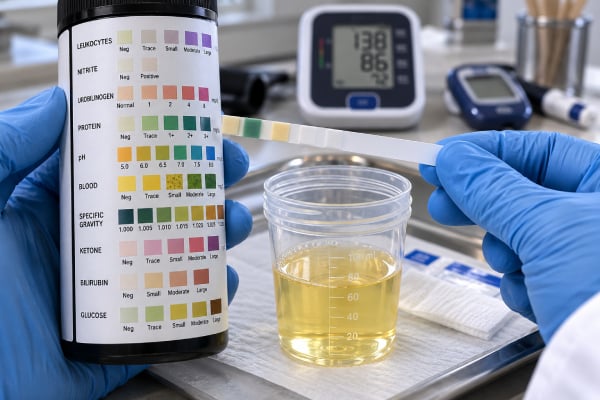
A urine protein test checks whether more protein than expected is passing into the urine. Proteinuria can be a temporary finding after exercise, fever, dehydration, or acute illness, but persistent protein loss can signal kidney disease. The phrase “urine protein test” can refer to a dipstick, a laboratory total protein measurement, a urine protein-creatinine ratio (UPCR), a urine albumin-creatinine ratio (uACR), or a 24-hour collection. These tests are not interchangeable. Dipsticks mainly detect albumin, while UPCR and total protein assays measure a broader range of proteins. uACR is generally preferred for CKD screening because low-level albumin leakage is an established marker of kidney and cardiovascular risk. A positive result therefore needs context: how the protein was measured, how much was present, whether blood or infection was present, whether the result persists, and whether eGFR or other kidney findings are abnormal. The cause determines how much concern and follow-up are needed.
- A positive urine protein test does not automatically mean chronic kidney disease; temporary proteinuria is common.
- Dipstick protein mainly detects albumin, while UPCR measures total protein and uACR measures albumin specifically.
- Persistent proteinuria is more important than one isolated positive sample and should be quantified when clinically relevant.
- Heavy protein loss can cause foamy urine and swelling, but many people with mild proteinuria have no symptoms.
- Proteinuria with declining kidney function, blood in urine, severe swelling, or very high blood pressure needs more urgent evaluation.
Table of Contents
- What protein in urine means
- Types of urine protein tests
- Normal and high urine protein results
- Common causes of proteinuria
- Temporary and benign proteinuria
- How an abnormal result is evaluated
- Treatment and follow-up
What protein in urine means
Healthy kidneys keep most blood proteins in the circulation. The glomeruli filter blood while retaining large molecules, and the kidney tubules reclaim many smaller proteins that do pass through. Proteinuria develops when one of these systems is overwhelmed or damaged, or when protein is added to urine farther down the urinary tract.
Albumin is the best-known urinary protein because increased albumin excretion is strongly linked with kidney and cardiovascular risk. However, urine can contain many other proteins. Immunoglobulin light chains, beta-2 microglobulin, alpha-1 microglobulin, and other low-molecular-weight proteins become important in specific diseases.
This is why “high urine protein” is a finding rather than a diagnosis. The result may reflect glomerular disease, tubular injury, excessive production of a circulating protein, urinary tract inflammation, or a transient physiologic response.
Proteinuria can be asymptomatic. Mild elevations do not necessarily change urine appearance. Heavy protein loss may produce persistent foam or froth, especially when the urine is concentrated, but bubbles alone are not a reliable diagnostic test. Edema in the legs, around the eyes, or elsewhere can occur when severe protein loss lowers serum albumin, as in nephrotic syndrome.
Types of urine protein tests
The word “protein test” can describe several different methods, and knowing the method prevents misinterpretation.
Urine dipstick protein is quick and inexpensive. It is most sensitive to albumin and less sensitive to many non-albumin proteins. A positive result is often graded trace, 1+, 2+, 3+, or 4+. The grade is semiquantitative and strongly affected by urine concentration. A concentrated sample may look more positive; very dilute urine may understate protein. A urine protein dipstick is best viewed as a screening tool.
uACR measures albumin relative to creatinine. It is the preferred routine test for CKD detection and risk staging in many adults, especially those with diabetes or hypertension. It can detect moderately increased albuminuria that a standard dipstick may miss.
UPCR measures total protein relative to creatinine. It is useful when total protein burden matters, including many glomerular diseases and situations where non-albumin protein may be present. The UPCR test can estimate daily protein loss from a spot specimen.
24-hour urine protein directly totals protein collected over a day. It can be valuable when precise quantification is needed but is vulnerable to missed or extra samples.
Protein electrophoresis and immunofixation are specialized tests used to identify monoclonal proteins rather than simply measure the total amount.
Normal and high urine protein results
Normal ranges depend on the test. A dipstick may simply report “negative” or “trace.” A total protein concentration may be listed in mg/dL or mg/L, which varies with hydration and cannot be translated directly into daily protein loss.
For a spot UPCR, many adult laboratories use an upper reference limit near 150–200 mg/g. For uACR, the standard kidney categories are below 30 mg/g, 30–299 mg/g, and 300 mg/g or more. Those numbers should not be applied to total protein concentration reports.
A 24-hour urine collection in healthy adults generally contains less than about 150 mg of total protein per day, although laboratories differ. Albumin accounts for only a portion of normal urinary protein.
Heavy protein loss is often discussed in terms of “nephrotic range.” Traditionally, total protein excretion around 3.5 g/day or more in adults has been used as a classic threshold, with spot UPCR values in roughly the 3–3.5 g/g range often used as an estimate. Yet nephrotic syndrome is more than a number: low serum albumin, edema, and metabolic changes are part of the syndrome. The nephrotic syndrome urine evaluation considers the wider pattern.
When a result is only mildly abnormal, confirmation matters. The difference between a temporary 1+ dipstick after a long run and persistent quantitative proteinuria over months is clinically substantial.
Common causes of proteinuria
Persistent proteinuria is commonly divided by mechanism.
Glomerular proteinuria occurs when the kidney’s filtration barrier allows excess protein—especially albumin—to pass. Common causes include diabetic kidney disease, hypertensive kidney disease, IgA nephropathy, focal segmental glomerulosclerosis, membranous nephropathy, lupus nephritis, and other forms of glomerulonephritis. Glomerular disease can range from low-level albuminuria to several grams of protein loss per day.
Tubular proteinuria develops when the proximal tubules do not reabsorb smaller filtered proteins effectively. Tubulointerstitial nephritis, some medications and toxins, inherited tubular disorders, and other forms of tubule injury can produce this pattern. Albumin may not dominate, so a standard dipstick can look relatively unimpressive despite increased total protein. A tubular proteinuria test may target low-molecular-weight proteins.
Overflow proteinuria occurs when the bloodstream contains an unusually high concentration of a small protein. Monoclonal immunoglobulin light chains in plasma cell disorders are an important example. Because dipsticks mainly detect albumin, light-chain proteinuria can be missed or underestimated. A large gap between UPCR and uACR can be a clue, but specialized electrophoresis, immunofixation, and serum free light-chain testing are needed.
Post-renal proteinuria results when protein enters urine from inflammation, bleeding, or infection in the urinary tract. Pyuria and hematuria on urinalysis can support that explanation.
Kidney disease itself is not the only issue. Heart failure, severe hypertension, systemic inflammatory diseases, pregnancy-related disorders, and acute illnesses can all increase urinary protein.
Temporary and benign proteinuria
Not every positive protein result represents structural kidney damage. Transient proteinuria can occur with fever, strenuous exercise, acute infection, dehydration, emotional or physical stress, seizures, and short-term severe blood pressure elevation. The protein often disappears after the trigger resolves.
Orthostatic proteinuria is another special pattern, most often seen in adolescents and young adults. Protein appears while the person is upright during the day but is low or absent in a first-morning sample after lying down overnight. It is usually benign once more serious causes have been excluded. A first-morning orthostatic proteinuria evaluation can help distinguish the pattern.
Exercise-related proteinuria is especially relevant for athletes. Long, intense training can temporarily increase glomerular permeability and urinary albumin. Testing immediately after a marathon, heavy lifting session, or strenuous endurance event may therefore overstate baseline protein excretion.
Urinary infection can cause protein to appear along with white blood cells, bacteria, and sometimes blood. Repeating the protein measurement after treatment may be more informative than diagnosing kidney disease from the acute sample.
Temporary causes matter because chronic kidney disease requires persistence or other evidence of lasting damage. A single isolated result should trigger judgment, not panic.
How an abnormal result is evaluated
The first question after a positive urine protein test is how much protein is present and what type of protein is it likely to be? A clinician may confirm a dipstick finding with uACR or UPCR. The choice depends on the clinical question.
For routine CKD screening, uACR is often preferred. For heavier proteinuria or suspected non-albumin protein, UPCR adds value. Comparing the two can be revealing. If UPCR is 1,500 mg/g but uACR is only 100 mg/g, much of the urine protein is not albumin and further investigation may be needed.
A complete urinalysis can identify blood, leukocytes, casts, glucose, and urine concentration. Active sediment with red blood cell casts or substantial hematuria raises concern for glomerulonephritis. Serum creatinine and eGFR show whether filtration is impaired. Blood pressure, diabetes control, medication history, systemic symptoms, family history, and duration of abnormality all help narrow the cause.
If monoclonal protein is suspected, serum protein electrophoresis, serum immunofixation, serum free light chains, urine electrophoresis, or urine immunofixation may be ordered. If glomerular disease remains unexplained and the result would affect treatment, a kidney biopsy may be required.
Repeated testing should usually occur after temporary factors resolve. First-morning urine can reduce variation and is particularly useful for mild abnormalities or suspected orthostatic proteinuria.
Treatment and follow-up
There is no single treatment for “protein in urine.” Treatment targets the underlying cause and the kidney risk associated with persistent protein loss.
For diabetic or hypertensive kidney disease, blood pressure control is central. ACE inhibitors and angiotensin receptor blockers can reduce albuminuria and slow progression in appropriate patients. SGLT2 inhibitors provide kidney and cardiovascular protection for many people with CKD and are now used beyond glucose lowering in selected patients. Other medicines may be indicated depending on diabetes status, potassium, eGFR, and cardiovascular risk.
Immune-mediated glomerular diseases may require disease-specific immunologic therapy. Monoclonal protein disorders require hematologic evaluation and sometimes clone-directed treatment. Infection-related proteinuria may resolve when the infection is treated.
General kidney-protective steps include avoiding smoking, limiting excess dietary sodium, maintaining healthy blood pressure and blood sugar, exercising appropriately, and avoiding unnecessary nephrotoxic medications. Protein intake should not be drastically restricted without professional advice; needs vary with CKD stage, nutritional status, and other conditions.
Referral to nephrology is reasonable for heavy or rapidly increasing proteinuria, nephrotic-range protein loss, unexplained non-albumin protein, active urine sediment, falling eGFR, resistant hypertension, or uncertain cause. Marked swelling, shortness of breath, visible blood in urine, very low urine output, or symptoms of severe acute illness deserve prompt medical care.
The most useful way to read a urine protein result is to ask four questions: Which test was used? How high is it? Does it persist? What other kidney findings are present? Those answers turn a nonspecific positive result into a clinically meaningful assessment.
Common result patterns and what they suggest
A trace or 1+ dipstick result in concentrated urine is a common reason for unnecessary alarm. Because dipstick color change reflects concentration, a dehydrated person can have a positive screening result with relatively little daily protein loss. Confirming with uACR or UPCR is usually more informative than repeatedly checking dipsticks under different hydration conditions.
The opposite problem can occur when the urine contains mainly non-albumin protein. A patient with monoclonal free light chains may have a large quantitative total protein result while the dipstick is only trace or 1+. That mismatch is a clue that albumin is not the dominant protein and that specialized monoclonal protein testing may be needed.
Another useful pattern is protein plus blood. Small amounts of both can occur with several conditions, but substantial proteinuria combined with dysmorphic red blood cells or red blood cell casts raises concern for glomerular inflammation. Protein plus white blood cells and bacteria may instead reflect urinary infection. The urinalysis sediment therefore adds diagnostic meaning that a protein number alone cannot provide.
Heavy proteinuria with low serum albumin and edema suggests nephrotic syndrome. Patients may notice swollen ankles, puffiness around the eyes, rapid weight gain from fluid, or persistent foamy urine. Blood clots and infection risk can also rise in some nephrotic conditions. This pattern needs timely medical assessment because the underlying causes range from primary glomerular diseases to diabetes, medications, infections, malignancy-associated conditions, and systemic autoimmune disease.
A less obvious pattern is persistent modest proteinuria with normal eGFR. It still matters. Structural kidney damage can precede a fall in filtration, and persistent albuminuria is itself a CKD marker. That is why screening programs often pair uACR with eGFR rather than waiting for serum creatinine to become abnormal.
Finally, a negative dipstick should not always end the evaluation. Standard reagent strips are relatively insensitive to low-level albumin and many non-albumin proteins. If someone has diabetes, unexplained CKD, suspected light-chain disease, or another strong reason to look for kidney damage, a quantitative test can be appropriate even when the routine dipstick is negative.
The practical rule is to match the assay to the suspected disease. Dipstick is convenient screening, uACR is strong for albuminuric CKD risk, UPCR measures total burden, and electrophoresis or immunofixation identifies abnormal protein types. Choosing the right test prevents both false reassurance and unnecessary investigation.
Medication review is another important part of evaluation. Nonsteroidal anti-inflammatory drugs, some antibiotics, lithium, and several other agents can contribute to kidney injury or proteinuria in susceptible people. A new protein finding after a medication change should be discussed with the prescriber rather than prompting abrupt self-discontinuation of essential treatment.
Family history can also change the threshold for investigation. Persistent proteinuria in a younger person with relatives who have kidney failure, hearing loss, cystic disease, or unexplained hematuria may point toward an inherited kidney disorder. In that setting, even modest proteinuria can be diagnostically useful when combined with imaging, blood tests, genetic evaluation, or specialist assessment.
For people with established CKD, proteinuria is not only a clue to cause but also a marker of future risk. Higher persistent albumin or total protein generally tracks with faster kidney decline and higher cardiovascular risk. Reducing proteinuria when possible is therefore part of kidney protection, not simply a way to improve a laboratory number.
For routine follow-up, ask which quantitative marker the clinician wants repeated. Rechecking a dipstick when the treatment target is uACR or UPCR can create confusion because the tests respond differently to concentration and protein type. Using the same quantitative marker over time makes meaningful improvement or worsening easier to recognize.
When a repeat quantitative result is normal and the rest of the kidney evaluation is reassuring, clinicians may return to routine risk-based screening rather than continuing an extensive workup.
References
- KDIGO 2024 Clinical Practice Guideline for the Evaluation and Management of Chronic Kidney Disease 2024 (Guideline)
- Methods for Diagnosing Proteinuria-When to Use Which Test and Why: A Review 2025 (Review)
- Measuring albuminuria or proteinuria: does one answer fit all? 2023 (Review)
- Protein handling in kidney tubules 2025 (Review)
- Proteinuria or Albuminuria as Markers of Kidney and Cardiovascular Disease Risk : An Individual Patient-Level Meta-analysis 2026 (Meta-Analysis)
Disclaimer
This article provides general education and cannot determine the cause of protein in an individual urine sample. Interpretation depends on the test method, amount, repeat results, eGFR, urinalysis, symptoms, medications, pregnancy status, and medical history. Seek prompt care for severe swelling, breathing difficulty, gross hematuria, very low urine output, or acute illness.