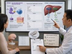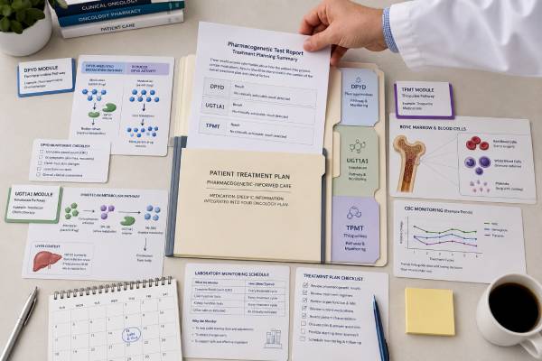
A chemotherapy pharmacogenetic test can identify inherited differences that affect how a patient handles particular anticancer drugs, but no single result predicts response to “chemotherapy” as a whole. DPYD is most relevant to fluoropyrimidines such as fluorouracil and capecitabine, UGT1A1 to irinotecan, and TPMT—together with NUDT15—to thiopurines such as mercaptopurine, thioguanine, and azathioprine. Clinically actionable variants can indicate a need for a lower starting dose, slower titration, an alternative drug, or intensified monitoring to reduce severe toxicity. The genes do not determine whether a cancer will be cured, and normal results do not eliminate adverse-effect risk. Interpretation must be tied to the exact drug, regimen, dose intensity, diagnosis, organ function, interacting medicines, and treatment goal. Because chemotherapy delays can matter, testing is most useful when ordered early enough for a validated result to reach the oncology team before the first relevant dose.
- DPYD results guide fluorouracil and capecitabine safety because reduced DPD activity can cause early, life-threatening toxicity.
- UGT1A1 poor-metabolizer status can increase irinotecan-related neutropenia and diarrhea, especially at higher doses.
- TPMT should be interpreted with NUDT15 before thiopurines because either gene can markedly increase myelosuppression risk.
- A normal pharmacogenetic result does not guarantee tolerability; blood counts, symptoms, and organ function still require close monitoring.
- Each gene result applies to defined drugs, not to every chemotherapy agent or to the cancer’s likelihood of responding.
Table of Contents
- One Panel, Three Different Drug Pathways
- DPYD Results for Fluorouracil and Capecitabine
- UGT1A1 Results for Irinotecan
- TPMT, NUDT15, and Thiopurine Results
- How Chemotherapy Pharmacogenetic Testing Is Performed
- Turning Results Into a Treatment Plan
- Limitations, Monitoring, and Urgent Toxicity
- Questions to Review With the Oncology Team
One Panel, Three Different Drug Pathways
DPYD, UGT1A1, and TPMT are often grouped on oncology pharmacogenetic panels, but they do not form one shared pathway. Each result must be matched to a medication that depends on the encoded enzyme. A report that lists all three genes can therefore contain one highly actionable result, one result that is irrelevant to the current regimen, and one result that may matter years later.
DPYD encodes dihydropyrimidine dehydrogenase, or DPD, the principal enzyme responsible for breaking down fluorouracil. Capecitabine is converted to fluorouracil in the body, so inherited DPD deficiency also affects capecitabine safety. Reduced clearance can expose tissues to excessive active drug and cause severe mucositis, diarrhea, neutropenia, hand-foot syndrome, neurotoxicity, cardiac toxicity, or death.
UGT1A1 encodes an enzyme that glucuronidates SN-38, the active metabolite of irinotecan. Reduced UGT1A1 activity can slow SN-38 inactivation, increasing exposure and the risk of dose-limiting neutropenia and delayed diarrhea. The clinical impact varies with irinotecan dose, schedule, regimen, ancestry, liver function, and other risk factors.
TPMT encodes thiopurine S-methyltransferase, which helps divert thiopurine metabolites away from active thioguanine nucleotides. Low TPMT activity can lead to excessive active metabolites and profound bone-marrow suppression at standard doses. Modern prescribing also considers NUDT15, which protects cells from incorporating toxic thiopurine metabolites into DNA. NUDT15 variants are especially important in many East Asian, Hispanic/Latino, and Indigenous American populations but occur across ancestries.
These are inherited, or germline, findings. They differ from tumor biomarker testing, which analyzes acquired changes in cancer cells to select targeted therapy or immunotherapy. A patient may need both. A pharmacogenetic test asks how the patient may process a medicine; a tumor test asks what biological features the cancer has.
The panel also does not replace routine clinical assessment. Kidney and liver function, age, nutritional status, previous treatment, infection, marrow reserve, drug interactions, and regimen intensity can alter toxicity independently of genotype. The value of pharmacogenetics is to identify a preventable component of risk before exposure, then integrate it into the complete plan.
DPYD Results for Fluorouracil and Capecitabine
Fluoropyrimidines are used in many gastrointestinal, breast, head and neck, and other cancers. Most administered fluorouracil is catabolized through DPD. When DPD activity is substantially reduced, even a standard first dose can produce unusually early and severe toxicity. Symptoms may begin during the first cycle or within days and can progress rapidly.
DPYD results are commonly translated into a DPD phenotype or activity score. Laboratories may report normal, intermediate, or poor metabolizer status, although terminology and score details should be checked against the laboratory’s report and the guideline used by the treating center.
A normal metabolizer result means the tested alleles predict typical DPD activity. Standard starting treatment may be considered, but this is not a guarantee of safety. Most patients who develop fluoropyrimidine toxicity do not have one of the limited variants on a small genotyping panel. Toxicity can also arise from dose, organ dysfunction, drug interactions, infection, or other genetic and non-genetic factors.
An intermediate metabolizer result indicates partial DPD deficiency. Current expert recommendations generally support reducing the initial fluoropyrimidine dose and then carefully titrating according to toxicity, efficacy, and—where available—therapeutic drug monitoring. The exact starting percentage depends on the activity score, variant evidence, regimen, and guideline. Dose reduction is a starting strategy, not a permanent assumption that the patient must receive less treatment throughout therapy.
A poor metabolizer result suggests complete or near-complete DPD deficiency. The risk from standard fluorouracil or capecitabine is extreme. Regulatory labeling advises avoiding these drugs in patients with certain biallelic no-function combinations associated with complete deficiency because no dose has been proven safe. If a fluoropyrimidine is considered despite very low activity, it requires exceptional specialist judgment, a markedly reduced exposure, and close pharmacokinetic and clinical monitoring.
Commonly tested variants include DPYD2A, DPYD13, c.2846A>T, and the HapB3-associated intronic or linked marker. Some panels also include c.557A>G, which is more relevant in people with African ancestry. The exact coverage matters because a “negative” four-variant assay does not mean the entire gene was normal. Rare deleterious variants occur throughout DPYD, and sequencing can find more of them, though it also creates uncertain findings.
Some health systems combine genotyping with phenotyping, such as measuring pretreatment plasma uracil or a uracil-to-dihydrouracil ratio. Genotype and phenotype are related but not identical. Phenotyping can be affected by sample handling, kidney function, illness, and laboratory methods. The oncology team should follow the locally validated protocol rather than combining cutoffs from different systems.
The U.S. Food and Drug Administration’s 2026 labeling update advises DPYD testing before capecitabine or fluorouracil unless treatment must begin immediately. This makes preemptive turnaround increasingly important. A result returned after the first infusion may still guide later cycles, but it cannot prevent first-dose toxicity.
UGT1A1 Results for Irinotecan
Irinotecan is converted to SN-38, a much more active topoisomerase I inhibitor. UGT1A1 attaches glucuronic acid to SN-38 so it can be eliminated. Reduced UGT1A1 activity raises active-metabolite exposure and can increase severe neutropenia and delayed diarrhea. The association is strongest in people with two reduced-function alleles and at moderate-to-high irinotecan doses.
The best-known allele is UGT1A128, which contains an additional TA repeat in the promoter and reduces gene expression. UGT1A16 is a reduced-function coding variant that is particularly important in East Asian populations. A test limited to *28 may therefore under-detect clinically important risk in a diverse patient population. Reports may identify normal, intermediate, or poor metabolizer status based on the two inherited alleles.
A normal metabolizer generally has two normal-function alleles and no genotype-based reason to change the starting dose. An intermediate metabolizer has one reduced-function allele. Many guidelines do not require routine preemptive dose reduction for intermediate metabolizers, but other clinical risk factors still matter. A poor metabolizer has two reduced-function alleles, such as 28/28, 6/6, or 6/28. The Dutch Pharmacogenetics Working Group recommends a reduced initial irinotecan dose for poor metabolizers, followed by escalation if tolerated.
There is no single universal percentage for every regimen. Irinotecan appears in schedules ranging from low weekly doses to high-dose intermittent therapy and in combinations such as FOLFIRI or FOLFIRINOX. The contribution of UGT1A1 is more pronounced as dose increases. Oncology guidelines, product labels, and regional pharmacogenetic groups may differ in thresholds and wording. The prescriber should use guidance applicable to the exact regimen rather than applying a result from one dose setting to another.
UGT1A1 also conjugates bilirubin. Reduced-function genotypes can cause Gilbert syndrome, a benign tendency toward unconjugated hyperbilirubinemia. A patient with 28/28 may have mildly elevated bilirubin without liver injury. However, bilirubin elevation during cancer treatment can also reflect obstruction, hepatic metastases, hemolysis, infection, or drug toxicity. The genotype provides context; it does not diagnose the cause of every abnormal liver test.
The result is mainly a toxicity marker, not a reliable predictor that irinotecan will or will not control the cancer. A reduced starting dose should be paired with a plan for reassessment and possible escalation, because excessive underdosing could compromise treatment intensity. Neutrophil counts, diarrhea, hydration, bilirubin, liver function, and co-medications remain central to safe care.
TPMT, NUDT15, and Thiopurine Results
Thiopurines include mercaptopurine and thioguanine, which are central to many leukemia protocols, and azathioprine, which is more often used for nonmalignant immune-mediated disease. Their dose-toxicity relationship differs from conventional short-course chemotherapy because treatment can continue for months or years and dosing is often adjusted repeatedly to blood counts and disease-specific targets.
TPMT and NUDT15 should be considered together. A normal TPMT result cannot compensate for a poor-function NUDT15 genotype, and the reverse is also true. The updated CPIC guideline provides combined recommendations, including for people who are intermediate metabolizers in both genes. Laboratories that report only TPMT may miss a major source of thiopurine intolerance.
A normal metabolizer for both genes can usually begin with the protocol-defined standard starting dose. This does not remove the need for complete blood counts and liver tests. Infection, adherence, drug interactions, treatment phase, kidney function, and other inherited factors can change tolerance.
A TPMT or NUDT15 intermediate metabolizer has reduced activity in one pathway. Standard doses can produce excessive thioguanine nucleotide exposure or DNA-incorporated metabolites, increasing leukopenia, neutropenia, and treatment interruptions. Guidelines generally recommend a reduced starting dose when the protocol uses a conventional or high starting dose, followed by adjustment to myelosuppression and therapeutic targets. Some patients eventually tolerate near-standard dosing; others need substantial continuing reduction.
A poor metabolizer in either gene is at very high risk of severe, potentially fatal myelosuppression with full-dose thiopurine therapy. For malignancy, expert guidance supports drastically reduced starting doses and reduced dosing frequency, with careful escalation only under protocol-specific supervision. For nonmalignant conditions, an alternative non-thiopurine therapy is often preferred. Recommendations differ by drug because mercaptopurine, thioguanine, and azathioprine produce different metabolite patterns.
A person who is intermediate for both TPMT and NUDT15 can have greater risk than a single-gene intermediate metabolizer. The 2025 CPIC update recognizes this compound phenotype and generally advises a larger starting reduction. The report must show both results clearly; a generic label such as “intermediate metabolizer” without naming the genes is inadequate.
Genotyping and enzyme phenotyping are different methods. TPMT enzyme activity can be measured in red blood cells, but recent transfusion can make the result reflect donor cells. Genotyping is not altered by transfusion, though it may not detect every rare functional allele. NUDT15 is usually assessed genetically. When genotype and phenotype disagree, the oncology pharmacist, laboratory, or genetics service should investigate before treatment decisions are finalized.
The genotype guides the initial dose. It does not replace ongoing titration, because effective leukemia therapy often intentionally produces a controlled degree of marrow suppression. The correct target is disease- and protocol-specific, not “no change in blood counts.”
How Chemotherapy Pharmacogenetic Testing Is Performed
Testing usually uses blood, saliva, or a cheek swab and can be ordered as a focused assay or a broader panel. Because the result is germline, it generally remains applicable throughout life. A properly documented result can be reused when the patient encounters the relevant drug again, although variant interpretation and dosing guidelines may be updated.
A focused genotyping assay looks for a defined list of common actionable alleles. It is usually fast and easy to interpret, but a negative result only excludes those alleles. Full-gene sequencing detects a wider range of rare variants, yet many rare changes lack validated functional evidence. Copy-number changes, complex haplotypes, and phasing can require additional methods.
The report should answer four practical questions:
- Which genes, variants, and regions were tested?
- What diplotype and predicted phenotype were assigned?
- Which drug-specific recommendation source was used?
- What limitations or ancestry-related gaps remain?
Sample source deserves attention in hematologic malignancy. Blood DNA can be complicated by donor-derived cells after allogeneic stem-cell transplantation and, in some settings, by high circulating tumor burden. Saliva can also contain donor leukocytes after transplantation. The laboratory and transplant team should choose a specimen that reflects the patient’s germline DNA, such as a validated pretransplant sample or another appropriate tissue.
Turnaround time can determine usefulness. DPYD testing is ideally completed before the first fluoropyrimidine dose. TPMT/NUDT15 results should be available before starting a thiopurine whenever the clinical situation permits. UGT1A1 testing may be ordered before irinotecan or after unexpected toxicity, but a pre-treatment result offers the best opportunity to prevent harm.
Direct-to-consumer raw data are not an adequate substitute for a clinical assay. Strand orientation, allele definition, phasing, quality control, and the set of interrogated variants all matter. A clinically important finding should be confirmed in an accredited laboratory before chemotherapy is altered. Broader genetic panel testing can be useful, but more genes do not automatically produce better oncology decisions.
Consent should include the possibility of incidental information. DPYD deficiency can have rare implications beyond medication exposure, UGT1A1 variants may explain Gilbert syndrome, and inherited results can be shared by relatives. However, these tests are not designed as hereditary cancer-risk panels and generally do not explain why the cancer developed.
Turning Results Into a Treatment Plan
The report is the beginning of a decision, not an automatic prescription. The oncology team first confirms that the gene-drug pair applies to the intended regimen. It then maps the diplotype to phenotype using a current, recognized guideline and considers whether the recommendation addresses a starting dose, an alternative drug, or intensified monitoring.
For DPYD, the decision may be to avoid fluoropyrimidines in complete deficiency or begin at a reduced dose in partial deficiency. Dose titration can use clinical tolerance, laboratory results, and fluorouracil therapeutic drug monitoring when available. For UGT1A1 poor metabolizers, the team weighs irinotecan dose and regimen and may reduce the starting dose, then escalate as tolerated. For TPMT/NUDT15, the starting thiopurine dose and frequency are adjusted, followed by repeated blood-count-guided titration.
The treatment goal matters. Curative leukemia maintenance, adjuvant colon-cancer therapy, and palliative metastatic treatment involve different consequences of toxicity and underexposure. A pharmacogenetic dose reduction should preserve the intended antitumor strategy whenever possible. It is not synonymous with withholding effective treatment.
Documentation should include the raw genotype, phenotype, interpretation date, guideline version, and final clinical action. Phenotype labels alone can change as allele function is reclassified. Storing the actual alleles allows later reinterpretation. The result should appear in the medication-safety section of the health record and be communicated to the patient, oncology pharmacist, and future treating teams.
For combination regimens, genotype explains only one component. A patient receiving FOLFIRINOX may have both DPYD-related fluoropyrimidine risk and UGT1A1-related irinotecan risk, while oxaliplatin toxicity follows different mechanisms. Reducing one component does not dictate changes to every drug. Similarly, TPMT/NUDT15 guidance does not determine dosing for methotrexate, vincristine, corticosteroids, or other agents used alongside mercaptopurine.
A useful plan states what happens after the first dose: when blood counts and chemistry are checked, which symptoms require a call, whether drug levels will be measured, how dose escalation will occur, and who will review an amended genetic report. Without that follow-through, testing can create false reassurance or unnecessary undertreatment.
Limitations, Monitoring, and Urgent Toxicity
Pharmacogenetic testing lowers preventable risk; it does not eliminate chemotherapy toxicity. Panels differ in allele coverage, especially across ancestries. Evidence is strongest for selected variants and drugs, while rare variants may remain uncertain. Genotype usually estimates enzyme function rather than measuring drug exposure directly. Cancer, organ dysfunction, inflammation, age, co-medications, and treatment intensity can override or amplify inherited effects.
A variant of uncertain significance should not be converted into a metabolizer phenotype unless the laboratory has sufficient evidence. Treating every rare variant as harmful can deny effective therapy. Ignoring a recognized no-function allele can be equally dangerous. Uncertain findings should be reviewed by the testing laboratory or a pharmacogenetics specialist and revisited as evidence changes.
Normal results require normal monitoring. Fluoropyrimidines can still cause diarrhea, mucositis, neutropenia, hand-foot syndrome, chest pain, or neurologic symptoms. Irinotecan can cause acute cholinergic symptoms and delayed diarrhea in any genotype. Thiopurines can cause myelosuppression, infection, liver injury, pancreatitis, or other adverse effects even with normal TPMT and NUDT15 function.
Patients should contact the oncology team immediately for severe or persistent diarrhea, inability to drink, extensive mouth sores, fever, confusion, unusual weakness, chest pain, shortness of breath, bleeding, or rapidly worsening symptoms. Fever during chemotherapy can represent neutropenic sepsis and requires urgent assessment. A patient should not wait for the next scheduled visit or attempt to self-correct toxicity by changing doses without instructions.
Early, unusually severe fluoropyrimidine toxicity is particularly concerning for DPD deficiency. Uridine triacetate is an emergency antidote for fluorouracil or capecitabine overdose and certain severe early-onset toxicities; benefit is time sensitive. The treating team should contact poison-control or emergency oncology resources promptly rather than wait for delayed DPYD testing.
Drug interactions also remain important. Strong inhibitors or inducers, altered bilirubin handling, allopurinol with thiopurines, and other regimen-specific factors can substantially change exposure. Pharmacogenetics and interaction checking are complementary safety steps.
Questions to Review With the Oncology Team
A patient does not need to memorize star alleles, but should leave the discussion knowing how the result changes care. Useful questions include whether the relevant drug is actually in the planned regimen, whether testing covered the variants most important for the patient’s ancestry, and whether the phenotype came from one gene or a combined interpretation.
For a DPYD result, ask whether the plan follows the current product label, CPIC guidance, a national oncology guideline, or a local protocol; what the starting dose will be; and whether therapeutic drug monitoring is available. For UGT1A1, ask how the planned irinotecan dose and schedule affect the recommendation. For thiopurines, confirm that both TPMT and NUDT15 were assessed and how frequently blood counts will be measured after initiation or dose changes.
Patients should also ask for a copy of the complete laboratory report rather than a portal message stating only “positive” or “normal.” The report should travel with them if care moves between hospitals. Because these results can remain relevant for life, they belong in medication history even after cancer treatment ends.
Relatives usually do not need testing solely because one family member has a common reduced-function pharmacogene allele. These variants often influence medication choice rather than indicate a disease. Sharing the result may still be useful if a relative is prescribed the same drug, but testing decisions should be made in that person’s clinical context.
Finally, confirm who owns follow-up. Oncology may act on the immediate regimen, while a clinical pharmacist, primary-care clinician, genetics service, or survivorship program preserves the result for future use. A well-managed pharmacogenetic finding becomes a durable safety tool: drug specific, evidence based, and connected to monitoring—not a broad prediction about cancer treatment.
References
- Safety labeling update for capecitabine and fluorouracil (5-FU) on risks associated with dihydropyrimidine dehydrogenase (DPD) deficiency 2026
- Clinical Pharmacogenetics Implementation Consortium guideline for thiopurine dosing based on TPMT and NUDT15 genotypes: 2025 update 2026
- Dutch Pharmacogenetics Working Group (DPWG) guideline for the gene-drug interaction between UGT1A1 and irinotecan 2023
- Guideline for DPYD and Fluoropyrimidines 2026 (Clinical Pharmacogenetics Implementation Consortium)
- Recommendations for Clinical DPYD Genotyping Allele Selection 2024 (Association for Molecular Pathology and CPIC)
Disclaimer
This article is for general education and does not replace an oncology prescription, laboratory interpretation, or individualized risk assessment. Chemotherapy doses should be selected and changed only by the treating oncology team using the exact regimen, current guidelines, laboratory monitoring, and the patient’s clinical condition. Severe symptoms during treatment require urgent contact with the oncology team or emergency services regardless of genetic test results.