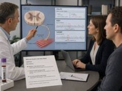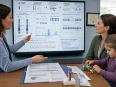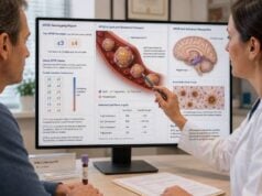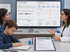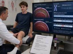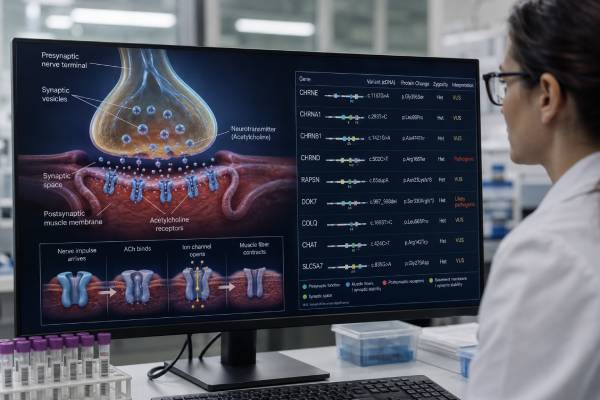
Congenital myasthenic syndrome genetic testing looks for inherited variants that disrupt communication between a motor nerve and muscle at the neuromuscular junction. These disorders can cause fluctuating or activity-related weakness, drooping eyelids, feeding problems, delayed motor milestones, and episodes of breathing failure. Symptoms may begin at birth, during childhood, or even in adulthood. A molecular diagnosis is unusually important because treatment that helps one genetic subtype can be ineffective or harmful in another. Modern panels examine genes involved before, within, and after release of acetylcholine, including CHRNE, RAPSN, CHAT, COLQ, DOK7, GFPT1, GMPPB, MUSK, AGRN, and others. Testing does not replace repetitive nerve stimulation, single-fiber electromyography, antibody testing, muscle studies, or evaluation for other neuromuscular conditions. A positive result can confirm the subtype, direct medication selection, identify respiratory risks, and clarify family recurrence. Negative and uncertain results require careful review because some panels omit rare genes, copy-number variants, deep intronic changes, or newly described causes.
- The test usually uses a multigene panel: more than 40 genes have been linked to congenital myasthenic syndromes.
- Genotype can change treatment: pyridostigmine may help many subtypes but can worsen DOK7-, COLQ-, and slow-channel disease.
- A normal antibody test does not exclude CMS: CMS is genetic rather than autoimmune.
- Breathing crises can occur during fever or infection: sudden shallow breathing, blue color, or inability to clear secretions needs emergency care.
- No fasting is required: blood or saliva DNA is commonly used, and medications do not alter the inherited result.
Table of Contents
- What Congenital Myasthenic Syndrome Is
- Genes and Neuromuscular Junction Defects
- When Genetic Testing Is Considered
- Testing Methods and Sample Process
- Positive, Negative, and Uncertain Results
- How Results Guide Treatment
- Inheritance, Family Testing, and Pregnancy
- Follow-Up, Safety, and Unresolved Cases
What Congenital Myasthenic Syndrome Is
Congenital myasthenic syndromes, or CMS, are inherited disorders of neuromuscular transmission. A motor nerve normally releases acetylcholine into the small space between nerve and muscle. Acetylcholine binds receptors on the muscle membrane, creating an electrical signal that triggers contraction. Proteins on both sides of the junction organize, release, clear, and respond to this chemical message.
A pathogenic variant can weaken any step in that process. The muscle itself may be structurally intact, yet repeated signals fail to produce reliable contraction. This creates fatigable weakness: strength decreases with activity and may improve after rest. The pattern is not always obvious. Some people have fixed limb-girdle weakness, childhood motor delay, recurrent apnea, or slowly progressive respiratory weakness rather than dramatic day-to-day fluctuation.
Possible features include:
- drooping eyelids or limited eye movements;
- facial weakness or a weak cry;
- poor suck, prolonged feeding, choking, or nasal speech;
- delayed sitting, standing, or walking;
- difficulty climbing stairs, running, rising from the floor, or lifting the arms;
- head drop, scoliosis, or axial weakness;
- reduced exercise tolerance;
- episodic respiratory arrest or worsening during infection; and
- improvement or deterioration with particular neuromuscular medications.
“Congenital” refers to a genetic origin, not necessarily symptoms visible at birth. DOK7-, GFPT1-, and some acetylcholine receptor–related forms may first become clear in later childhood or adulthood. Adults can be misdiagnosed with seronegative myasthenia gravis, muscular dystrophy, mitochondrial disease, motor neuron disease, or an unexplained limb-girdle myopathy for years.
CMS differs from autoimmune myasthenia gravis. In myasthenia gravis, antibodies attack proteins at the neuromuscular junction, and immune therapies may help. In CMS, inherited variants alter the junction itself; immunosuppressive therapy usually does not correct the cause. Antibody tests are often negative, although a person could rarely have both an inherited and an autoimmune disorder.
Genes and Neuromuscular Junction Defects
CMS genes are often grouped by where their proteins act: presynaptic, synaptic, postsynaptic, or in broader pathways that support the junction.
Presynaptic genes
Presynaptic proteins help make, package, release, and recycle acetylcholine. Important genes include CHAT, SLC18A3, SLC5A7, SYT2, VAMP1, SNAP25, and UNC13A.
CHAT-related CMS can cause episodic apnea, especially during fever, infection, stress, or prolonged crying. A child may appear relatively well between attacks. SLC18A3 and SLC5A7 variants can produce severe neonatal weakness, feeding problems, breathing difficulties, or developmental features. SYT2 can cause dominant or recessive presynaptic disease and may resemble a motor neuropathy or Lambert-Eaton–like syndrome.
Synaptic genes
The synaptic space contains acetylcholinesterase, which rapidly breaks down acetylcholine after a signal. COLQ anchors acetylcholinesterase at the endplate. Biallelic COLQ variants cause endplate acetylcholinesterase deficiency, allowing acetylcholine to remain too long and repeatedly stimulate the receptor. Patients may have marked axial, limb, respiratory, and eye movement weakness.
This mechanism explains why an acetylcholinesterase inhibitor such as pyridostigmine can worsen COLQ-related disease: the drug further slows acetylcholine breakdown.
Postsynaptic acetylcholine receptor genes
The adult muscle acetylcholine receptor contains subunits encoded by CHRNA1, CHRNB1, CHRND, and CHRNE. Variants can reduce the number of receptors, shorten channel opening, or keep channels open too long.
- Acetylcholine receptor deficiency often involves biallelic CHRNE variants and commonly responds to medicines that increase acetylcholine signaling.
- Fast-channel syndrome shortens receptor opening and usually follows recessive inheritance.
- Slow-channel syndrome prolongs receptor opening, damages the endplate, and usually follows dominant inheritance.
These are not interchangeable. A report naming an acetylcholine receptor gene should specify the variant mechanism when possible because treatment can move in opposite directions.
Receptor clustering and maintenance genes
RAPSN anchors acetylcholine receptors. AGRN, LRP4, MUSK, and DOK7 form a signaling pathway that organizes receptor clusters at the muscle endplate. DOK7-related CMS often produces a limb-girdle pattern with proximal weakness, waddling gait, and respiratory involvement; eye movement limitation may be less prominent than in receptor deficiency.
COL13A1 helps mature and maintain the neuromuscular junction. Variants can cause neonatal feeding and breathing problems, facial weakness, scoliosis, and delayed motor milestones.
Glycosylation and structural pathway genes
Genes such as GFPT1, DPAGT1, ALG2, ALG14, and GMPPB affect glycosylation, a cellular process that modifies proteins. These forms may look like limb-girdle muscular dystrophy and can include elevated creatine kinase or tubular aggregates on muscle biopsy. GMPPB can also cause muscular dystrophy, so the phenotype and electrophysiology determine whether a variant explains a myasthenic syndrome.
Other genes, including PLEC, PREPL, LAMB2, SCN4A, DES, and MYO9A, can produce additional structural, metabolic, kidney, eye, skin, or developmental findings. A current genetic panel should reflect the patient’s full phenotype rather than contain only the historically common CMS genes.
When Genetic Testing Is Considered
Genetic testing is appropriate when clinical and electrophysiologic findings suggest impaired neuromuscular transmission, particularly when autoimmune testing is negative or the history began early in life.
Clues that raise suspicion include:
- weakness from birth or childhood;
- recurrent unexplained apnea, especially during infections;
- fatigable eye, facial, swallowing, neck, respiratory, or limb weakness;
- siblings with similar symptoms and unaffected parents;
- consanguinity or ancestry associated with a founder variant;
- a limb-girdle syndrome with preserved sensation and normal or mildly elevated creatine kinase;
- a decrement on repetitive nerve stimulation;
- increased jitter or blocking on single-fiber electromyography;
- absent acetylcholine receptor, MuSK, and related autoimmune antibodies; or
- a medication response that is atypical for autoimmune myasthenia.
The diagnostic evaluation should remain broad. Congenital myopathies, muscular dystrophies, spinal muscular atrophy, mitochondrial disorders, channelopathies, motor neuropathies, brainstem conditions, botulism, and metabolic disease can overlap. In infants, sepsis, airway problems, heart disease, and central apnea must also be considered.
Electrodiagnostic testing may show a drop in muscle response after repetitive nerve stimulation. Testing the correct muscles matters: a proximal or facial muscle may reveal a defect that distal testing misses. Single-fiber electromyography is sensitive but not specific; abnormal jitter can occur in other nerve and muscle disorders.
Serum creatine kinase is often normal or mildly elevated, though glycosylation-related forms can have higher values. Autoantibody testing helps separate autoimmune myasthenia, but negative antibodies alone do not prove CMS.
Genetic testing may be urgent in a newborn or child with recurrent breathing crises because the subtype can influence emergency prevention and medication. In adults, testing can end a long diagnostic delay and prevent unnecessary immunosuppression.
Testing Methods and Sample Process
Most laboratories begin with a next-generation sequencing panel using blood or saliva. No fasting is needed, and the inherited DNA sequence is not altered by pyridostigmine, albuterol, immunotherapy, or other medication.
A useful panel should assess:
- single-nucleotide substitutions;
- small insertions and deletions;
- exon-level deletions and duplications when validated;
- genes for presynaptic, synaptic, postsynaptic, clustering, glycosylation, and structural subtypes; and
- relevant overlapping neuromuscular conditions.
The number of genes varies widely. A panel with 20 genes and one with 100 genes are not equivalent, but more genes are not automatically better. Coverage, copy-number sensitivity, variant interpretation, and phenotype matching are as important as panel size.
For a child with a classic recessive presentation, sequencing may find two pathogenic variants in the same gene. When only one is found, the laboratory should consider whether a second variant could be a deletion, duplication, deep intronic change, promoter variant, or structural rearrangement not detected by the assay.
If panel testing is negative, whole-exome sequencing or genome sequencing may be appropriate, often as a trio with samples from the patient and both parents. Trio analysis improves detection of de novo variants and helps determine whether two recessive variants lie on opposite parental copies.
RNA studies from muscle or cultured cells can sometimes reveal abnormal splicing that blood DNA analysis could not interpret. Muscle biopsy is less commonly the first test but may show tubular aggregates, endplate abnormalities, or a myopathy that redirects genetic analysis. Specialized in vitro studies may be needed to distinguish fast-channel, slow-channel, receptor-deficiency, or uncertain receptor variants.
Before ordering, ask whether the laboratory:
- includes recently established CMS genes;
- detects copy-number variants;
- reports coverage gaps;
- offers parental testing for uncertain or recessive findings;
- reanalyzes negative exome or genome data; and
- provides variant-level evidence relevant to treatment.
The test should be ordered with detailed clinical information. “Weakness” is less useful than “neonatal apnea, ptosis, bulbar weakness, decrement on repetitive stimulation, negative AChR antibodies, and improvement with pyridostigmine.” Phenotype information helps the laboratory prioritize variants and avoid reporting an unrelated rare change as the diagnosis.
Positive, Negative, and Uncertain Results
CMS results require both variant classification and inheritance analysis.
| Result | What it can mean | Next step |
|---|---|---|
| Pathogenic or likely pathogenic variants consistent with inheritance | A molecular diagnosis is established when the gene, variant mechanism, and phenotype fit | Use gene-specific treatment guidance and test relatives as appropriate |
| One pathogenic variant in a recessive gene | The person may be a carrier, or a second disease-causing variant may have been missed | Review copy-number, intronic, structural, and parental testing |
| Variant of uncertain significance | Evidence is insufficient to call the change disease-causing or benign | Do not base high-risk treatment or reproductive testing on it alone |
| Negative | No reportable cause was found within the assay’s scope | Reassess the diagnosis and consider broader testing or reanalysis |
Positive result
A convincing positive result matches the inheritance model. Most CMS forms are autosomal recessive, so two pathogenic or likely pathogenic variants are generally required, one on each copy of the gene. A single heterozygous variant is not enough unless the gene and mechanism are known to cause dominant disease, as in many slow-channel receptor syndromes or selected SYT2-related conditions.
“Likely pathogenic” usually carries the same clinical weight as pathogenic when the phenotype fits. The label refers to confidence in disease causation, not expected symptom severity.
A result can establish the diagnosis without predicting an exact course. People with the same gene, and sometimes the same variant, may differ in respiratory risk, eye involvement, walking ability, and medication response. Clinical monitoring remains essential.
Variant of uncertain significance
A variant of uncertain significance is not a positive diagnosis. Evidence may improve through parental testing, affected-relative testing, population data, RNA analysis, functional experiments, or later case reports.
An uncertain finding in a receptor gene should not be assumed to be slow-channel or fast-channel disease. Giving a channel blocker or avoiding a useful acetylcholinesterase inhibitor based only on a VUS can cause harm. Treatment decisions may still be made from the clinical syndrome, but the uncertainty should be explicit.
Negative result
A negative test does not exclude CMS. The disease may involve a gene not on the panel, a variant type the assay cannot detect, or a mechanism that remains undiscovered. The patient may also have autoimmune myasthenia, congenital myopathy, muscular dystrophy, neuropathy, or another diagnosis.
Reviewing the raw phenotype after a negative result is often more productive than immediately ordering the same type of larger panel. A specialist may repeat electrophysiology, test a clinically affected muscle, obtain exome or genome sequencing, pursue RNA studies, or reconsider a metabolic or mitochondrial cause.
How Results Guide Treatment
CMS is one of the clearest examples of why a genetic diagnosis can directly change medication choice. Treatment should be supervised by a neuromuscular specialist because responses vary and some drugs can worsen weakness or cause serious cardiac, psychiatric, or respiratory effects.
| Genetic mechanism or subtype | Common treatment direction | Important caution |
|---|---|---|
| Many receptor-deficiency, RAPSN, and presynaptic forms | Pyridostigmine may improve transmission; selected patients may also use 3,4-diaminopyridine | Response is subtype- and age-dependent |
| DOK7 or COLQ | Albuterol/salbutamol or ephedrine may help | Pyridostigmine may be ineffective or worsen weakness |
| Slow-channel acetylcholine receptor syndrome | Open-channel blockers such as fluoxetine or quinidine may be considered by specialists | Cardiac and psychiatric risks require close oversight; wrong mechanism can worsen disease |
| CHAT or RAPSN with episodic apnea | Continuous or preventive therapy may reduce crisis risk | Families still need an emergency respiratory plan |
Pyridostigmine inhibits acetylcholinesterase, leaving acetylcholine available longer. It helps many CMS subtypes, but not all. DOK7-related disease, COLQ-related acetylcholinesterase deficiency, and slow-channel syndrome can deteriorate with it. A prior worsening response is therefore diagnostically useful, though it is not sufficient by itself to identify the gene.
3,4-diaminopyridine increases acetylcholine release from the nerve terminal. It may be used alone or with pyridostigmine in selected subtypes. Dosing and age require caution, especially in children and in fast-channel disease.
Beta-2 adrenergic agonists such as albuterol or salbutamol can produce major improvement in DOK7, COLQ, and some other forms, but benefits may develop over weeks or months. Heart rate, tremor, potassium, and other adverse effects may need monitoring. Ephedrine is another option in selected patients.
Slow-channel syndrome requires a different approach aimed at shortening excessive receptor activation. Fluoxetine or quinidine may be considered in expert care, but neither is a casual trial. Fluoxetine carries psychiatric and drug-interaction risks; quinidine can cause dangerous arrhythmia and worsen weakness. The precise receptor mechanism should be established whenever possible.
Immune therapies such as corticosteroids, intravenous immunoglobulin, plasma exchange, or B-cell depletion do not treat the inherited defect and are generally ineffective in isolated CMS. A brief response can be nonspecific and should not override genetic and electrophysiologic evidence.
Supportive care may include respiratory monitoring, noninvasive ventilation, cough assistance, feeding support, speech therapy, physical and occupational therapy, orthotics, scoliosis care, and an individualized activity plan. During surgery, anesthesia teams need the diagnosis and medication list because neuromuscular blockers and other agents can have exaggerated effects.
Inheritance, Family Testing, and Pregnancy
Most CMS subtypes are autosomal recessive. An affected person has pathogenic variants in both copies of the same gene, while each parent usually carries one variant and has no symptoms. When both parents are carriers, each pregnancy has:
- a 25% chance of an affected child;
- a 50% chance of an unaffected carrier child; and
- a 25% chance of a child who inherited neither familial variant.
This calculation resets with every pregnancy. It does not mean that one of every four children in a small family must be affected.
Some CMS forms are autosomal dominant. A person with a dominant pathogenic variant has a 50% chance of passing it to each child. Dominant slow-channel receptor syndromes and selected presynaptic disorders are examples. A variant may be inherited from an affected parent or arise de novo.
Once the family variants are confirmed, relatives can receive a familial variant test. Testing apparently healthy newborn siblings can be medically useful in families with severe apnea-prone CMS because early treatment and emergency planning may prevent life-threatening events.
Carrier testing is meaningful only after the familial pathogenic variants are known. A broad carrier panel that includes one CMS gene may identify one variant but cannot necessarily establish the family’s full risk.
Reproductive options include natural conception, prenatal diagnosis, in vitro fertilization with preimplantation genetic testing, donor gametes, or adoption. Prenatal testing can determine whether a fetus inherited the familial variants but usually cannot predict exact severity. Counseling should discuss the known range of the specific genotype rather than describe all CMS as equally severe.
Pregnancy in an affected adult may worsen, improve, or leave weakness unchanged. Respiratory reserve, swallowing, medication safety, labor planning, and anesthesia should be reviewed by neuromuscular, maternal-fetal medicine, and anesthesia teams.
Follow-Up, Safety, and Unresolved Cases
Follow-up should track strength, falls, feeding, swallowing, speech, scoliosis, respiratory function, sleep-related symptoms, and medication response. Children often need review at least every six months, while stable adults may be seen yearly, with more frequent visits for respiratory disease or treatment changes.
Signs of nighttime hypoventilation include morning headaches, restless sleep, daytime sleepiness, weak cough, recurrent chest infections, or difficulty breathing when lying flat. Normal daytime appearance does not exclude nocturnal respiratory weakness.
Families at risk of episodic apnea need a written emergency plan. Fever, infection, prolonged crying, surgery, and missed medication can trigger deterioration in some subtypes. Caregivers may need cardiopulmonary resuscitation training, airway-clearance equipment, or home ventilation support. Emergency clinicians should be told that the condition is congenital myasthenic syndrome, not simply “muscle weakness.”
Medicines that impair neuromuscular transmission can worsen symptoms. Certain antibiotics, beta-blockers, magnesium, antiarrhythmics, anesthetic agents, and other drugs deserve review, but they are not universally forbidden. The treating team should weigh the medical need, gene-specific risk, dose, alternatives, and monitoring rather than stopping essential therapy without advice.
For an unresolved case, preserve the complete laboratory report, raw-data access information when available, electrophysiology results, antibody results, medication-response history, and a detailed family tree. Reanalysis after one to three years can identify newly established gene-disease relationships or reclassify a prior finding. A broader whole-genome sequencing test, RNA analysis, or a research referral may be appropriate when clinical evidence remains strong.
A negative genetic result should never delay treatment of respiratory failure or severe bulbar weakness. Conversely, a positive result should not cause every new symptom to be attributed to CMS. Aspiration, infection, heart disease, medication toxicity, autoimmune myasthenia, and other acquired problems can occur in a genetically affected person and may need separate treatment.
References
- Review of 40 genes causing congenital myasthenic syndromes 2025 (Review)
- Guideline for the management of myasthenic syndromes 2023 (Guideline)
- Congenital myasthenic syndromes in adults: clinical features, diagnosis and long-term prognosis 2024 (Research Study)
- Screening for Congenital Myasthenic Syndromes in Adults With Seronegative Myasthenic Syndromes 2025 (Research Study)
- Congenital Myasthenic Syndromes Overview 2021 (GeneReviews)
Disclaimer
Congenital myasthenic syndrome testing and treatment require specialist neuromuscular and genetics care because medication effects differ by gene and mechanism. Do not start, stop, or change pyridostigmine, albuterol, 3,4-diaminopyridine, fluoxetine, quinidine, or other neuromuscular medicines based only on general information or an uncertain variant. Call emergency services for apnea, blue color, severe breathing difficulty, inability to swallow secretions, or rapidly worsening weakness.