
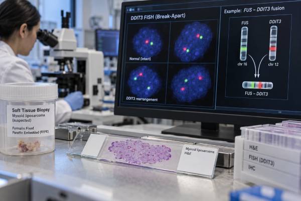
A DDIT3 fusion test looks for a characteristic gene rearrangement that strongly supports the diagnosis of myxoid liposarcoma. Most myxoid liposarcomas contain a FUS::DDIT3 fusion created by the translocation t(12;16), while a smaller minority contain EWSR1::DDIT3. Because these fusions are highly characteristic, molecular testing can be especially useful when the tumor is high grade, the biopsy is small, or the microscopic appearance overlaps with other myxoid or round-cell sarcomas. A positive DDIT3 rearrangement is a tumor finding, not an inherited mutation, and it does not imply that relatives are at increased hereditary cancer risk. Testing may use FISH, RT-PCR, or RNA-based next-generation sequencing. The result helps classify the tumor, but it does not by itself determine stage, metastatic risk, or treatment. Those depend on tumor size, location, round-cell component, imaging, resectability, and other clinical factors.
- A DDIT3 rearrangement strongly supports myxoid liposarcoma when the tumor’s morphology and clinical setting are compatible.
- FUS::DDIT3 is the dominant fusion, found in the great majority of cases; EWSR1::DDIT3 is much less common.
- DDIT3 testing is most useful in difficult biopsies or high-grade round-cell tumors that resemble other sarcomas.
- A positive fusion is somatic, not hereditary: it does not usually create a family-testing indication.
- A negative test does not always exclude myxoid liposarcoma: assay type, tissue quality, rare fusion partners, and sampling can matter.
Table of Contents
- What DDIT3 testing detects
- Why the fusion defines myxoid liposarcoma
- When the test is ordered
- How DDIT3 testing is performed
- How to interpret results
- Prognosis and treatment context
- Limitations and next steps
What DDIT3 testing detects
DDIT3 encodes a transcription factor also known as CHOP. In myxoid liposarcoma, chromosomal rearrangement places DDIT3 next to another gene, creating an abnormal fusion protein that changes gene regulation and helps drive the tumor.
The classic event is t(12;16)(q13;p11), which creates FUS::DDIT3. More than 90% of myxoid liposarcomas contain this fusion. A smaller group carries t(12;22) and EWSR1::DDIT3.
The fusion is not merely a passenger mutation. Experimental studies show that FUS::DDIT3 alters transcriptional programs and chromatin regulation and interferes with normal adipocytic differentiation. This molecular identity is one reason myxoid liposarcoma is considered a translocation-defined sarcoma.
DDIT3 testing should not be confused with CDK4 amplification testing or MDM2 amplification testing. Those are characteristic of atypical lipomatous tumor/well-differentiated and dedifferentiated liposarcoma, not myxoid liposarcoma.
Why the fusion defines myxoid liposarcoma
Low-grade myxoid liposarcoma has a recognizable combination of bland spindle cells, myxoid matrix, delicate branching capillaries, and variable lipoblasts. High-grade disease, historically called round-cell liposarcoma, can lose much of that classic appearance and resemble other round-cell malignancies.
In these difficult cases, detecting a DDIT3 rearrangement provides powerful confirmation that the tumor belongs to the myxoid liposarcoma family.
The fusion also helps distinguish myxoid liposarcoma from:
- myxofibrosarcoma;
- extraskeletal myxoid chondrosarcoma;
- Ewing sarcoma and other round-cell sarcomas;
- dedifferentiated liposarcoma with myxoid areas;
- low-grade fibromyxoid sarcoma; and
- benign myxoid soft-tissue lesions.
Pathology still matters. Molecular results are interpreted together with morphology because some assays can detect a rearrangement without identifying the exact partner, and rare technical artifacts are possible.
When the test is ordered
A pathologist may order DDIT3 testing when myxoid liposarcoma is suspected but not unequivocal. It is particularly useful when:
- a core biopsy contains only a small amount of tumor;
- the lesion is predominantly high-grade round-cell tissue;
- prior radiation or chemotherapy has altered the morphology;
- the differential diagnosis includes another translocation-associated sarcoma; or
- the tumor occurs in an unusual site.
Testing may also be done as part of a broad RNA fusion panel that simultaneously evaluates many sarcoma-associated rearrangements.
Myxoid liposarcoma most often arises in deep soft tissues of the extremities, especially the thigh, but it can metastasize to unusual extrapulmonary sites. Molecular confirmation can be helpful when a metastatic lesion appears in bone, soft tissue, or another location and the origin is uncertain.
How DDIT3 testing is performed
FISH with a DDIT3 break-apart probe is a common method. It detects separation of fluorescent signals around the DDIT3 locus, indicating that the gene has been rearranged. FISH works on formalin-fixed paraffin tissue and does not require knowing the fusion partner.
RT-PCR detects a specific fusion transcript. It can be highly specific but may miss unusual breakpoint variants if the primers do not match the transcript present.
RNA-based next-generation sequencing is increasingly useful because it can identify both the rearranged gene and the fusion partner, including uncommon fusion forms. It may also evaluate many sarcoma fusions at once.
DDIT3 immunohistochemistry can show strong nuclear expression and has demonstrated high sensitivity and specificity in studies, particularly for high-grade myxoid liposarcoma. It can serve as a useful screening or diagnostic adjunct, but molecular confirmation remains valuable when the diagnosis is uncertain.
No fasting or medication changes are required for the molecular assay itself. Preparation relates only to how the biopsy or surgery is obtained.
How to interpret results
| Finding | Meaning | Caution |
|---|---|---|
| FUS::DDIT3 detected | Strong molecular confirmation of myxoid liposarcoma | Final diagnosis still integrates morphology and site |
| EWSR1::DDIT3 detected | Rare but recognized myxoid liposarcoma fusion | Do not confuse with other EWSR1-rearranged sarcomas |
| DDIT3 break-apart positive by FISH | Shows DDIT3 rearrangement even if partner is not identified | Additional RNA testing may clarify the partner if needed |
| No rearrangement detected | Makes classic myxoid liposarcoma less likely | False negatives can occur with poor tissue, assay limits, or rare events |
A positive result does not measure tumor burden and does not determine whether the tumor has metastasized. It is a classification marker.
A negative result should be reviewed against the pathology. If the morphology is highly convincing, the pathologist may repeat testing with another method, test RNA, or review tissue quality.
Prognosis and treatment context
DDIT3 fusion status confirms tumor type but is not used as a simple high-risk versus low-risk scale. Prognosis depends more on tumor size, depth, surgical margins, metastatic disease, and the proportion of high-grade round-cell component.
Myxoid liposarcoma has several unusual clinical features. It is generally more sensitive to radiation than many other soft-tissue sarcomas, and it has a tendency to metastasize outside the lungs, including to soft tissue and bone. Imaging strategy may therefore differ from that used for some other sarcomas.
Surgery remains central for localized disease, often combined with radiotherapy depending on tumor location and resection planning. Systemic therapy may be considered for advanced or high-risk disease. Trabectedin has notable activity in myxoid liposarcoma, and other therapies are selected according to sarcoma guidelines and clinical context.
The FUS::DDIT3 protein and its downstream biology are active therapeutic research areas. However, a positive DDIT3 fusion is not currently a stand-alone companion diagnostic for a specific fusion-targeted drug.
Limitations and next steps
The main limitations are tissue quality, assay design, and sampling. Formalin can degrade RNA, small biopsies may contain few tumor cells, and RT-PCR assays can miss uncommon transcript variants. FISH can show rearrangement but may not identify the partner.
After a positive result, the next step is to finalize sarcoma subtype and grade, review imaging for local and distant disease, and plan care with a sarcoma multidisciplinary team.
After a negative result in a highly suspicious case, reasonable options include RNA-based sequencing, repeat FISH, DDIT3 immunohistochemistry, or expert pathology review.
The fusion is not a hereditary finding, so routine family testing is not indicated simply because FUS::DDIT3 or EWSR1::DDIT3 is detected.
A myxoid appearance is not enough for diagnosis
Many soft-tissue tumors can contain abundant myxoid, or gelatin-like, extracellular material. Myxoid liposarcoma is one specific sarcoma entity, not a general name for every tumor with myxoid stroma. The pathologist looks for characteristic architecture, delicate branching capillaries, lipoblasts in appropriate cases, and the overall cellular pattern. Molecular confirmation becomes especially valuable when the biopsy is small, the morphology is unusual, or the differential diagnosis includes other round-cell or myxoid neoplasms.
The defining event is usually a fusion of DDIT3 with FUS and less commonly with EWSR1. Demonstrating a DDIT3 rearrangement or the specific fusion transcript strongly supports myxoid liposarcoma when the morphology and clinical setting fit. Because the alteration is highly characteristic, it can resolve cases that would otherwise remain uncertain. It should still be interpreted with the pathology rather than as a stand-alone label detached from tissue appearance and site.
Round-cell change affects grade and risk
Myxoid liposarcoma can contain areas with increased cellularity and less obvious myxoid background, historically described as round-cell components. The proportion and extent of this higher-grade morphology are clinically important because they are associated with more aggressive behavior. Molecular detection of DDIT3 confirms lineage, but it does not measure the amount of high-grade component. That assessment depends on adequate tissue sampling and pathology review.
A core biopsy may under-sample a heterogeneous mass. If imaging shows distinct regions, the most solid or metabolically active area may need targeted sampling. Surgical specimens can also reveal a higher-grade component that was not represented in the original needle biopsy. This is one reason molecular confirmation, imaging, and morphology are complementary rather than interchangeable.
Choosing between FISH, RNA sequencing, RT-PCR, and immunohistochemistry
Break-apart FISH asks whether the DDIT3 locus is rearranged. It works on routinely processed tissue and can be useful when RNA quality is poor, but it usually does not identify the fusion partner or exact transcript. RNA-based next-generation sequencing can identify the fusion partners and breakpoint at transcript level and can survey many sarcoma fusions at once. It is especially useful when the differential diagnosis is broad. Its limitation is that formalin-fixed tissue may yield degraded RNA and a poor-quality specimen can produce a false-negative result.
Targeted RT-PCR can be sensitive when the expected fusion transcript is known, but assays designed for common breakpoints may miss uncommon transcript variants. DDIT3 immunohistochemistry can provide supportive evidence and may be useful as a screening tool, particularly when molecular testing is limited. Immunostaining is not identical to demonstrating the genetic fusion, so discordant cases may require an orthogonal molecular method.
When a test is negative despite classic morphology, the right response is not to assume the diagnosis is excluded. The team should check tumor content and assay quality, confirm what alteration types the method could detect, and consider a second method or a better specimen.
Imaging and metastatic pattern are part of the clinical picture
Myxoid liposarcoma has a metastatic pattern that can differ from many other soft-tissue sarcomas. Metastases may occur in extrapulmonary soft tissue, bone, or other sites, not only the lungs. Staging and follow-up strategies therefore need to reflect the sarcoma subtype and the patient’s individual risk. A DDIT3 fusion result helps establish the subtype but does not replace imaging.
This tumor is also recognized as relatively radiosensitive compared with many other sarcomas, which can influence multidisciplinary treatment planning in selected localized cases. Surgery remains central when feasible, while radiation and systemic therapy decisions depend on location, size, grade, resectability, metastatic status, prior treatment, and specialist sarcoma guidance. The presence of the fusion does not by itself prescribe one drug or radiation regimen.
The fusion is usually stable and acquired, not hereditary
FUS::DDIT3 or EWSR1::DDIT3 is a somatic rearrangement acquired by the tumor cells. It is not the kind of germline finding that is ordinarily passed from parent to child. Family members therefore do not need predictive testing for this fusion simply because it was found in a tumor.
Because the fusion is a defining driver, it is generally retained as the cancer evolves. If a later lesion has compatible morphology and the same defining fusion, that can support a relationship to the original myxoid liposarcoma. In routine care, however, the fusion is mainly a diagnostic marker rather than a standard blood marker for surveillance or response.
Important diagnostic look-alikes
The differential diagnosis depends on age, site, and morphology. Other myxoid sarcomas, low-grade fibromyxoid sarcoma, extraskeletal myxoid chondrosarcoma, and tumors with round-cell morphology can enter the discussion in difficult cases. Each has a different molecular signature. A broad RNA fusion panel can be efficient when the biopsy does not clearly favor one entity because it can look for DDIT3 fusions and alternative defining rearrangements in the same test.
This is particularly helpful when immunohistochemistry is nonspecific. Many sarcomas do not have one protein stain that proves the diagnosis. Molecular testing is strongest when it resolves a focused pathology differential rather than when it is ordered without morphologic context.
What a positive fusion report should contain
Ideally, the report states whether it detected a DDIT3 rearrangement only or identified a specific partner such as FUS or EWSR1. RNA sequencing may also provide the exact fusion junction. The distinction matters because a break-apart result confirms disruption of the DDIT3 locus, while a partner-specific result provides more direct molecular definition. Both can be diagnostically useful when technically valid.
A positive fusion does not provide the tumor size, margin status, percent round-cell component, or metastatic burden. Those features come from pathology and imaging and remain necessary for staging and prognosis. Molecular certainty about tumor type should not be confused with certainty about clinical outcome.
Why specialist pathology review can matter
Soft-tissue sarcomas are uncommon and encompass many molecularly distinct entities. When a biopsy is small, morphology is unusual, or molecular and histologic findings conflict, review by a pathologist who routinely evaluates sarcoma can prevent both overdiagnosis and underdiagnosis. The reviewer may recommend a different tissue block, additional immunostains, or an orthogonal fusion assay.
This is especially important before major surgery or systemic treatment. A defining DDIT3 fusion can make the diagnosis much more secure, but an unexpected positive result still deserves correlation with the tumor’s location and morphology. Good molecular pathology is not merely finding a rearrangement; it is showing that the rearrangement explains the specimen being evaluated.
Preanalytic quality can determine whether fusion testing succeeds
Fusion assays depend on what happened to the specimen before it reached the molecular laboratory. Prolonged fixation, extensive necrosis, very small biopsies, and low tumor percentage can reduce the amount or quality of usable nucleic acid. Decalcification is particularly problematic for some molecular methods when a suspected metastasis is sampled from bone. The pathology team may select a different block or reserve unstained slides to preserve material for RNA testing.
A failed assay is not the same as a negative assay. “Quantity not sufficient,” “RNA quality inadequate,” or “no result” means the test could not answer the question. In a case where confirming myxoid liposarcoma matters, another block, another biopsy, or an alternative method such as FISH may be appropriate.
Fusion nomenclature can look different across reports
Older reports may use a hyphen, such as FUS-DDIT3, while newer standardized nomenclature often uses a double colon, FUS::DDIT3. Both can refer to the same biologic fusion. The report may also describe the historical translocation associated with the fusion. Patients should focus on the genes involved and the laboratory interpretation rather than assuming different punctuation means a different disease.
References
- Diagnosis and Treatment of Myxoid Liposarcoma 2024 (Review)
- FUS::DDIT3 Fusion Protein in the Development of Myxoid Liposarcoma and Possible Implications for Therapy 2024 (Review)
- The FUS::DDIT3 fusion oncoprotein inhibits BAF complex targeting and activity in myxoid liposarcoma 2022
- Nuclear expression of DDIT3 distinguishes high-grade myxoid liposarcoma from other round cell sarcomas 2021
- DDIT3 Immunohistochemistry Is a Useful Tool for the Diagnosis of Myxoid Liposarcoma 2021
- DDIT3 gene break-apart as a molecular marker for diagnosis of myxoid liposarcoma–assay validation and clinical experience. 2012
Disclaimer
DDIT3 fusion testing is a tumor-classification tool and should be interpreted by a pathologist experienced in soft-tissue tumors. A positive result does not by itself determine stage, prognosis, or treatment, and a negative result may require another testing method when morphology remains strongly suspicious.





