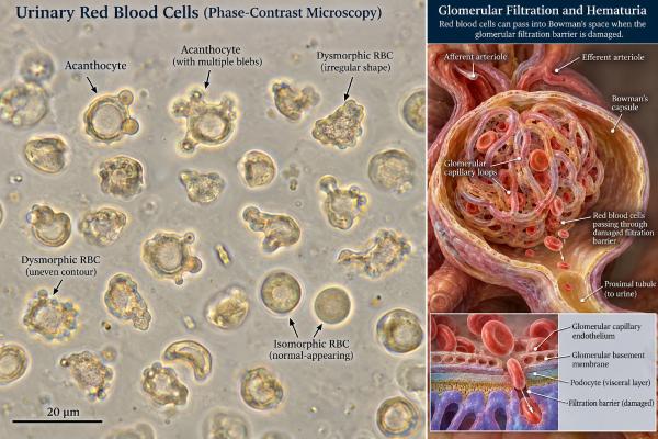
Dysmorphic red blood cells in urine are abnormally shaped RBCs that can point to bleeding from the kidney’s glomeruli—the microscopic filters that normally keep blood cells inside the circulation. Their value is highest when a fresh urine sample is examined carefully, especially with phase-contrast microscopy, and when the result is interpreted alongside protein or albumin in urine, kidney function, blood pressure, and other sediment findings. Acanthocytes, a distinctive form of dysmorphic RBC with one or more blister-like membrane projections, are particularly suggestive of glomerular bleeding. However, there is no single percentage cutoff that works for every laboratory, and dysmorphic cells do not identify the exact kidney disease by themselves. They are a localization clue. Persistent hematuria with dysmorphic RBCs, RBC casts, significant proteinuria, reduced kidney function, or high blood pressure generally warrants medical evaluation for glomerular disease rather than being dismissed as a lower urinary tract source.
- Dysmorphic RBCs suggest a kidney-filter source: they are more consistent with glomerular bleeding than with bleeding from the bladder or urethra.
- Acanthocytes are especially useful: a meaningful proportion of acanthocytes increases the likelihood of glomerular hematuria, but laboratory methods and thresholds vary.
- A positive dipstick is not enough: hematuria should be confirmed with microscopic RBC counting in a properly collected sample.
- Proteinuria and RBC casts raise concern: these findings strengthen the case for glomerular inflammation or other medical kidney disease.
- Urgent assessment may be needed: visible blood with reduced urine, swelling, shortness of breath, rapidly rising creatinine, or severe high blood pressure can signal significant kidney injury.
Table of Contents
- What Dysmorphic Red Blood Cells Mean
- Why Glomerular Bleeding Changes RBC Shape
- Microscopy, Acanthocytes, and Thresholds
- Causes of Glomerular Hematuria
- Dysmorphic vs Isomorphic RBCs
- How Abnormal Results Are Worked Up
- Common Result Patterns and What They Suggest
- Limitations and Common Interpretation Mistakes
- When to Seek Medical Care
What Dysmorphic Red Blood Cells Mean
Dysmorphic RBCs are red blood cells with abnormal size, contour, membrane projections, or loss of the usual smooth disc shape. In urine microscopy, these changes can indicate that RBCs have passed through a damaged glomerular filtration barrier and then traveled through the nephron before being excreted.
The finding helps answer where the blood is probably coming from. Hematuria from the bladder, ureter, prostate, or urethra more often contains relatively uniform, isomorphic RBCs. Hematuria arising from a glomerulus is more likely to contain dysmorphic cells, especially acanthocytes, and may also include red blood cell casts.
This distinction matters because the diagnostic pathways differ. A stone, urinary infection, prostate bleeding, and bladder lesion are evaluated differently from IgA nephropathy, vasculitis, lupus nephritis, or inherited glomerular disorders. Dysmorphic RBCs do not replace imaging, urine culture, serology, or kidney biopsy when those tests are indicated, but they can direct attention toward medical kidney disease.
Hematuria itself is commonly defined microscopically. Current urologic guidance uses at least 3 RBCs per high-power field in a properly collected specimen as the threshold for microhematuria in adults. A urine dipstick detects heme activity and can be positive from intact RBCs, free hemoglobin, or myoglobin, so microscopic confirmation remains important. A detailed red blood cells in urine test result is more informative than a dipstick label of “blood” alone.
Why Glomerular Bleeding Changes RBC Shape
The glomerulus is a high-pressure capillary tuft surrounded by a specialized filtration barrier. That barrier includes endothelial cells, the glomerular basement membrane, and podocytes with slit diaphragms. It allows water and small solutes to pass while retaining most blood cells and large proteins.
When the barrier is inflamed, structurally abnormal, or damaged, RBCs can squeeze through defects they normally could not cross. Mechanical stress during this passage may deform the membrane. The cells then encounter changes in osmolality and pH as they move through the tubules. Repeated swelling, shrinking, and membrane stress can produce irregular forms by the time the cells reach the final urine.
Not every oddly shaped RBC is evidence of glomerular disease. Cells can crenate in concentrated urine, swell or lyse in dilute urine, and change after a specimen sits too long. For that reason, laboratories distinguish clinically useful dysmorphism from nonspecific artifacts.
The most important morphology is the acanthocyte. In urinary microscopy, an acanthocyte is typically ring-shaped with one or more rounded, bubble-like protrusions or invaginations. Acanthocytes are considered more specific for glomerular bleeding than a broad category of “dysmorphic RBCs.” The related acanthocytes in urine finding can therefore carry substantial diagnostic weight when identified by an experienced observer.
Microscopy, Acanthocytes, and Thresholds
Urinary RBC morphology is best assessed in a fresh specimen. Phase-contrast microscopy makes transparent cell outlines easier to see and is often preferred for recognizing acanthocytes and other dysmorphic forms. Bright-field microscopy can also be useful when performed carefully, while automated analyzers may provide RBC counts and pattern information but may not classify every abnormal morphology reliably.
There is no universally accepted dysmorphic-RBC percentage that diagnoses glomerular disease in every setting. Published studies have used different definitions, microscopes, observers, patient groups, and cutoffs, producing wide ranges of sensitivity and specificity. This is one reason a report should be read in the context of the laboratory’s own method.
Some useful reference points illustrate the issue rather than create universal rules:
- A 2025 multicenter study found higher dysmorphic-RBC percentages in biopsy-confirmed glomerular disease and used an 18% cutoff in its study analysis, but that value should not be treated as a universal clinical threshold.
- Pediatric and young-adult guidance identifies at least 5% acanthocytes as a strong reason for nephrologic evaluation when microhematuria persists.
- Some laboratory frameworks use much higher proportions of broadly dysmorphic RBCs to increase specificity for glomerular bleeding.
The overall sediment pattern is often more meaningful than one percentage. Dysmorphic RBCs plus albuminuria, RBC casts, or reduced kidney function form a stronger glomerular pattern than dysmorphic RBCs alone. A comprehensive urine microscopy assessment for kidney disease looks at these components together.
Specimen handling also matters. Cells may lyse in very dilute or alkaline urine, potentially lowering the visible dysmorphic-RBC count. Delayed examination can alter morphology. Menstrual or genital blood can add RBCs that do not reflect urinary tract bleeding, so proper collection and timing are important.
Causes of Glomerular Hematuria
Dysmorphic RBCs do not tell you which glomerular disorder is present. They indicate a likely location, after which the clinical pattern narrows the cause.
Important possibilities include:
- IgA nephropathy: can cause microscopic hematuria or episodes of visible blood, often around respiratory infections.
- Thin basement membrane disorders and Alport spectrum disease: inherited abnormalities of glomerular basement membrane collagen can cause persistent hematuria, sometimes with a family history of kidney disease or hearing problems.
- Post-infectious or infection-related glomerulonephritis: may produce hematuria, proteinuria, edema, high blood pressure, and reduced kidney function.
- ANCA-associated vasculitis: can cause rapidly progressive glomerulonephritis and may be accompanied by systemic symptoms involving the lungs, skin, nerves, or other organs.
- Anti-GBM disease: can produce severe glomerular injury, sometimes with lung bleeding.
- Lupus nephritis: can cause hematuria, proteinuria, cellular casts, and changing kidney function in people with systemic lupus erythematosus.
- Other immune-complex glomerulonephritides: a range of systemic and kidney-limited disorders can produce a nephritic urine sediment.
The amount of hematuria does not always match disease severity. A person can have significant glomerular disease with only microscopic blood, while striking red urine can sometimes occur with preserved filtration. Protein excretion, serum creatinine, blood pressure, edema, and systemic findings help establish urgency.
Importantly, glomerular and urologic causes can coexist. An adult with dysmorphic RBCs and proteinuria may need nephrologic evaluation, but age, smoking history, visible hematuria, or other risk factors can still justify a separate urologic assessment. Current microhematuria guidance specifically recognizes that suspected medical kidney disease does not automatically eliminate the need to evaluate relevant urologic risk.
Dysmorphic vs Isomorphic RBCs
The terms “dysmorphic” and “isomorphic” describe appearance, not severity.
| Feature | Dysmorphic RBC pattern | Isomorphic RBC pattern |
|---|---|---|
| Cell appearance | Variable size and shape, irregular membranes, possible acanthocytes | Relatively uniform round cells |
| More suggestive source | Glomerular kidney bleeding | Post-glomerular urinary tract bleeding |
| Associated sediment clues | RBC casts, proteinuria, sometimes other cellular casts | May occur with stones, tumors, infection, trauma, or prostate bleeding |
| Important limitation | Artifacts and threshold variation can reduce accuracy | A uniform pattern does not completely exclude kidney disease |
This is not a binary test. Mixed populations occur. A sample can contain both isomorphic and dysmorphic cells, especially when more than one process is present or when specimen conditions alter cell morphology.
RBC casts are generally more specific for bleeding within the nephron because casts form in renal tubules. Yet casts can be sparse and easy to miss, so their absence does not exclude glomerulonephritis. Acanthocytes may be present when no RBC casts are seen.
The safest interpretation is probabilistic: a strong dysmorphic/acanthocytic pattern raises the probability of glomerular hematuria; a strongly isomorphic pattern shifts attention toward a nonglomerular source; neither result should be separated from the rest of the urinalysis and clinical history.
How Abnormal Results Are Worked Up
When dysmorphic RBCs are reported, follow-up typically begins by confirming that hematuria is persistent and the sample is valid. A clinician may repeat a clean-catch urinalysis after menstruation, strenuous exercise, an infection, or another temporary source has resolved.
The next step is to assess whether the urine pattern looks glomerular. Common tests include a urine albumin-to-creatinine ratio or protein-to-creatinine ratio, serum creatinine with estimated GFR, and blood pressure measurement. The combination of hematuria and meaningful proteinuria is more concerning for glomerular disease than isolated microscopic blood.
Additional testing depends on the presentation. Complement levels, ANA and other lupus tests, ANCA, anti-GBM antibodies, hepatitis testing, streptococcal serologies, serum protein studies, or genetic testing may be appropriate in selected patients. These are not routine tests for every person with a few dysmorphic RBCs.
Kidney ultrasound is often used to evaluate kidney structure and exclude obstruction or major anatomic abnormalities. Urologic imaging or cystoscopy may be considered based on age and malignancy risk. A kidney biopsy is reserved for situations where tissue diagnosis is likely to change management—for example, significant proteinuria, worsening kidney function, an active nephritic sediment, or suspected systemic glomerulonephritis.
The most actionable finding is not “dysmorphic RBCs” in isolation but a pattern plus trajectory. Stable kidney function and trace hematuria over years is a different problem from new dysmorphic hematuria with edema, hypertension, heavy proteinuria, and creatinine rising over several days.
Common Result Patterns and What They Suggest
Dysmorphic RBCs become more useful when they are grouped with the rest of the urinalysis rather than interpreted as a stand-alone flag. Several common patterns illustrate how clinicians think about the result.
Dysmorphic RBCs plus albuminuria or proteinuria strongly raises suspicion for glomerular disease because both findings point toward the filtration barrier. Protein amount matters. Trace protein on a concentrated urine sample is much less informative than persistent albuminuria or a clearly elevated protein-to-creatinine ratio. If protein excretion is substantial, clinicians usually assess kidney function and blood pressure even when the person has no symptoms.
Dysmorphic RBCs plus RBC casts is an even more glomerular pattern. RBC casts form within renal tubules, so they indicate that bleeding occurred upstream in the nephron. This combination can occur in inflammatory glomerulonephritis and should not be dismissed as contamination from menstruation or a lower urinary tract source.
Dysmorphic RBCs with normal creatinine and little or no protein can occur in milder or inherited glomerular conditions. Persistent isolated microscopic hematuria still deserves follow-up because some disorders, such as thin basement membrane or Alport-spectrum disease, may initially preserve filtration. Family history, hearing findings, and long-term urine-protein trends can become important.
Isomorphic RBCs with urinary symptoms shifts attention toward nonglomerular causes such as infection, stones, prostate bleeding, or bladder pathology. Leukocyte esterase, nitrites, white blood cells, bacteriuria, flank pain, dysuria, or stone symptoms help refine that differential. The RBC shape does not replace culture or imaging when those tests are indicated.
Mixed RBC morphology is common and can reflect technical factors or more than one source of bleeding. A person with underlying glomerular hematuria can also have a urinary infection or stone. In these cases, the proportion of acanthocytes, the presence of protein or casts, and whether hematuria persists after the temporary problem resolves are more informative than a simple “mixed” label.
A positive blood dipstick with few or no RBCs is a different pattern. It can occur with free hemoglobin or myoglobin rather than true hematuria. That distinction is important after severe muscle injury or hemolysis because the work-up targets those processes rather than glomerular bleeding.
These patterns also explain why repeat testing can be useful. If hematuria disappears after a urinary infection is treated or after menstruation ends, the significance changes. If dysmorphic hematuria persists across properly collected samples, particularly with albuminuria, the likelihood of a kidney-filter disorder increases and specialist evaluation becomes more appropriate.
Limitations and Common Interpretation Mistakes
Urinary RBC morphology is valuable but operator-dependent. Results can vary between laboratories, microscopy methods, and observers. Automated systems improve standardization of particle counts but may still require manual confirmation when morphology is clinically important.
Common mistakes include treating any misshapen RBC as “glomerular,” using a percentage cutoff from one research study as a universal diagnostic rule, or assuming a negative dysmorphic-RBC result excludes glomerulonephritis. Dilute urine can lyse abnormal cells, and an old specimen can create misleading shapes. Conversely, a few dysmorphic cells in a poorly collected sample may not be meaningful.
Another mistake is to focus on RBC shape while ignoring protein. Albuminuria, especially when persistent or substantial, provides independent evidence of glomerular barrier dysfunction. Likewise, rising creatinine or new hypertension can make a modest sediment abnormality clinically important.
Finally, do not assume glomerular localization identifies the exact disease. The same sediment pattern can occur in several glomerulopathies. The microscopy finding guides the next step; it does not replace disease-specific testing.
When to Seek Medical Care
A routine appointment is appropriate for persistent microscopic hematuria, especially when dysmorphic RBCs or acanthocytes are repeatedly reported. Prompt medical review is more important when hematuria is accompanied by proteinuria, new hypertension, swelling, reduced kidney function, or a known systemic autoimmune disease.
Seek urgent evaluation for markedly reduced urine output, rapidly increasing swelling, shortness of breath, coughing blood, severe weakness, confusion, severe headache with very high blood pressure, or a rapid rise in creatinine. Visible blood with clots and inability to urinate is also urgent, although clots usually suggest a nonglomerular source.
Dysmorphic RBCs are best viewed as a localization marker for glomerular bleeding. When the finding is persistent and supported by proteinuria, casts, or kidney-function changes, it can help identify kidney disease earlier and focus the work-up where it matters.
References
- The EFLM European Urinalysis Guideline 2023 2024 (Guideline)
- Glomerular Hematuria for the Diagnosis of Glomerulonephritis 2025
- Comparison of Urinary Red Blood Cell Distribution (URD) and Dysmorphic Red Blood Cells for Detecting Glomerular Hematuria: A Multicenter Study 2025
- Updates to Microhematuria: AUA/SUFU Guideline (2025) 2025 (Guideline)
- Clinical Practice Guideline: Microhematuria in Children and Young Adults 2024 (Guideline)
- Urine Microscopy: The Enduring Value of a Classic Test. 2025
Disclaimer
Dysmorphic RBCs can suggest glomerular bleeding, but they do not diagnose a specific kidney disease and should be interpreted with the complete urinalysis, kidney function, blood pressure, symptoms, and clinical history. Persistent hematuria or hematuria with proteinuria, reduced kidney function, or systemic symptoms needs professional evaluation. Seek urgent care for sharply reduced urine output, severe swelling or breathlessness, coughing blood, or rapidly worsening illness.