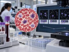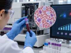
An exosome cancer test analyzes tiny membrane-bound particles released by cells into blood or other body fluids for molecular clues associated with cancer. These particles can carry RNA, DNA, proteins, lipids, and other cargo from their cells of origin, which makes them attractive candidates for liquid biopsy. Tumor-derived extracellular vesicles may contain signals that help researchers detect cancer, classify tumor type, estimate prognosis, or monitor treatment without repeatedly sampling tumor tissue.
The science is promising, but the term “exosome test” can imply more clinical certainty than the field currently supports. Exosomes are one subtype of extracellular vesicle, and proving that a particle came through the endosomal exosome pathway is difficult in routine samples. Isolation methods, measurement platforms, and biomarker panels vary widely between studies. As a result, there is no universal exosome cancer reference range, no single positive cutoff across tumor types, and no general blood exosome test that can replace pathology, imaging, or established cancer screening.
- Exosome cancer testing looks for tumor-associated molecular cargo—often RNA, microRNA, proteins, or DNA—inside or associated with extracellular vesicles in blood or other fluids.
- There is no universal “normal exosome level” for cancer because laboratories measure different vesicle populations, cargo molecules, and signal thresholds.
- An abnormal research result can suggest a cancer-associated molecular pattern, but it does not by itself diagnose cancer or identify its stage.
- Exosome and extracellular-vesicle assays are still limited by differences in isolation, purity, nomenclature, and analytical standardization.
- Established imaging, pathology, and guideline-recommended cancer tests should not be delayed or replaced by an investigational exosome assay.
Table of Contents
- What Exosomes and Extracellular Vesicles Are
- What an Exosome Cancer Test Can Measure
- How Exosome Testing Is Performed
- What Positive or Abnormal Results Mean
- Where Exosome Biomarkers May Help
- Why Standardization Is Difficult
- How to Evaluate an Exosome Test
What Exosomes and Extracellular Vesicles Are
Cells release membrane-bound particles called extracellular vesicles (EVs) into blood, urine, saliva, cerebrospinal fluid, and other biological fluids. EVs contain selected proteins, nucleic acids, lipids, and metabolites. Because their cargo can reflect the cell that produced them, researchers are studying EVs as potential biomarkers of cancer and many other diseases.
“Exosome” is often used as a catch-all term, but scientifically it has a narrower meaning. Exosomes are generally described as small EVs formed inside multivesicular endosomes and released when those compartments fuse with the cell membrane. Other EVs can bud directly from the plasma membrane or arise through different processes.
This distinction matters because a particle’s size alone does not prove that it is an exosome. Current extracellular-vesicle guidance encourages researchers to describe particles according to measurable characteristics—such as size, density, biochemical markers, or separation method—unless their cellular origin has been demonstrated. Many studies marketed or discussed as “exosome” research would more precisely be called small extracellular-vesicle research.
Cancer cells release EVs into the circulation, but so do normal blood cells, immune cells, platelets, endothelial cells, and other tissues. A blood sample therefore contains a mixed population. The analytical challenge is not simply counting vesicles; it is finding cancer-specific information within a large background of normal biological material.
This is one reason exosome testing differs from a cell-free DNA cancer test. cfDNA assays analyze DNA fragments freely circulating in plasma, while EV assays attempt to isolate vesicles or vesicle-associated cargo before measuring tumor-related signals.
What an Exosome Cancer Test Can Measure
An exosome or EV-based cancer assay can target several types of biological information. There is no standard panel used for all cancers.
RNA and microRNA
RNA is one of the most studied EV cargos. Researchers can measure messenger RNA, microRNA, long non-coding RNA, and other RNA species. A tumor-associated RNA signature may reflect altered gene expression even when a specific DNA mutation is not present. Vesicle membranes can also protect RNA from rapid degradation in circulation, which is one reason EV RNA is attractive for liquid biopsy.
DNA
EV-associated DNA can contain tumor mutations or other genomic abnormalities. However, determining whether DNA is truly inside vesicles, attached to their outer surface, or co-isolated as non-vesicular material requires careful methodology. A conventional ctDNA mutation panel is currently much more established for clinical genomic profiling in many advanced cancers.
Proteins
Tumor-derived EVs can carry membrane proteins and internal proteins associated with tumor lineage, signaling pathways, immune interactions, and metastatic behavior. Protein panels can potentially improve specificity when several markers are combined rather than relying on a single molecule.
Lipids and multi-omic signatures
Vesicle membranes and internal cargo contain lipid patterns that may also change in cancer. Research platforms increasingly combine RNA, protein, lipid, and clinical features with machine learning to build multi-marker classifiers.
The key point is that “exosome level” is usually not the result. A meaningful assay typically measures a defined molecular signature in a defined EV population. Two tests that both use the word exosome may be measuring completely different biology.
How Exosome Testing Is Performed
The process usually begins with blood, urine, or another body fluid. For a blood-based test, plasma or serum is prepared and then processed to enrich or isolate extracellular vesicles. Researchers may use ultracentrifugation, size-exclusion chromatography, precipitation, filtration, immunoaffinity capture, microfluidic devices, or combinations of methods.
Each method has tradeoffs. High-purity separation can be time-consuming and may lose vesicles. Fast commercial precipitation can recover more material but may also pull down lipoproteins, protein complexes, and non-vesicular nucleic acids. Immunocapture can enrich vesicles carrying selected surface markers but may miss tumor vesicles that lack those markers.
After enrichment, the laboratory may characterize particles using techniques such as nanoparticle tracking, electron microscopy, flow-based methods, or protein-marker analysis. Molecular cargo is then measured using PCR, sequencing, mass spectrometry, immunoassays, biosensors, or other platforms.
A rigorous workflow has to address several questions:
- What biological fluid was used, and how was it collected and stored?
- What EV population was separated, and how pure was it?
- Which markers confirm that the preparation contains EVs?
- Is the measured RNA, DNA, or protein inside vesicles or merely co-isolated?
- What cancer-associated signature is being tested?
- What population was used to establish the cutoff and validate performance?
- Has the classifier been tested independently and prospectively?
These details are why the MISEV guidance is important. It does not validate a particular cancer test; it provides standards that improve rigor, reproducibility, and transparency in EV research.
Specimen type can also change what a test is capable of seeing. Plasma gives access to vesicles circulating throughout the body, but it contains an enormous background from blood and vascular cells. Urine may be useful for cancers of the urinary tract or prostate because material from those organs can enter the urinary space more directly. Saliva, pleural fluid, cerebrospinal fluid, and other samples may be attractive for particular tumors, but evidence from one fluid cannot automatically be applied to another. A biomarker panel has to be validated using the same specimen type, collection tube, processing interval, storage conditions, and laboratory workflow intended for clinical use.
EV testing also creates an important interpretation problem when the molecular target could exist outside vesicles. For example, an RNA molecule or DNA mutation detected after an EV-enrichment step may come from a true vesicle, a protein-RNA complex, a lipoprotein particle, or free nucleic acid carried through the preparation. Studies that compare EV-enriched material with unprocessed plasma can help establish whether vesicle isolation actually adds diagnostic information. Without that comparison, it can be difficult to know whether an assay is measuring a distinct exosome signal or simply another form of circulating biomarker.
For this reason, the strongest studies separate analytical validation from clinical validation. Analytical validation asks whether the assay measures its intended target reliably, including precision, detection limits, interference, specimen stability, and reproducibility. Clinical validation asks whether the measured signature accurately distinguishes the clinical states of interest. Clinical utility goes one step further: it asks whether using the result changes management in a way that improves outcomes. All three layers matter before a research biomarker becomes a dependable cancer test.
What Positive or Abnormal Results Mean
There is no single interpretation that applies to every exosome cancer test. A result may be a concentration, expression value, ratio, multi-marker score, machine-learning probability, or classification such as positive/negative. The meaning depends entirely on the validated assay.
A positive research result usually means the sample matched a predefined molecular pattern associated with cancer in the study population. It does not automatically establish that a tumor is present. False positives can occur because inflammatory states, benign diseases, age-related changes, or technical contamination can alter extracellular-vesicle abundance and cargo.
A negative result also cannot reliably rule out cancer unless the assay has been validated for that use. Early-stage tumors may release few tumor-derived vesicles, and different cancers may produce different cargo. Sample handling and isolation efficiency can further reduce signal.
The setting in which a test is used changes the meaning of the same sensitivity and specificity. In a screening population where cancer is uncommon, even a highly specific assay can generate more false-positive than true-positive results if the prevalence is low enough. Positive predictive value therefore cannot be inferred from a case-control study that deliberately includes many cancer samples. Conversely, a negative result in a person with a suspicious mass or persistent warning symptoms should not override diagnostic imaging or biopsy merely because a research classifier returned a low-risk score.
Some reports may also give a probability rather than a binary answer. A score such as 0.8 or “high likelihood” is not a standardized cancer probability unless the test developer has calibrated and validated it that way in the intended population. The appropriate response depends on the assay’s prespecified cutoff, the person’s symptoms and baseline risk, and the recommended confirmatory pathway. Results from different exosome platforms should not be trended as though they were interchangeable laboratory values.
Unlike many routine blood tests, there is no universal range such as “0–X exosomes/mL is normal.” Total EV concentration is influenced by many non-cancer processes and by the measurement method. A diagnostic claim must be based on a specific validated biomarker signature, not merely on a high vesicle count.
| Reported output | Potential meaning | Main limitation |
|---|---|---|
| Total EV or exosome count | Amount of measured vesicle-like particles | Not cancer-specific |
| RNA or microRNA signature | Pattern associated with tumor biology | Platform and cohort dependent |
| Protein marker panel | May suggest tumor type or activity | Marker overlap with normal or inflammatory cells |
| Mutation detected in EV-associated DNA/RNA | Potential tumor-derived genomic signal | Requires proof of analytical validity and source |
| Machine-learning cancer score | Probability or classification based on multiple features | Performance may fall outside the training population |
Where Exosome Biomarkers May Help
Research spans the full cancer journey, from early detection to recurrence monitoring.
Early detection is attractive because EVs may contain amplified biological information. A tumor cell can release many vesicles, each carrying molecular cargo, potentially providing signal even when ctDNA is scarce. Researchers have reported promising signatures in pancreatic, breast, ovarian, lung, liver, prostate, colorectal, brain, and other cancers. The challenge is proving high performance in representative screening populations rather than small case-control cohorts.
Tumor classification is another possibility. Surface proteins and RNA expression patterns may help identify the tissue or cell type that released an EV. In principle, this could complement imaging when the primary tumor is uncertain.
Treatment monitoring may be useful because EV cargo can change as tumor signaling changes. Serial sampling could potentially reveal response or emerging resistance. This resembles the logic of a liquid biopsy resistance mutation test, although clinical validation for EV-based monitoring is less mature than for many ctDNA applications.
Prognosis and recurrence risk are also active research areas. Studies have associated specific EV RNAs or proteins with stage, metastatic behavior, treatment response, and recurrence. These associations can be biologically interesting without yet being strong enough to guide an individual patient’s treatment.
EVs may eventually complement rather than replace other liquid-biopsy analytes. A future assay could combine ctDNA mutations, circulating tumor cells, EV RNA, proteins, methylation, and fragmentomic features to improve sensitivity and specificity. Multi-analyte designs are appealing because no single biomarker captures every cancer equally well.
Why Standardization Is Difficult
The central challenge is heterogeneity at every step. Blood contains multiple types of EVs and non-vesicular particles with overlapping sizes and densities. Lipoproteins are especially abundant and can contaminate preparations. Different separation methods therefore produce different biological mixtures even when researchers start with the same specimen type.
Pre-analytical factors also matter. Fasting status, exercise, time to processing, storage temperature, freeze-thaw cycles, anticoagulant, platelet contamination, and hemolysis can change the measured EV population or cargo. Without consistent procedures, apparent biological differences can reflect sample handling.
Nomenclature is another issue. Calling every small particle an exosome can obscure what was actually measured. MISEV2023 emphasizes transparent reporting of separation and characterization methods and encourages cautious terminology when vesicle biogenesis has not been established.
Clinical validation presents an even higher bar. A useful cancer diagnostic must perform in the population where it will be used. Comparing known cancer cases with young healthy controls can exaggerate accuracy because the groups differ in age, comorbidities, inflammation, and other variables. Screening tests need large prospective cohorts with appropriate controls and adequate follow-up of negative results.
A test also needs a defined clinical role. Even excellent discrimination in a study does not prove that using the test improves outcomes. Researchers must show what happens after a positive result, how many people undergo imaging or invasive procedures, how often false positives occur, whether early detection changes treatment, and whether benefits outweigh harms and cost.
How to Evaluate an Exosome Test
Before relying on an exosome-based test, identify exactly what it measures and what claim has been validated. Marketing language such as “tumor exosome profile” is not enough to judge clinical usefulness.
Useful questions include:
- Is the test intended for screening, diagnosis, prognosis, treatment selection, or monitoring?
- Does it measure blood, urine, or another fluid?
- Are the particles actually characterized as EVs, and how were they isolated?
- Which RNA, DNA, protein, or multi-omic biomarkers are included?
- Was the cutoff established before independent validation?
- Was the test studied prospectively in people similar to the intended users?
- What are sensitivity, specificity, positive predictive value, and negative predictive value in that population?
- What follow-up is recommended after an abnormal result?
- Is the assay part of an established guideline or approved companion-diagnostic pathway, or is it investigational?
- Would an established liquid biopsy test, tissue biopsy, imaging study, or standard screening test answer the clinical question more directly?
Patients should be especially cautious when a broad exosome test promises to detect many cancers but provides little information about independent validation, false-positive workup, or clinical utility. A research assay can be scientifically interesting without being ready to guide medical decisions.
The field is advancing rapidly because extracellular vesicles carry rich biological information that other analytes may miss. The most credible progress will come from standardized sample handling, transparent EV characterization, reproducible multi-center validation, and studies that show not only that a cancer signal can be measured, but that acting on it improves care.
References
- Minimal information for studies of extracellular vesicles (MISEV2023): From basic to advanced approaches 2024 (Position Statement)
- The new advance of exosome-based liquid biopsy for cancer diagnosis 2024 (Review)
- Tumor-Derived Extracellular Vesicles as Liquid Biopsy for Diagnosis and Prognosis of Solid Tumors: Their Clinical Utility and Reliability as Tumor Biomarkers. 2024 (Review)
- Diagnostic potential of exosomal extracellular vesicles in oncology 2024 (Review)
- Extracellular Vesicles as Liquid Biopsy Biomarkers across the Cancer Journey: From Early Detection to Recurrence 2024 (Review)
Disclaimer
Exosome and extracellular-vesicle cancer assays remain an evolving area, and a research or commercial result should not be used to diagnose, exclude, or treat cancer without appropriate clinical validation and specialist interpretation. Established screening, imaging, pathology, and molecular testing remain necessary when medically indicated. Discuss any abnormal or experimental liquid-biopsy result with a qualified clinician before making care decisions.