An insulin blood test measures the amount of insulin in serum or plasma at a specific moment. Insulin is released by pancreatic beta cells and helps move glucose into cells, restrain liver glucose production, store nutrients, and limit fat breakdown. The result is meaningful only beside the glucose level, fasting status, meal timing, symptoms, medications, and assay method. High insulin with normal glucose may reflect compensation for insulin resistance, while high insulin during true hypoglycemia can indicate excessive endogenous secretion, an insulin-releasing medicine, or injected insulin. Low insulin may be appropriate during fasting or hypoglycemia, but low insulin with high glucose can point to inadequate beta-cell function. Reference ranges vary substantially among laboratories, and insulin assays are not fully standardized. The test does not by itself diagnose diabetes, type 1 diabetes, type 2 diabetes, insulin resistance, or an insulinoma; each question requires a different collection setting and supporting tests.
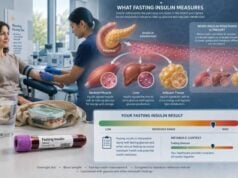
- Fasting insulin is usually collected after an 8- to 12-hour fast and must be interpreted with fasting glucose from the same draw.
- A typical adult fasting reference interval may span roughly 2–25 µIU/mL, but the reporting laboratory’s range controls interpretation.
- High insulin with normal glucose often suggests compensation, while high insulin during low glucose is abnormal.
- Low insulin with high glucose may indicate reduced pancreatic insulin production.
- Injected insulin, insulin antibodies, biotin, kidney disease, and assay cross-reactivity can distort results.
Table of Contents
- What an Insulin Blood Test Measures
- Why Insulin Testing Is Ordered
- Preparation and Test Procedure
- Normal Range and Result Timing
- High Insulin Results
- Low Insulin Results
- Insulin Resistance, Diabetes, and Hypoglycemia
- Limitations, Follow-Up, and Next Steps
What an Insulin Blood Test Measures
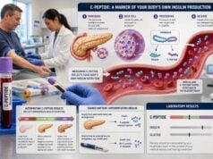
Insulin is a peptide hormone made in the beta cells of the pancreatic islets. Beta cells first produce proinsulin. Enzymes split proinsulin into insulin and C-peptide, which enter the bloodstream in approximately equal molar amounts. The liver removes a large portion of insulin during its first pass, while C-peptide circulates longer and is cleared mainly by the kidneys.
An insulin assay measures immunoreactive insulin in a blood sample. Depending on the method, that signal may include native human insulin, varying amounts of proinsulin, and some—but not all—pharmaceutical insulin analogs. The result is usually reported in µIU/mL, mcIU/mL, mIU/L, or pmol/L. The first three units are numerically equivalent in common laboratory reporting, while conversion to pmol/L depends on the factor used by the laboratory.
Insulin changes quickly. It rises after carbohydrate and mixed meals, falls during fasting and exercise, and responds to stress hormones, sleep, illness, and medications. A random insulin value without a simultaneous glucose concentration often has little meaning.
Several test formats are used:
- Fasting insulin: A single baseline value after an overnight fast
- Random insulin: Collected at the time of symptoms or another clinically important event
- Stimulated insulin: Timed values after oral glucose or a mixed meal
- Critical-sample insulin: Collected during confirmed hypoglycemia before treatment
- Free and total insulin: Used when insulin-binding antibodies may interfere
The clinical question determines which format is appropriate. A fasting sample can support an insulin-resistance estimate, while suspected insulinoma requires insulin during low plasma glucose. A normal fasting insulin cannot exclude inappropriate insulin secretion during a later episode.
Insulin is not the same as insulin-like growth factor 1, or IGF-1. Despite the similar name, IGF-1 is primarily used in growth hormone evaluation and has different physiology and reference ranges.
Why Insulin Testing Is Ordered
Clinicians may order insulin testing to investigate several distinct problems.
Possible insulin resistance
Fasting insulin may be paired with fasting glucose to show how much insulin the pancreas needs to maintain glucose overnight. A high insulin requirement can precede elevated glucose. The two values can be used to calculate HOMA-IR, an estimate used frequently in research and selected clinical settings.
Insulin resistance is not diagnosed from one universal fasting-insulin cutoff. Waist circumference, triglycerides, HDL cholesterol, blood pressure, glucose, A1C, fatty liver, sleep apnea, polycystic ovary syndrome, medications, and family history all contribute to the assessment.
Hypoglycemia
Insulin measurement is central when a person without diabetes has documented low plasma glucose with compatible symptoms. The sample must be drawn during the low glucose episode and paired with C-peptide, proinsulin, beta-hydroxybutyrate, and a screen for sulfonylureas and meglitinides. Detectable insulin can be abnormal even if it falls within the laboratory’s ordinary fasting range because normal beta cells should nearly stop insulin secretion during hypoglycemia.
Diabetes classification or beta-cell function
Insulin may be low in type 1 diabetes and in advanced type 2 diabetes after beta-cell function declines. However, C-peptide is usually more useful for estimating endogenous insulin production, especially in a person receiving injected insulin. Diabetes type is classified using clinical features, autoantibodies when appropriate, C-peptide in selected cases, and the disease course—not a single serum insulin value.
Rare beta-cell tumors or disorders
An insulinoma secretes insulin inappropriately and causes hypoglycemia. Biochemical proof comes before imaging. High insulin while glucose is normal does not diagnose an insulinoma because many people with insulin resistance have fasting or post-meal hyperinsulinemia without a tumor.
The test may also be used in metabolic studies, bariatric-surgery evaluations, research glucose-tolerance tests, and selected assessments of insulin antibodies or insulin clearance.
Preparation and Test Procedure
Preparation depends on the purpose of testing. A fasting insulin test commonly requires 8 to 12 hours without calories. Water is generally allowed. Coffee with milk or sugar, sweetened drinks, gum with carbohydrate, and snacks break the fast.
Before a scheduled fasting draw:
- Eat normally the day before unless given a specific protocol.
- Avoid heavy alcohol intake and unusually intense exercise.
- Try to obtain a normal night of sleep.
- Do not smoke or use nicotine immediately before collection.
- Sit quietly before the blood draw.
- Follow individualized medication instructions.
Many laboratories advise stopping high-dose biotin supplements for a defined period before immunoassay testing because biotin can interfere with some platforms. The exact hold depends on dose, kidney function, and the assay. Do not stop prescribed biotin used for a medical disorder without clinical advice.
Medication review should include:
- Injected insulin and the exact analog
- Sulfonylureas and meglitinides
- Metformin and other glucose-lowering drugs
- Glucocorticoids
- Antipsychotic medicines
- Weight-management and incretin-based therapies
- Beta-blockers and drugs that may mask hypoglycemia symptoms
For a glucose challenge or mixed-meal test, several samples may be collected over two to five hours. The laboratory should label each tube with the exact time. Meal composition and timing need to be standardized if results will be compared.
For suspected spontaneous hypoglycemia, do not fast at home to provoke an episode. A supervised fast may last up to 72 hours and requires clinical monitoring. During severe symptoms, immediate treatment takes priority. Clinicians obtain the critical sample before glucose treatment only when it can be done safely without delay.
The sample is usually serum or plasma. Hemolysis can affect some related tests, and delayed separation may compromise specimen quality. If insulin antibodies are suspected, the laboratory may measure free insulin after separating antibody-bound hormone from the biologically available fraction.
Normal Range and Result Timing
Insulin has no single universal normal range. A major reference laboratory, for example, lists an adult fasting serum interval of 2.6–24.9 µIU/mL for one electrochemiluminescence assay. Other laboratories use narrower, wider, age-specific, or method-specific intervals. A value should be compared only with the range printed on that report.
A broad reference interval is not the same as an optimal metabolic target. The interval generally describes the central distribution in a selected reference population; it does not guarantee ideal insulin sensitivity or predict individual risk.
Timing changes interpretation:
| Collection setting | Expected physiology | Main interpretation question |
|---|---|---|
| Overnight fasting | Relatively low, stable insulin | How much insulin is needed to maintain fasting glucose? |
| 30–60 minutes after carbohydrate | Rapid insulin rise | Did beta cells respond to the glucose increase? |
| Later after a meal | Insulin declines as glucose falls | Is secretion prolonged or delayed? |
| During hypoglycemia | Insulin should be nearly suppressed | Is insulin inappropriately present? |
A post-meal value that would appear high for a fasting specimen may be completely expected. Conversely, an insulin value inside the fasting reference range may be dangerously inappropriate when plasma glucose is 45 mg/dL.
Insulin also varies from pulse to pulse. A single value can differ from another sample collected minutes later. Research protocols sometimes average multiple fasting samples or use dynamic modeling to improve precision.
When following a trend, use the same laboratory and similar fasting duration, draw time, medication timing, illness status, and exercise conditions. Assay changes can create an apparent change even when physiology is stable.
High Insulin Results
High insulin is called hyperinsulinemia. The likely cause depends first on the simultaneous glucose level.
High insulin with normal glucose
This pattern often reflects compensation for insulin resistance. Muscle, liver, or fat tissue responds less efficiently, so beta cells release more insulin to keep glucose within range. Common associations include:
- Visceral obesity
- Physical inactivity
- Genetic susceptibility to type 2 diabetes
- Polycystic ovary syndrome
- Fatty liver disease
- Sleep apnea and chronic sleep loss
- Glucocorticoid therapy
- Some antipsychotic or immunosuppressive medicines
- Puberty or pregnancy
Normal glucose does not erase the high insulin requirement. However, one elevated result can also reflect incomplete fasting, stress, recent exercise, or assay variation.
High insulin with high glucose
This commonly indicates insulin resistance with beta-cell compensation that is no longer sufficient to normalize glucose. It can occur in prediabetes or type 2 diabetes. Markedly high glucose is evaluated with validated diabetes criteria, not insulin concentration alone.
High insulin with low glucose
This is potentially dangerous and requires a complete critical-sample interpretation. Causes include endogenous hyperinsulinism, injected insulin, sulfonylurea or meglitinide exposure, and insulin autoimmune hypoglycemia. C-peptide and proinsulin show whether the pancreas is secreting hormone; beta-hydroxybutyrate shows whether insulin action has suppressed ketone production.
An insulinoma is uncommon. It should not be inferred from elevated fasting insulin unless low plasma glucose and an appropriate biochemical pattern are documented. The hypoglycemia hormone panel is designed for that distinction.
Other causes of a high reported insulin value include insulin antibodies, assay interference, reduced kidney clearance, and cross-reactivity with an injected analog. A result that conflicts with glucose, C-peptide, or symptoms should be investigated before treatment decisions are made.
Low Insulin Results
Low insulin may be normal or abnormal. During fasting, vigorous activity, a low-carbohydrate intake, or hypoglycemia, healthy beta cells reduce secretion. The concern arises when insulin is too low for the glucose concentration.
Low insulin with high glucose may occur with:
- Type 1 diabetes caused by autoimmune beta-cell destruction
- Advanced type 2 diabetes with substantial beta-cell failure
- Pancreatitis, pancreatic surgery, pancreatic cancer, or cystic fibrosis-related diabetes
- Certain monogenic forms of diabetes
- Severe metabolic stress, depending on timing
- An assay that does not detect the insulin analog being used
Low insulin by itself does not diagnose type 1 diabetes. Autoantibodies, C-peptide with concurrent glucose, age, body habitus, ketosis, family history, and clinical course help classify diabetes. Some adults with type 1 diabetes initially retain meaningful insulin secretion, while some people with type 2 diabetes eventually have very low secretion.
Low insulin with normal glucose may indicate good insulin sensitivity, particularly in a healthy person with normal C-peptide and no symptoms. It may also follow prolonged fasting or recent exercise.
Low insulin during true hypoglycemia is usually appropriate. In that setting, elevated beta-hydroxybutyrate suggests the body has activated fasting fuel pathways. Clinicians then investigate non-insulin causes such as alcohol effects, adrenal insufficiency, liver failure, kidney disease, malnutrition, critical illness, or an IGF-2-producing tumor.
C-peptide often clarifies a low insulin result. A low C-peptide with high glucose supports reduced endogenous production. A normal or high C-peptide with unexpectedly low insulin may reflect differential clearance, assay issues, or timing.
Insulin Resistance, Diabetes, and Hypoglycemia
An insulin test interacts with three different clinical concepts that are often confused.
Insulin resistance describes reduced tissue response. It may be present while fasting glucose and A1C remain normal because the pancreas compensates. Fasting insulin can support the assessment, but there is no globally accepted diagnostic cutoff. HOMA-IR, triglyceride-glucose indices, oral tolerance-derived measures, or research clamp studies may be used depending on the purpose.
Diabetes describes chronic hyperglycemia due to insufficient insulin action, insulin secretion, or both. Diagnostic criteria rely on A1C and plasma glucose. Insulin concentration does not define diabetes because early type 2 diabetes can have high insulin, advanced type 2 diabetes can have low insulin, and type 1 diabetes progresses over time.
Hyperinsulinemic hypoglycemia describes insulin activity that fails to shut off during low glucose. Here, an insulin value does not need to exceed the ordinary reference interval to be abnormal. Context-specific suppression is the issue.
A useful four-quadrant view is:
| Glucose | Insulin | Possible interpretation |
|---|---|---|
| Normal | High | Compensated insulin resistance, nonfasting sample, or assay effect |
| High | High | Insulin resistance with inadequate compensation |
| High | Low | Insulin deficiency or beta-cell failure |
| Low | Detectable or high | Inappropriate insulin action requiring critical-sample evaluation |
This framework is a starting point, not a diagnosis. Meal timing, injected insulin, kidney function, medication exposure, and C-peptide can reverse or blur the expected pattern.
Limitations, Follow-Up, and Next Steps
Insulin testing has important analytical limitations. Commercial assays are not fully harmonized, so the same specimen can produce different values on different platforms. Antibodies recognize different parts of the insulin molecule and vary in cross-reactivity with proinsulin and analogs.
Common sources of misleading results include:
- A nonfasting sample labeled as fasting
- Glucose and insulin collected at different times
- High-dose biotin interference
- Hemolysis or specimen-processing errors
- Anti-insulin or heterophile antibodies
- Kidney dysfunction that alters clearance
- An injected analog that the assay detects weakly or strongly
- Recent dextrose treatment before a hypoglycemia sample
- Comparing different laboratories or methods
When results are discordant, the clinician may repeat simultaneous glucose, insulin, and C-peptide; request free and total insulin; ask the laboratory about analog cross-reactivity; test insulin antibodies; or use mass spectrometry in specialized situations.
Follow-up for possible insulin resistance may include A1C, fasting glucose, lipid profile, blood pressure, waist circumference, liver enzymes, and assessment for sleep apnea or polycystic ovary syndrome. Treatment targets the overall risk profile through sustainable nutrition, regular aerobic and resistance activity, sleep, weight management when appropriate, and evidence-based medication—not a pursuit of the lowest possible insulin number.
Follow-up for high glucose follows diabetes diagnostic and classification standards. Follow-up for hypoglycemia requires immediate safety planning and properly timed testing. Confusion, seizure, loss of consciousness, or inability to swallow requires emergency treatment. Do not delay rescue to obtain a laboratory value.
A useful insulin result always answers a defined question. “Is fasting insulin elevated for this glucose?” is different from “Is insulin suppressed during hypoglycemia?” and different again from “Does this person still make endogenous insulin?” Matching the test conditions to the question prevents most interpretation errors.
Common Interpretation Scenarios
High insulin with normal fasting glucose often means the pancreas is compensating successfully for reduced insulin sensitivity. This pattern may accompany central adiposity, high triglycerides, fatty liver, polycystic ovary syndrome, sleep apnea, or a strong family history of type 2 diabetes. It does not establish a diagnosis by itself. A1c, blood pressure, lipids, waist circumference, liver assessment, and sometimes an oral glucose tolerance test provide the risk context.
High insulin with high glucose suggests that compensation is no longer sufficient for the degree of resistance, although recent food intake, stress, steroids, and assay timing can contribute. Diabetes is diagnosed with validated glucose or A1c criteria, not an insulin threshold. The insulin result can help explain physiology but should not delay treatment of established hyperglycemia.
High insulin with low glucose is a different and potentially urgent problem. The sample must be interpreted with C-peptide, proinsulin, beta-hydroxybutyrate, and a secretagogue screen obtained during the same episode. High C-peptide supports endogenous secretion or an insulin-releasing drug; suppressed C-peptide may support injected insulin, subject to analog detection and assay interference.
Low insulin with high glucose can reflect beta-cell failure, advanced type 2 diabetes, autoimmune diabetes, pancreatic disease, or a sample collected after insulin secretion has waned. C-peptide is generally more useful for estimating endogenous production because injected insulin does not contain C-peptide and the peptide has a longer half-life. Ketones and symptoms determine urgency.
Low insulin with normal or low glucose may be appropriate during fasting, after exercise, or in a highly insulin-sensitive person. During true hypoglycemia, insulin should be nearly suppressed. A printed “normal” insulin concentration can therefore be abnormally high for the glucose level even when it falls inside the laboratory reference interval.
Assay and Timing Questions to Ask
Before comparing results, confirm whether the sample was fasting, how long the fast lasted, and whether insulin or an insulin secretagogue was taken beforehand. Different immunoassays recognize pharmaceutical insulin analogs to different degrees. Hemolysis, insulin antibodies, heterophile antibodies, kidney dysfunction, and delayed processing can also distort the concentration.
Repeat testing is most useful when it answers a defined question and collection conditions can be reproduced. For metabolic follow-up, established outcomes such as glucose, A1c, lipids, blood pressure, liver health, and weight trajectory usually matter more than frequent insulin measurements.
An insulin test should also be separated from an insulin-antibody test. The former measures circulating hormone; the latter looks for antibodies that may alter insulin action or interfere with measurement. Ordering the wrong test can produce an apparently reassuring result while leaving the clinical question unanswered.
References
- INS – Overview: Insulin, Serum 2026
- The Current Status of Serum Insulin Measurements and the Need for Standardization 2025 (Review)
- Evaluation of insulin secretion and insulin sensitivity in pregnant women: Application value of simple indices 2024 (Review)
- Non-Diabetic Hypoglycemia: Evaluation and Management in Adults 2025 (Review)
- 2. Diagnosis and Classification of Diabetes: Standards of Care in Diabetes—2026 2026 (Guideline)
Disclaimer
This article provides general information and cannot diagnose insulin resistance, diabetes, insulinoma, or the cause of hypoglycemia. Insulin results require interpretation with simultaneous glucose, fasting or meal timing, medications, kidney function, and the laboratory method. Severe hypoglycemia is an emergency; do not delay treatment to obtain testing.