
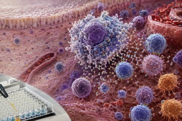
An interleukin-18 test measures an inflammasome-related cytokine that can become markedly elevated in several hyperinflammatory disorders. Interleukin-18, abbreviated IL-18, is made as an inactive precursor and activated by caspase-1, much like IL-1β. It is stored in many cells and can be released rapidly after inflammasome activation or cell injury. In the presence of IL-12, IL-18 strongly promotes interferon-gamma production by natural killer cells and T cells. Its activity is normally controlled by IL-18 binding protein, which captures much of the cytokine in blood. This creates an important testing distinction: most clinical and research assays report total IL-18, while free IL-18 better represents unbound biologically available cytokine but is harder to measure. Very high total IL-18 can support suspicion for Still disease, macrophage activation syndrome, selected monogenic autoinflammatory conditions, and certain inflammasome disorders. It is not diagnostic alone, and dangerous hyperinflammation must be recognized from fever, blood counts, ferritin, coagulation changes, liver injury, organ dysfunction, and the overall clinical course.
- Total IL-18 includes cytokine bound to IL-18 binding protein; free IL-18 is the unbound active fraction.
- Markedly high IL-18 can be a useful clue to Still disease, macrophage activation syndrome, and selected autoinflammatory pathways.
- Moderate elevation is nonspecific and can occur in infection, autoimmune disease, malignancy, metabolic disease, and tissue injury.
- A normal result does not exclude early or treated macrophage activation syndrome.
- IL-18 should never delay urgent treatment when a patient has rapidly worsening fever, cytopenias, coagulopathy, neurologic change, or organ failure.
Table of Contents
- How IL-18 Is Activated and Regulated
- Total IL-18, Free IL-18, and IL-18 Binding Protein
- When IL-18 Testing Is Most Useful
- What a High IL-18 Result May Mean
- IL-18 in Still Disease and Macrophage Activation Syndrome
- Very High IL-18 in Rare Autoinflammatory Disorders
- Meaning of a Normal or Low Result
- Assay Limitations, Urgent Clues, and Follow-Up
How IL-18 Is Activated and Regulated
IL-18 belongs to the interleukin-1 family. Many immune and tissue cells contain inactive pro-IL-18 before inflammation begins. Macrophages, monocytes, dendritic cells, epithelial cells, and other populations can be sources. When an inflammasome activates caspase-1, the enzyme cuts pro-IL-18 into its mature form. Gasdermin pores and inflammatory cell death can help release the cytokine.
Several inflammasomes can participate. NLRP3 responds to varied cellular stress, while NLRC4, pyrin, AIM2, and other sensors recognize different microbial or damage-related changes. This means elevated IL-18 does not identify one inflammasome automatically. The trigger may be infection, genetic dysregulation, tissue damage, or another inflammatory process.
IL-18 was originally described as an interferon-gamma–inducing factor. Its strongest type 1 effect occurs with IL-12, which prepares natural killer cells and T cells to respond. Together, the cytokines promote IFN-γ production, macrophage activation, and cytotoxic immune activity. In other cytokine environments, IL-18 can support different responses, including type 2 inflammation. Its action is therefore context-dependent rather than fixed.
The IL-18 receptor contains alpha and beta chains and signals through MyD88, IRAK proteins, TRAF6, NF-κB, and other pathways. This links IL-18 to a broader family of innate immune signaling systems. Target cells must express the receptor and be in the appropriate activation state for a blood concentration to have a biological effect.
IL-18 is regulated differently from many cytokines because a high-affinity soluble protein, IL-18 binding protein, or IL-18BP, captures it. IFN-γ can increase IL-18BP production, creating a negative feedback loop: IL-18 promotes IFN-γ, and IFN-γ promotes a protein that neutralizes IL-18. Disease can overwhelm or disrupt this buffer.
This biology explains why total concentration is only part of the story. Two patients with the same total IL-18 may have different amounts of free cytokine depending on IL-18BP. It also explains why a dramatic inflammatory phenotype can emerge when total production rises beyond the binding capacity.
IL-18 and IL-1β are both processed by inflammasomes but are not interchangeable. IL-1β strongly drives fever and local inflammatory recruitment, while IL-18 is closely linked to IFN-γ and cytotoxic lymphocyte activation. The IL-1β test answers a related but distinct question.
Total IL-18, Free IL-18, and IL-18 Binding Protein
Most commercially available or research assays measure total IL-18. This includes free cytokine and cytokine bound in complexes with IL-18BP. Total IL-18 is technically easier to measure, relatively stable in serum, and can reach striking concentrations in selected diseases.
Free IL-18 is the fraction available to bind cell-surface receptors. It may be measured directly with specialized methods or calculated from total IL-18 and IL-18BP concentrations using binding assumptions. Calculated values depend on assay accuracy and mathematical constants, so they are not automatically equivalent to direct measurement.
IL-18BP can also be measured. A high binding-protein concentration may be an appropriate counter-response to high IL-18. If IL-18 rises faster than IL-18BP, free cytokine can increase. If both rise but binding capacity remains sufficient, total IL-18 may look dramatic while biologically active free IL-18 is less extreme.
Reports should therefore state whether the result is total IL-18, free IL-18, or IL-18BP. Units also matter. Picograms per milliliter and nanograms per liter are numerically equivalent, but other conversions are not. Reference ranges differ by platform and population.
Some cytokine panels measure IL-18 along with IL-6, IL-10, IFN-γ, CXCL9, soluble IL-2 receptor, and other markers. Patterns may help distinguish sources of cytokine storm or identify a macrophage-activation profile. A cytokine storm blood test panel remains supportive; clinical severity and organ dysfunction determine urgency.
Tissue, cerebrospinal fluid, synovial fluid, or other local specimens are used in specialized contexts. Local levels cannot be interpreted with serum reference ranges. Dilution, cell content, and sampling technique can strongly affect nonblood specimens.
Genetic tests, inflammasome-function assays, and cell-stimulation studies examine upstream causes of IL-18 production. They do not substitute for a concentration test. Conversely, a very high concentration can suggest a pathway but does not identify the causative gene.
When IL-18 Testing Is Most Useful
IL-18 testing is most useful when the differential diagnosis includes diseases known to produce disproportionately high concentrations. Pediatric rheumatologists, adult rheumatologists, immunologists, and hematologists may use it when fever and systemic inflammation do not fit a routine infection or autoimmune pattern.
Still disease is a major indication. This name includes systemic juvenile idiopathic arthritis in children and adult-onset Still disease in adults. Features can include daily spiking fever, evanescent rash, arthritis, neutrophilic leukocytosis, liver abnormalities, lymph-node enlargement, sore throat, and high ferritin. IL-18 may help distinguish Still disease from other systemic inflammatory disorders, especially when markedly elevated.
Macrophage activation syndrome is another important indication. MAS is a life-threatening hyperinflammatory complication often associated with Still disease but possible in lupus, infection, malignancy, and other conditions. Total IL-18 can be very high, yet it is not a stand-alone criterion and may already be elevated before overt MAS in a patient with Still disease.
A specialist may order IL-18 when a monogenic autoinflammatory disorder is suspected. Very high concentrations are reported in NLRC4-associated disease, XIAP deficiency, and selected pyrin-inflammasome disorders. Age of onset, recurrent attacks, bowel inflammation, cytopenias, infection history, family history, and genetic testing guide interpretation.
IL-18 can also be investigated in lupus nephritis, inflammatory bowel disease, infection, cancer, metabolic disease, lung inflammation, and cardiovascular research. In these settings, moderate elevation is less specific and the assay may not have a validated clinical decision threshold.
Monitoring is a developing use. Serial IL-18 may help describe disease course or response to targeted therapy in selected programs. However, ferritin, CRP, blood counts, liver tests, coagulation markers, clinical symptoms, and organ function remain more available and often more immediately actionable.
The test is generally not appropriate as a broad screen for chronic fatigue, nonspecific pain, or “hidden inflammation.” A mildly high result can lead to extensive low-yield testing if ordered without a defined clinical hypothesis.
What a High IL-18 Result May Mean
The degree of elevation matters. Mild or moderate increases occur across many conditions, while exceptionally high values narrow the pathway-level differential. Exact cutoffs remain assay- and study-specific.
Acute infection can increase IL-18 through inflammasome activation and tissue injury. Viral, bacterial, fungal, and parasitic infections may be involved. The cytokine does not identify the pathogen or distinguish infection from sterile inflammation. Cultures, molecular testing, imaging, and clinical examination are required.
Still disease often produces much higher total IL-18 than many other inflammatory conditions. The value may support the diagnosis when the fever pattern, rash, arthritis, neutrophilia, ferritin, and exclusion of infection or malignancy are compatible. It cannot replace clinical criteria.
Macrophage activation syndrome or HLH can involve high IL-18, IFN-γ-related chemokines, soluble IL-2 receptor, ferritin, and broad immune activation. The macrophage activation syndrome blood test panel evaluates the syndrome as a whole.
Monogenic inflammasome disorders can cause persistent or attack-related extreme elevation. NLRC4 gain-of-function can produce early-onset enterocolitis, recurrent MAS-like inflammation, and very high IL-18. XIAP deficiency can cause HLH, inflammatory bowel disease, and severe immune dysregulation. Pyrin-associated disorders may also show elevated total IL-18.
Autoimmune disease, including lupus, may increase IL-18. In lupus nephritis, serum or urine-associated findings have been studied as markers of kidney inflammation. The test does not replace urinalysis, urine protein measurement, complement, anti-double-stranded DNA, kidney function, or biopsy when needed.
Malignancy and cancer therapy can alter IL-18 through tumor production, immune activation, tissue damage, infection, or treatment. A high result is not a cancer screening test.
Metabolic, cardiovascular, and pulmonary disease may show group-level elevation. These associations do not create reliable individual diagnostic cutoffs. Standard disease-specific assessment remains necessary.
A surprising result can also reflect analytical interference or specimen problems. Heterophile antibodies, rheumatoid factor, calibration differences, and delayed processing may affect immunoassays. Repeating a discordant result with an alternate method may be appropriate.
IL-18 in Still Disease and Macrophage Activation Syndrome
Still disease and MAS overlap biologically but are not identical. A patient with active Still disease may have high IL-18 and ferritin without meeting criteria for MAS. MAS represents a dangerous escalation with uncontrolled activation of macrophages and lymphocytes, falling blood counts, liver dysfunction, coagulation disturbance, neurologic changes, and organ injury.
Early MAS can be difficult to recognize because several routine markers may initially resemble an ordinary Still flare. A falling platelet count, decreasing white-cell count after prior leukocytosis, rising AST, rapidly increasing ferritin, higher triglycerides, lower fibrinogen, prolonged coagulation tests, and worsening mental or respiratory status are concerning trends.
IL-18 can contribute to the IFN-γ-rich inflammatory circuit. IL-18 plus IL-12 stimulates T cells and natural killer cells to produce IFN-γ. IFN-γ activates macrophages and induces chemokines such as CXCL9. This network can amplify tissue injury and additional cytokine release.
Total IL-18 may remain high even when immediate disease activity changes, especially in Still disease. Free IL-18 or a ratio involving IL-18BP may track biological activity differently, but these measurements are not universally available. Trends should be interpreted with the same assay.
Ferritin is far more widely accessible. Very high or rapidly rising ferritin supports concern but is not specific. Liver injury, infection, malignancy, iron overload, and other inflammatory states can elevate it. A ferritin blood test is most useful as part of a time-dependent pattern.
Soluble IL-2 receptor reflects T-cell activation and is used in HLH evaluation. Fibrinogen may fall as consumption and liver dysfunction develop, even though it is usually an acute-phase protein. Bone marrow hemophagocytosis can be absent early and is neither required nor specific by itself.
Treatment decisions are based on the clinical syndrome and may involve high-dose glucocorticoids, IL-1 blockade, calcineurin inhibition, JAK inhibition, IFN-γ-directed therapy, chemotherapy-based HLH regimens, treatment of infection, or other targeted approaches. The choice depends on the trigger, age, organ involvement, and severity. Waiting for IL-18 results can be dangerous when MAS is strongly suspected.
Very High IL-18 in Rare Autoinflammatory Disorders
A persistent extreme IL-18 elevation, especially from infancy or childhood, can point toward a narrower group of disorders. The phenotype is essential because different pathways can produce overlapping cytokine results.
NLRC4-associated autoinflammation may present with neonatal or infantile enterocolitis, severe diarrhea, failure to thrive, recurrent fever, splenomegaly, cytopenias, and MAS. Gain-of-function variants activate the NLRC4 inflammasome and can drive extraordinary IL-18 production. Some affected people have milder phenotypes.
XIAP deficiency is an X-linked inborn error that can cause recurrent HLH, inflammatory bowel disease, splenomegaly, cytopenias, unusual infection responses, and immune dysregulation. IL-18 may be elevated, but diagnosis requires protein expression, functional, and genetic evaluation.
CDC42-related disorders can involve cytopenias, autoinflammation, immunodeficiency, developmental findings, hearing loss, or abnormal blood-cell morphology depending on the variant. Certain phenotypes show high IL-18 and MAS-like inflammation.
Pyrin inflammasome disorders include familial Mediterranean fever and other autoinflammatory syndromes. Recent data suggest that total IL-18 may be especially elevated in several pyrin-related diseases, although levels overlap and are not a replacement for clinical criteria or genetic interpretation.
Aicardi-Goutières syndrome, type I interferonopathies, and other immune dysregulation conditions can also enter the differential when neurologic, skin, vascular, or developmental features accompany inflammation. The cytokine pattern may guide pathway-focused testing but does not identify the gene.
Evaluation commonly includes complete blood counts, immunoglobulins, lymphocyte subsets, ferritin, triglycerides, fibrinogen, liver tests, soluble IL-2 receptor, CXCL9, natural killer-cell function, perforin and degranulation studies, and genetic sequencing. Not every patient needs every test; the clinical team selects them according to age, severity, and phenotype.
Family counseling is important. A variant of uncertain significance should not be declared causal based solely on a high IL-18 concentration. Functional evidence, inheritance, population frequency, and phenotype matching determine classification.
Meaning of a Normal or Low Result
A normal IL-18 result makes an extreme IL-18-associated phenotype less likely but does not exclude hyperinflammation. MAS can arise through different cytokine pathways, and timing or treatment may lower the value.
Sampling early in an episode may precede the peak. Glucocorticoids, IL-1 blockade, JAK inhibitors, chemotherapy, infection treatment, or other immunomodulation can alter IL-18 production and the surrounding network. Total IL-18 may also lag behind clinical change.
Assay sensitivity and reference intervals differ. A value called normal by one platform may not align with another. Age-specific information is limited in some laboratories, and pediatric inflammatory diseases may require disease-specific comparators rather than a general adult range.
Low free IL-18 can coexist with high total IL-18 if IL-18BP is abundant. That pattern does not mean production is low; it indicates substantial neutralization. Conversely, free IL-18 can rise when binding capacity is exceeded even if total values are similar to those of another patient.
A low concentration does not show that inflammasomes are defective. Functional assays would be needed to assess cytokine processing. Nor does it establish weak natural killer or T-cell activity. Those cells respond to many signals beyond IL-18.
There is no general health goal to maximize IL-18. Excessive cytokine can be dangerous, while an appropriate local response supports host defense. Treatment is directed at a diagnosed disorder rather than at raising or lowering a number in isolation.
Assay Limitations, Urgent Clues, and Follow-Up
IL-18 is often measured by immunoassay in serum or plasma. The selected matrix should remain consistent because clotting and anticoagulants can influence results. Samples should be processed, aliquoted, frozen, and thawed according to the performing laboratory’s validation.
Total and free assays require different antibodies and standards. A report should not be interpreted unless the analyte is clear. Treatment with an IL-18-binding agent may also change assay recovery by hiding or stabilizing epitopes.
Reference ranges are not universal. Research studies may define cutoffs optimized for their own cohort. Applying a published threshold to another laboratory can misclassify patients. Serial values are most credible on one platform, collected at comparable times relative to symptoms and medication. The laboratory should also state how values below or above the measuring range are handled, whether samples were diluted, and whether the assay has been evaluated for interference from rheumatoid factor, heterophile antibodies, hemolysis, or high lipid concentrations. When free IL-18 is calculated, clinicians should confirm which binding equation and IL-18BP assay were used. A calculated free value can change substantially when either input is near its analytical limit. These technical details are especially important when a result will influence genetic testing or expensive targeted therapy.
When IL-18 is high, follow-up is driven by urgency and phenotype. Routine studies include serial complete blood counts, ferritin, CRP, liver enzymes, bilirubin, triglycerides, fibrinogen, D-dimer, coagulation times, kidney function, and urinalysis. Infection testing, imaging, bone marrow evaluation, soluble IL-2 receptor, CXCL9, and immune-function studies are added as indicated.
Danger signs include persistent high fever with deterioration, confusion, seizures, severe sleepiness, low blood pressure, breathing difficulty, jaundice, unusual bleeding, rapidly falling blood counts, reduced urine output, or multi-organ dysfunction. These require emergency assessment regardless of the IL-18 result.
For a stable patient with recurrent inflammation, useful questions include whether the value was total or free, whether it was drawn during an attack, whether ferritin and blood counts changed at the same time, and whether the pattern suggests Still disease or a monogenic disorder. Rheumatology, hematology, immunology, infectious disease, and genetics may collaborate.
The best use of IL-18 is as a discriminating pathway marker within a carefully defined clinical problem. It can sharpen suspicion and guide additional testing, but it cannot safely replace syndrome recognition, organ monitoring, or rapid treatment of hyperinflammation. When repeating the test, clinicians should compare the result with the same-day ferritin, platelet count, AST, fibrinogen, and clinical state. A cytokine trend that moves opposite to the patient’s condition deserves review for timing, assay variation, treatment effects, or a different inflammatory driver rather than automatic escalation of therapy. Clinical context remains the final safeguard against biomarker-driven unnecessary overdiagnosis and overtreatment.
References
- Biologic and clinical roles of IL-18 in inflammatory diseases 2023 (Review)
- IL-18 and IL-18BP: A Unique Dyad in Health and Disease 2024 (Review)
- Hyperferritinemia Screening to Aid Identification and Differentiation of Patients With Systemic Autoinflammatory Disorders 2024 (Clinical study)
- Elevated serum levels of interleukin-18 discriminate Still’s disease from other systemic autoinflammatory diseases 2025 (Clinical study)
- Serum interleukin-18 levels are specifically elevated in autoinflammatory diseases involving the pyrin inflammasome 2025 (Cohort study)
- Interleukin-18 in lupus nephritis: a meta-analysis of clinical studies and mechanistic insights 2025 (Meta-analysis)
Disclaimer
This article is for general education and does not diagnose Still disease, macrophage activation syndrome, HLH, infection, or a genetic disorder. IL-18 results are specialized and must be interpreted with the exact assay, symptoms, serial blood counts, ferritin, coagulation findings, organ tests, and expert clinical assessment. Seek emergency care for confusion, breathing difficulty, shock, seizures, unusual bleeding, or rapidly worsening fever.





