
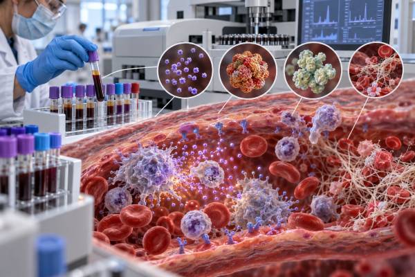
A “cytokine storm blood test panel” is not one standardized laboratory test. It is a group of tests used to look for severe, dysregulated inflammation and its effects on blood cells, clotting, the liver, kidneys, lungs, and other organs. Common measurements include IL-6, ferritin, C-reactive protein, D-dimer, complete blood count, fibrinogen, triglycerides, liver enzymes, lactate dehydrogenase, and coagulation studies. The pattern can support concern for cytokine release syndrome after immune therapy, hemophagocytic lymphohistiocytosis, macrophage activation syndrome, severe infection, or another hyperinflammatory state. No single value confirms a cytokine storm, and similar abnormalities occur in sepsis, cancer, liver disease, clotting disorders, and tissue injury. Diagnosis depends on the trigger, symptoms, vital signs, organ dysfunction, trends, and syndrome-specific criteria. Because true hyperinflammation can worsen rapidly, the panel is a hospital assessment tool—not a home screening test or a reason to delay urgent care.
- There is no universal cytokine storm panel or single diagnostic cutoff.
- IL-6 and CRP reflect signaling; ferritin and D-dimer reflect different downstream effects.
- Falling platelets or fibrinogen can be more concerning than one isolated high value.
- Cytokine release syndrome, HLH, MAS, and sepsis overlap but are not interchangeable.
- Severe fever, low blood pressure, breathing difficulty, or confusion needs urgent assessment.
Table of Contents
- What “cytokine storm” means clinically
- What the core blood tests show
- Other tests that complete the picture
- How the major hyperinflammatory syndromes differ
- How clinicians interpret the pattern and trend
- Why abnormal results do not always mean cytokine storm
- Symptoms and findings that require urgent care
- What happens after an abnormal panel
What “cytokine storm” means clinically
Cytokine storm is an informal umbrella term for a dangerous immune response in which inflammatory signaling becomes excessive, self-amplifying, and damaging to organs. It describes a mechanism rather than one disease. The same broad mechanism can emerge from very different triggers, including infection, cancer, autoimmune or autoinflammatory disease, inherited immune dysfunction, and treatments that activate T cells.
Cytokines are normally protective. IL-1, IL-6, TNF-alpha, interferon-gamma, and many other signals help control infection, recruit cells, produce fever, repair tissue, and coordinate adaptive immunity. Problems arise when activation becomes disproportionate or fails to shut down. Blood vessels can become leaky, clotting can become disordered, blood pressure can fall, oxygen exchange can worsen, and the liver, kidneys, brain, heart, or bone marrow can be affected.
Not every severe inflammatory illness is accurately described by the same syndrome. Cytokine release syndrome (CRS) usually refers to a recognized toxicity after CAR T-cell therapy, bispecific antibodies, and certain other immune treatments. Hemophagocytic lymphohistiocytosis (HLH) is a syndrome of uncontrolled immune activation that may be genetic or secondary to infection, malignancy, or other triggers. Macrophage activation syndrome (MAS) is an HLH-like hyperinflammatory complication most often discussed in rheumatic and autoinflammatory disease. Sepsis is life-threatening organ dysfunction caused by a dysregulated response to infection and can include both hyperinflammation and immune suppression.
These distinctions affect testing and treatment. In treatment-related CRS, clinicians grade severity mainly from fever, blood pressure, and oxygen support after establishing the appropriate clinical context. In suspected HLH or MAS, the evaluation weighs ferritin, cytopenias, liver injury, triglycerides, fibrinogen, soluble IL-2 receptor, spleen enlargement, immune-cell function, genetics, and the underlying trigger. In sepsis, rapid infection evaluation and organ support remain central even when inflammatory markers are extremely high.
The phrase “cytokine storm panel” is therefore best understood as a practical collection of tests. It can reveal inflammation, coagulation activation, tissue injury, and organ dysfunction. It cannot identify the syndrome without the bedside picture.
What the core blood tests show
The commonly named components—IL-6, ferritin, CRP, and D-dimer—measure different parts of the inflammatory response. They should not be treated as four versions of the same test.
Interleukin-6 (IL-6) is an immune-signaling protein involved in fever, acute-phase responses, blood-vessel activation, and communication among innate and adaptive immune cells. It can rise quickly during infection, immune therapy, trauma, surgery, autoimmune inflammation, and cancer. In some forms of CRS, IL-6 signaling is an important driver. However, the measured level may not arrive fast enough for emergency decisions, and there is no single IL-6 cutoff that diagnoses all cytokine storm syndromes. A very high IL-6 result supports strong pathway activation but must be tied to timing and cause.
IL-6 values can also behave unexpectedly after treatment. Blocking the IL-6 receptor may reduce signaling while temporarily increasing measurable circulating IL-6 because receptor-mediated clearance changes. A post-treatment rise does not automatically mean worsening disease.
Ferritin is an iron-storage protein and an acute-phase reactant. It may rise because macrophages and liver cells respond to inflammation, cells are damaged, iron handling changes, or clearance is impaired. Marked hyperferritinemia can strengthen concern for HLH or MAS, especially when it rises rapidly and accompanies fever, falling blood counts, liver dysfunction, high triglycerides, or low fibrinogen. Yet ferritin is not specific. Infection, malignancy, liver injury, kidney disease, repeated transfusions, iron overload, and other inflammatory disorders can also produce high values. The trajectory and surrounding findings matter more than calling one number diagnostic. A ferritin blood test measures both iron-storage and inflammatory biology.
C-reactive protein (CRP) is made mainly by the liver in response to inflammatory signaling, particularly IL-6-related pathways. It rises in many infections, inflammatory diseases, injuries, and cancers. CRP is widely available and useful for following trends, but it cannot distinguish cytokine storm from ordinary inflammation. Severe liver dysfunction can blunt CRP production. Drugs that block IL-6 signaling can also lower CRP even when another inflammatory pathway remains active. The CRP test is therefore a downstream marker, not a direct cytokine measurement.
D-dimer is produced when cross-linked fibrin is broken down. A high result indicates increased clot formation and breakdown somewhere in the body. Severe inflammation can activate clotting and damage endothelium, raising D-dimer. The result may support concern for coagulopathy or microvascular injury, but it is also elevated in venous thromboembolism, surgery, trauma, pregnancy, cancer, infection, older age, and liver disease. It cannot show whether a clot is present or where it is located. Imaging and clinical probability are needed when pulmonary embolism or deep-vein thrombosis is suspected.
Together, these markers answer four different questions: Is a major cytokine pathway active? Is there a strong acute-phase and macrophage-associated response? Is the liver producing an inflammatory protein? Is coagulation being activated and fibrin broken down? Their agreement—or disagreement—helps define the pattern.
Other tests that complete the picture
A useful hyperinflammation evaluation extends beyond the four headline markers. The additional tests often reveal whether inflammation is damaging organs or producing a syndrome-specific pattern.
A complete blood count with differential checks hemoglobin, platelets, neutrophils, lymphocytes, and other cells. HLH and MAS commonly raise concern when two or more blood-cell lines are low or declining. A falling platelet count can be an early warning even when it remains technically within the reference range. Cytopenias may arise from marrow suppression, immune destruction, consumption, infection, chemotherapy, bleeding, or an underlying cancer, so cause still requires investigation.
Fibrinogen is a clotting protein and an acute-phase reactant. Many inflammatory states raise it. In HLH or MAS, consumption and liver dysfunction may produce an inappropriately normal or low result despite intense inflammation. That contrast—very high ferritin with falling fibrinogen—can be more informative than either value alone.
Triglycerides may rise because inflammatory signaling reduces normal lipid clearance. Hypertriglyceridemia is part of established HLH criteria, but fasting status, diabetes, medications, nutrition, and liver disease can also affect it.
Liver tests include alanine aminotransferase, aspartate aminotransferase, bilirubin, alkaline phosphatase, albumin, and sometimes ammonia. Hyperinflammatory syndromes can cause hepatitis, cholestasis, impaired protein production, or liver failure. The pattern can also point toward viral infection, medication toxicity, biliary disease, or malignancy.
Lactate dehydrogenase (LDH) rises with tissue injury and rapid cell turnover. It is nonspecific but can help show the scale of organ damage, hemolysis, tumor burden, or hepatic stress. Creatinine, electrolytes, urinalysis, and urine output assess kidney involvement. Troponin, natriuretic peptides, electrocardiography, and echocardiography may be used if cardiac injury is suspected.
Coagulation tests such as prothrombin time, international normalized ratio, activated partial thromboplastin time, fibrinogen, and platelet count help identify disseminated intravascular coagulation or liver-related clotting problems. D-dimer alone is not enough.
Soluble IL-2 receptor (sCD25) reflects T-cell activation and is included in HLH evaluation. A high soluble IL-2 receptor test can support the picture, but it also rises in lymphoma, infection, autoimmune disease, and other T-cell activation states. Availability and turnaround time vary.
Natural killer-cell function, perforin expression, degranulation testing, and genetic studies may be important when inherited HLH is possible or when a specialist needs mechanistic evidence. IL-18, CXCL9, interferon-gamma, and other specialized biomarkers may help characterize selected cases, but many are not rapid routine tests. A dedicated IL-18 test is supportive rather than independently diagnostic.
Cultures, pathogen molecular tests, imaging, bone marrow examination, and cancer evaluation may be essential because finding the trigger is as important as naming the inflammatory syndrome.
How the major hyperinflammatory syndromes differ
The laboratory overlap is substantial, but the setting and dominant features help clinicians separate the major syndromes.
Immune effector cell-associated CRS generally occurs after a treatment known to activate immune cells. Fever of at least 38°C without another explanation is central at onset. Severity grading then depends on hypotension and hypoxia: whether blood pressure responds to fluids, whether vasopressors are needed, and what level of oxygen support is required. CRP, ferritin, IL-6, liver tests, and coagulation markers may be followed, but they do not determine the standard CRS grade by themselves. Infection must be evaluated because neutropenic sepsis can look nearly identical and may occur at the same time.
HLH can occur at any age. Primary forms result from genetic defects affecting cytotoxic lymphocyte control. Secondary forms may be triggered by Epstein-Barr virus and other infections, lymphoma and leukemia, rheumatic disease, immune therapy, or transplantation. Typical clues include persistent fever, enlarged spleen, cytopenias, high ferritin, high triglycerides, low fibrinogen, liver dysfunction, high soluble IL-2 receptor, impaired NK-cell activity, and hemophagocytosis. Hemophagocytosis is neither required early nor specific; its absence on one marrow sample does not rule out HLH.
Formal HLH-2004 criteria were developed mainly in children, while the HScore was developed for adults with secondary HLH. Specialists use these tools as frameworks rather than letting a score replace judgment. A critically ill patient may need treatment before every specialized result returns.
MAS describes HLH-like immune activation in rheumatic and autoinflammatory disease, especially systemic juvenile idiopathic arthritis and adult-onset Still disease. Persistent fever, rising ferritin, unexpectedly falling platelets or white cells, liver abnormalities, high D-dimer, prolonged clotting tests, falling fibrinogen, spleen enlargement, and neurologic changes are important warnings. Because active Still disease can already produce fever and high inflammatory markers, a change from the patient’s baseline may be more revealing than an isolated cutoff. A macrophage activation syndrome blood test panel integrates these dynamic changes.
Sepsis begins with suspected or confirmed infection and organ dysfunction. It can cause high IL-6, CRP, ferritin, D-dimer, liver enzymes, and lactate, as well as low platelets and disordered coagulation. Some patients develop an HLH-like phenotype; others shift toward immune exhaustion. Because suppressing inflammation without controlling infection can be dangerous, antimicrobial treatment, source control, cultures, imaging, and organ support remain priorities.
Other conditions can mimic these patterns: severe drug reactions, acute liver failure, catastrophic antiphospholipid syndrome, thrombotic microangiopathy, malignancy, transfusion reactions, pancreatitis, major trauma, and heat illness. The trigger and timeline are part of the diagnosis.
How clinicians interpret the pattern and trend
Interpretation starts with the patient, not a threshold. Clinicians first assess temperature, blood pressure, heart rate, oxygen level, mental status, urine output, rash, bleeding, enlarged liver or spleen, and the timing of infection, immune therapy, medication exposure, or rheumatic symptoms.
They then look for coherence among the tests. A rising ferritin with falling platelets, falling fibrinogen, increasing triglycerides, worsening liver enzymes, and persistent fever is more concerning for HLH or MAS than an isolated ferritin elevation. A high CRP and IL-6 shortly after CAR T-cell infusion with fever, low blood pressure, and oxygen need fits CRS more strongly than the same values months later in an untreated person. A high D-dimer with prolonged clotting times, low fibrinogen, thrombocytopenia, and bleeding raises concern for consumptive coagulopathy.
Trends are often more important than reference flags. A ferritin increase from 500 to 5,000 over a short period may carry more weight than a stable chronic value at 2,000. A platelet count falling from 250 to 120 can be clinically meaningful even if the laboratory’s lower limit is 150. The direction of CRP after IL-6 blockade needs special interpretation because the drug directly suppresses its production.
The laboratory’s units and method must be confirmed. Ferritin may be reported in nanograms per milliliter or micrograms per liter, which are numerically equivalent, but other analytes can use different units. IL-6 reference intervals vary widely by assay. D-dimer may be reported as fibrinogen-equivalent units or D-dimer units; cutoffs are not interchangeable.
Clinicians also account for baseline disease and treatment. A person with chronic liver disease may have elevated ferritin, low platelets, and abnormal coagulation before acute illness. A patient with leukemia may already have cytopenias. Steroids may suppress fever. Tocilizumab may reduce CRP. Transfusions can affect ferritin. Anticoagulation changes clotting interpretation.
Scoring systems and criteria help organize evidence, but they do not make all syndromes equivalent. An HScore supports adult secondary HLH probability; it is not a CRS grade. ASTCT CRS grading applies in the immune-effector therapy setting; it is not designed for infection-associated hyperinflammation. MAS criteria are most reliable in the disease population for which they were developed.
The best interpretation is repeated and multidisciplinary. Hematology, rheumatology, infectious disease, oncology, critical care, neurology, and laboratory medicine may each contribute a necessary piece.
Why abnormal results do not always mean cytokine storm
Every major component is nonspecific. High results deserve explanation, but the most dramatic explanation is not always the correct one.
Ferritin may be high because of chronic liver disease, alcohol-related injury, iron overload, kidney failure, repeated transfusion, malignancy, or ordinary infection. Very high values narrow the differential but still do not prove HLH. A ferritin test also cannot tell how much stored iron is safely available during active inflammation.
CRP rises after many bacterial and viral infections, surgery, trauma, inflammatory arthritis, vasculitis, and tissue necrosis. It may stay low in severe inflammation when liver synthetic function is poor or an IL-6 pathway drug is working. A normal CRP therefore cannot rule out every hyperinflammatory syndrome.
D-dimer is especially sensitive to context. It often rises with hospitalization, age, pregnancy, cancer, surgery, and infection. A high result does not diagnose disseminated intravascular coagulation or pulmonary embolism. Conversely, a low result is used to exclude venous thromboembolism only in appropriately selected patients with a validated assay and low or intermediate clinical probability.
IL-6 is transient and method-dependent. A sample taken before the peak, after treatment, or after the cytokine has moved into tissue may be unremarkable. Some assays measure total immunoreactive protein without showing how much is biologically active. Receptor-blocking therapy can alter measured concentrations.
Preanalytic problems can also distort the panel. Delayed processing, hemolysis, clotting, platelet activation, wrong collection tubes, improper transport, or repeated freeze-thaw cycles may change cytokines. D-dimer and coagulation tests can be affected by underfilled citrate tubes or anticoagulants.
The phrase “cytokine storm” is sometimes used too broadly for any high inflammatory marker. That can lead to inappropriate immune-suppressing treatment or missed infection. Laboratory abnormalities should prompt a differential diagnosis, not a predetermined conclusion.
Commercial wellness panels are not suitable for diagnosing this emergency. True cytokine storm syndromes are evaluated in a clinical setting where vital signs, organ function, rapid repeat testing, cultures, imaging, and treatment can occur together.
Symptoms and findings that require urgent care
Possible hyperinflammation can progress quickly. Seek emergency medical care for difficulty breathing, blue or gray lips, chest pain, new confusion, inability to stay awake, seizure, fainting, severe weakness, very low blood pressure, rapidly spreading rash, uncontrolled bleeding, or markedly reduced urine output.
People who recently received CAR T cells, a bispecific antibody, or another therapy with known CRS risk should follow the treatment center’s instructions. Fever may require immediate contact even when the person otherwise feels stable. Do not self-treat with leftover steroids or anti-inflammatory drugs unless the treating team has provided a specific plan; doing so can mask infection or alter grading.
Persistent high fever with bruising, bleeding, jaundice, abdominal swelling, severe headache, neurologic changes, or rapid deterioration also warrants urgent evaluation for HLH, MAS, sepsis, liver failure, or another critical illness. Children with systemic inflammatory disease may deteriorate despite a previously reassuring visit, so changes from baseline deserve attention.
Laboratory findings that commonly trigger escalation include falling blood pressure, rising oxygen requirement, rapidly increasing ferritin, worsening cytopenias, falling fibrinogen, increasing D-dimer with clotting abnormalities, rising lactate, increasing creatinine, severe liver injury, or altered mental status. No one laboratory threshold replaces the clinical emergency signs.
A person should not wait for an outpatient cytokine level if symptoms are severe. Treatment decisions are often based on the recognized syndrome and organ dysfunction before specialized cytokine results return.
What happens after an abnormal panel
The first goals are stabilization and identification of the trigger. Clinicians support breathing and circulation, monitor organs, obtain cultures and pathogen tests when infection is possible, review recent treatments, and involve the relevant specialists. Hyperinflammation and infection may be treated in parallel because waiting to distinguish them perfectly can be unsafe.
Repeat testing is usually targeted. Blood counts, ferritin, fibrinogen, liver tests, coagulation studies, creatinine, CRP, and other markers may be checked frequently in an unstable patient. The interval depends on severity and the expected speed of change. A broad cytokine panel is not necessarily repeated as often because turnaround time and clinical utility may be limited.
Further evaluation may include:
- Blood, urine, respiratory, or cerebrospinal fluid cultures and molecular tests
- Imaging to identify infection, cancer, organ enlargement, or thrombosis
- Bone marrow examination when malignancy, marrow failure, or HLH is suspected
- Soluble IL-2 receptor and specialized cytokine markers
- NK-cell function, degranulation, perforin expression, or genetic testing
- Autoimmune and autoinflammatory testing guided by symptoms
- Tissue biopsy when the underlying disease remains unclear
Treatment is syndrome- and trigger-specific. CRS after immune therapy may require supportive care, an IL-6 receptor blocker, corticosteroids, or other protocol-directed therapy depending on grade. HLH may require urgent immune suppression, treatment of infection or malignancy, targeted cytokine therapy, chemotherapy-based regimens, or stem-cell transplantation in selected genetic disease. MAS treatment is coordinated with rheumatology and may use corticosteroids and pathway-targeted agents. Sepsis requires prompt antimicrobial therapy when indicated, source control, fluids, vasopressors, respiratory support, and other critical care.
The purpose of monitoring is not merely to normalize a number. Clinicians look for recovery of blood pressure and oxygenation, improving mental status, restored urine output, reversal of organ injury, stabilization of blood counts, and a downward inflammatory trend. Some markers lag behind recovery; ferritin may remain elevated after the patient improves.
After discharge, follow-up depends on the cause. Survivors may need review of organ function, infection prevention, medication effects, genetic counseling, cancer treatment, rheumatologic control, or immune-therapy complications. The panel is one part of that story. The diagnosis and outcome are determined by the entire clinical course.
References
- Deep insight into cytokine storm: from pathogenesis to treatment 2025 (Review)
- EULAR/PReS recommendations for the diagnosis and management of Still’s disease, comprising systemic juvenile idiopathic arthritis and adult-onset Still’s disease 2024 (Clinical Guideline)
- ASTCT Consensus Grading for Cytokine Release Syndrome and Neurologic Toxicity Associated with Immune Effector Cells 2019 (Consensus Guideline)
- Hemophagocytic lymphohistiocytosis: current treatment advances, emerging targeted therapy and underlying mechanisms 2024 (Review)
- Pediatric CAR T-Cell Therapy (PDQ®)–Health Professional Version 2024 (Clinical Reference)
- Histiocyte Society blueprint for hemophagocytic lymphohistiocytosis research: urgent priorities and future directions 2025 (Consensus Review)
Disclaimer
This article is for general education and is not a diagnostic tool or a substitute for urgent medical care. Cytokine storm, CRS, HLH, MAS, and sepsis are potentially life-threatening and require interpretation by clinicians using symptoms, vital signs, trends, and syndrome-specific criteria. Seek emergency help for breathing difficulty, confusion, fainting, severe weakness, low blood pressure, bleeding, or rapid deterioration.





