
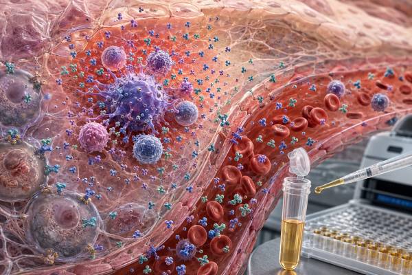
An interleukin-6 (IL-6) test measures a rapidly changing cytokine involved in infection defense, inflammation, antibody responses, blood-cell production, metabolism, and tissue repair. IL-6 can rise within hours after infection, injury, surgery, immune therapy, or an inflammatory flare and can stimulate the liver to produce C-reactive protein, fibrinogen, and other acute-phase proteins. Because many unrelated conditions increase IL-6, the test is not a stand-alone diagnosis for sepsis, autoimmune disease, cancer, or “cytokine storm.” It is most useful in selected hospital, immunology, oncology, rheumatology, and research settings when interpreted with symptoms, vital signs, organ function, cultures, blood counts, CRP, ferritin, procalcitonin, and treatment timing. Direct IL-6 assays differ in sensitivity and may measure free cytokine differently after receptor-blocking medicines. A high value supports active cytokine signaling but does not identify the cause. A low value may be normal, may reflect timing or treatment, and does not rule out localized or intermittent inflammation. The exact specimen, assay, units, reference interval, and clinical question determine what the number means.
- IL-6 is an early inflammatory signal, while CRP is a downstream liver response that often changes more slowly.
- Classic IL-6 signaling and trans-signaling affect different cell populations and can have different biological effects.
- High IL-6 is nonspecific and can occur with infection, immune therapy, trauma, autoimmune disease, and malignancy.
- A normal or low result does not exclude sepsis, tissue inflammation, or a cytokine-driven disorder.
- IL-6 receptor blockade can make circulating IL-6 rise even while signaling and inflammation improve.
Table of Contents
- IL-6 Biology and Signaling
- What the IL-6 Test Measures
- When IL-6 Testing May Help
- Collection, Timing, and Assay Limitations
- How to Interpret the Result
- Causes and Meaning of High IL-6
- Low IL-6 and False Reassurance
- Follow-Up Tests and IL-6-Targeted Treatment
IL-6 Biology and Signaling
IL-6 is produced by monocytes, macrophages, dendritic cells, endothelial cells, fibroblasts, adipose tissue, skeletal muscle, and many other cell types. Microbial products, tissue injury, IL-1, tumor necrosis factor, immune complexes, and cellular stress can trigger production. This broad source list explains why IL-6 is a sensitive sign of disturbance but a poor identifier of one specific disease.
The cytokine binds IL-6 receptor alpha, also called CD126, and then recruits the signal-transducing protein gp130, or CD130. The receptor complex activates JAK-STAT, MAP kinase, and PI3K pathways. In classic signaling, IL-6 binds membrane IL-6 receptor on a limited group of cells, including hepatocytes and selected leukocytes. Classic signaling contributes to acute-phase responses, immune defense, regeneration, and metabolic effects.
In trans-signaling, IL-6 binds a soluble form of IL-6 receptor. The complex can then activate gp130 on many cells that do not express membrane IL-6 receptor. This expands the range of responsive tissues and is strongly associated with leukocyte recruitment and chronic inflammation. Soluble gp130 naturally buffers trans-signaling. These details matter scientifically, but a routine IL-6 concentration does not distinguish classic from trans-signaling.
The liver is a major downstream target. IL-6 stimulates production of CRP, serum amyloid A, fibrinogen, hepcidin, and other acute-phase proteins. Hepcidin reduces iron availability and can contribute to anemia of inflammation. Fibrinogen affects clotting and the erythrocyte sedimentation rate. CRP is commonly used because it is easier to measure and remains elevated longer than IL-6.
IL-6 also supports B-cell differentiation, influences T-cell pathways, helps mobilize neutrophils, affects platelet production, and communicates with nervous and endocrine systems. During exercise, muscle-derived IL-6 can rise without indicating infection and participates in metabolic regulation. The meaning of a value therefore depends on whether the person is septic, recovering from surgery, receiving immunotherapy, exercising intensely, or living with chronic inflammatory disease.
A controlled IL-6 response can be protective. Excessive or prolonged signaling can contribute to capillary leakage, coagulation abnormalities, fever, fatigue, tissue damage, and chronic immune disease. The test measures concentration, not whether the signaling is beneficial or harmful.
IL-6 also changes over a normal day and with physiologic stress. Adipose tissue can contribute to low-grade production, while contracting skeletal muscle can release large amounts during prolonged exercise. In that setting, IL-6 helps mobilize energy substrates and can be followed by anti-inflammatory signals. This does not resemble the sustained inflammatory environment of sepsis or active autoimmune disease. Recent exercise, obesity, sleep loss, and metabolic illness can therefore shift a result without providing a single explanation for symptoms. The size and duration of the rise, accompanying markers, and recovery pattern are more informative than labeling every elevation as pathological.
What the IL-6 Test Measures
Most clinical IL-6 tests are immunoassays performed on serum or plasma. Enzyme-linked, chemiluminescent, electrochemiluminescent, bead-based, and digital platforms use antibodies to capture and quantify the cytokine. Results are commonly reported in picograms per milliliter. Laboratories use different calibrators, antibody pairs, detection limits, and specimen requirements, so numbers from different platforms may not match.
Serum forms after blood clots; plasma is separated from anticoagulated blood. Clotting can activate platelets and leukocytes, while anticoagulants can influence assay chemistry. The same patient may therefore have different serum and plasma results. Reference information must match the specimen used.
A direct IL-6 result is a snapshot of circulating protein. It does not reveal the source, receptor pathway, tissue concentration, or biological activity. Some research assays separately measure soluble IL-6 receptor, soluble gp130, phosphorylated STAT3, or IL-6-responsive gene signatures. These are not interchangeable with a routine IL-6 concentration.
Cell-stimulation assays can measure IL-6 production after blood cells are exposed to microbial ligands, mitogens, antigens, or medicines. Such tests ask how cells respond under controlled conditions. They may be used in research or rare immune-function evaluations, but their culture concentrations should not be compared with serum values.
IL-6 may be included in a cytokine panel. A panel can provide pattern information in severe immune dysregulation, but it also creates multiple comparisons and method-specific interpretation. One isolated elevation among many analytes does not automatically identify a syndrome.
A cytokine storm blood test panel often combines IL-6 with ferritin, CRP, D-dimer, blood counts, liver tests, and coagulation studies. These companion tests usually provide more immediate information about organ risk and the trajectory of illness than IL-6 alone.
When IL-6 Testing May Help
IL-6 testing is used selectively because the result is nonspecific and may not return quickly enough for urgent decisions. Its strongest roles are in specialized hospital algorithms, clinical trials, immune-therapy monitoring, and difficult inflammatory syndromes where a cytokine pattern adds information to standard testing.
After chimeric antigen receptor T-cell therapy or certain antibody treatments, fever and systemic inflammation may represent cytokine release syndrome. IL-6 often rises, but diagnosis and grading are clinical and depend on fever, blood pressure, oxygen requirement, and organ function. Treatment should not be delayed while waiting for a cytokine value when the syndrome is otherwise clear.
Sepsis can also produce high IL-6. The cytokine may rise earlier than CRP, but it cannot reliably distinguish bacterial infection from viral infection, sterile inflammation, trauma, pancreatitis, or immune therapy in every patient. Cultures, source evaluation, lactate, organ function, and prompt clinical treatment remain central. A procalcitonin test can add evidence for bacterial infection in selected settings, yet it also has limitations.
Distinguishing sepsis from immune-therapy toxicity is especially difficult because both can produce fever, low blood pressure, low oxygen, coagulation changes, and high inflammatory markers. They can also occur together. Treatment teams use the timing after infusion, cultures, imaging, clinical examination, and response to therapy rather than assuming that a high IL-6 proves one syndrome. Broad-spectrum antimicrobials may be started while cytokine release syndrome is treated when infection cannot be safely excluded. The danger lies in a false either-or decision: immune suppression can worsen an untreated infection, while delaying cytokine-directed treatment can permit rapid inflammatory deterioration.
Rheumatologists and immunologists may order IL-6 in Still disease, Castleman disease, rheumatoid arthritis, juvenile idiopathic arthritis, vasculitis, or undifferentiated hyperinflammation. In most of these conditions, diagnosis relies on clinical criteria and established laboratory patterns. IL-6 may support pathway activity but does not establish the condition.
Hematology and oncology teams may evaluate IL-6 when malignancy-associated inflammation, treatment toxicity, infection, or immune dysregulation is suspected. Some tumors and tumor-associated cells produce IL-6, but the test is not a general cancer screen and cannot locate a tumor.
Pediatric immune centers sometimes use cytokine panels to help separate familial hemophagocytic lymphohistiocytosis, cytokine release syndrome, systemic inflammatory disease, and sepsis. Interpretation requires age-appropriate reference data and expert review. Patterns observed in severely ill cohorts do not create a self-diagnostic test for less specific symptoms.
IL-6 testing is generally not useful as a wellness marker, a routine explanation for fatigue, or a way to choose supplements. Common inflammatory tests and focused evaluation usually provide more actionable information.
Collection, Timing, and Assay Limitations
Fasting is usually not required. The patient should report fever onset, surgery, strenuous exercise, trauma, recent vaccination, immune therapy, corticosteroids, anti-inflammatory drugs, antibiotics, and IL-6-pathway medicines. Sampling time relative to these events can change the result substantially.
IL-6 can rise and fall over hours. A sample obtained early in an illness may be high before CRP peaks; one drawn after treatment may already be falling. Conversely, ongoing tissue production may produce repeated peaks. A single value cannot reconstruct the full time course.
Blood should be collected into the specified tube and processed promptly. Cells left in contact with serum or plasma can release cytokines after collection. Hemolysis, lipemia, prolonged storage, and repeated freezing can affect some assays. In research, strict handling protocols are necessary for meaningful comparisons.
The lower end of the range is especially method-dependent. “Below detection” is not zero, and a small number just above the limit may be imprecise. At the high end, dilution and the assay’s reportable range matter. Laboratories may use different upper limits and may report “greater than” a threshold rather than an exact value.
Heterophile antibodies, rheumatoid factor, therapeutic antibodies, and other substances can interfere with immunoassays. An unexpected result that conflicts with the clinical picture may require repeat testing, dilution studies, or another platform. Repeat testing is most useful when it can change care.
IL-6 receptor blockers complicate interpretation. Tocilizumab and sarilumab prevent receptor-mediated clearance and signaling, so free circulating IL-6 may rise after treatment even as CRP and symptoms improve. A post-treatment increase should not automatically be interpreted as worsening inflammation. Direct IL-6-neutralizing antibodies can create different assay effects.
Venipuncture itself carries ordinary minor risks: brief pain, bruising, dizziness, and rarely infection or prolonged bleeding. The test does not provoke inflammation or expose the patient to cytokine.
How to Interpret the Result
Begin with the laboratory’s reference interval and method. There is no universal normal cutoff across assays, ages, specimens, and clinical settings. A research study’s threshold should not be applied to an unrelated commercial test.
Next, ask whether the result fits the timing. IL-6 is an upstream signal with rapid kinetics. CRP, ferritin, fibrinogen, and blood-cell changes are downstream and may lag. A high IL-6 with a modest CRP can occur early; a falling IL-6 with persistently high CRP can occur during recovery.
Then assess severity through the patient, not the cytokine number. Blood pressure, mental status, breathing, urine output, lactate, kidney and liver function, coagulation, and oxygen need determine urgency. Extremely high IL-6 can accompany severe illness, but no single concentration substitutes for clinical grading.
Evaluate the pattern. High IL-6 with neutrophilia, positive cultures, rising lactate, and organ dysfunction suggests a different problem from high IL-6 after CAR T-cell infusion or with quotidian fever, rash, and very high ferritin. Similar numbers can arise from different mechanisms.
A trend may help when samples are collected consistently on the same assay. Falling values can support response, while persistent elevation may prompt reassessment. However, receptor blockade, steroids, source control, and assay timing can uncouple IL-6 from clinical improvement.
Interpretation should also distinguish association from causation. Obesity, aging, chronic kidney disease, smoking, and other conditions can be associated with low-grade IL-6 elevation. This does not prove that IL-6 is the sole cause of symptoms or that direct cytokine blockade is appropriate.
Reference populations matter as well. Age, pregnancy, chronic illness, and intensive-care status can shift expected distributions, and pediatric values should not be judged by an adult research cutoff. A “high” flag simply means the result exceeded the laboratory’s comparison rule. The degree of elevation may help describe risk in a validated setting, but it does not create a universal severity scale. Clinicians should ask whether the cutoff was designed for diagnosis, prognosis, trial enrollment, or analytical reporting before using it in an individual treatment or diagnostic decision for that specific individual patient.
Causes and Meaning of High IL-6
High IL-6 means more cytokine was detected than expected for that assay. The differential diagnosis is broad and should be narrowed by context rather than by the magnitude alone.
Infection is common. Bacterial sepsis, viral infections, fungal disease, and severe localized infections can raise IL-6. Because sterile injury can produce the same response, cultures and source-directed testing are essential. Antibiotics should be based on clinical suspicion and microbiology, not an IL-6 number alone.
Cytokine release syndrome after cellular or antibody-based therapy is another important cause. Fever is usually the first sign; low blood pressure, low oxygen, and organ dysfunction indicate greater severity. IL-6 receptor blockade can be lifesaving in appropriate cases, but grading and treatment follow clinical protocols.
Autoimmune and autoinflammatory diseases may elevate IL-6 during active inflammation. Rheumatoid arthritis, Still disease, giant cell arteritis, polymyalgia rheumatica, and systemic juvenile idiopathic arthritis are examples of IL-6-associated conditions. Disease-specific symptoms and criteria remain necessary.
Macrophage activation syndrome and hemophagocytic lymphohistiocytosis can include high IL-6, but ferritin, cytopenias, triglycerides, fibrinogen, liver injury, soluble IL-2 receptor, natural killer-cell function, and genetic or tissue studies may be more discriminating. An MAS blood test panel is interpreted as a syndrome pattern, not a single-marker test.
Trauma, burns, major surgery, pancreatitis, ischemia, and strenuous exercise can produce sterile IL-6 release. The level may reflect tissue stress without infection. Clinical trajectory and source evaluation prevent unnecessary conclusions.
Some cancers and inflammatory tumor environments produce IL-6. Persistent unexplained fever, weight loss, night sweats, lymph-node enlargement, anemia, or abnormal imaging requires standard diagnostic workup. IL-6 is not specific enough for cancer screening or surveillance by itself.
Low IL-6 and False Reassurance
Low or undetectable IL-6 is common in healthy people. The cytokine is normally present at very low concentrations and rises transiently when needed. A low result does not indicate weak immunity or require treatment.
A normal value cannot exclude infection. Sampling may occur before the rise, after the peak, after treatment, or while inflammation remains localized. Immunosuppressed patients may also produce muted cytokine responses despite serious infection.
Corticosteroids, JAK inhibitors, IL-6-pathway drugs, and effective treatment of the underlying cause can lower signaling or downstream markers. The meaning depends on timing and expected drug action. After receptor blockade, IL-6 itself may rise while CRP becomes very low, so neither marker should be interpreted without the medication history.
Low serum IL-6 does not exclude tissue inflammation in joints, bowel, brain, skin, or tumors. Local production may not reach measurable systemic concentrations. Imaging, biopsy, organ-specific biomarkers, and examination can be more informative.
There is no clinical reason to “boost” IL-6 because a test is low. Excessive IL-6 can cause inflammation, anemia, coagulation changes, and systemic illness. Immune support should focus on vaccination, nutrition, sleep, management of chronic disease, and treatment of proven deficiencies rather than manipulating one cytokine.
Follow-Up Tests and IL-6-Targeted Treatment
Follow-up depends on the suspected cause. Common tests include a complete blood count, metabolic panel, liver tests, lactate, blood and site-specific cultures, urinalysis, imaging, CRP, ESR, ferritin, procalcitonin, coagulation studies, fibrinogen, triglycerides, and D-dimer. Not every patient needs every test.
CRP is often the most practical downstream companion. A CRP test is standardized and widely available, while an inflammatory marker panel can show whether several systems move together. These markers still do not identify the cause without clinical evaluation.
IL-6 receptor inhibitors and direct IL-6 inhibitors are used for specific diseases and emergencies. Approved indications and dosing differ by drug and country. Screening for infection, tuberculosis risk, liver abnormalities, blood-count changes, lipid changes, and gastrointestinal risk may be required. These medicines can suppress fever and CRP, so infection may be less obvious during therapy.
The absence of a CRP rise during IL-6 receptor blockade deserves particular caution. A patient can develop a serious bacterial infection while CRP remains low because the liver is no longer receiving the usual IL-6 signal. New localized pain, cough, urinary symptoms, confusion, or unexplained decline should be evaluated on their own merits. Neutropenia, elevated liver enzymes, lipid changes, and rare bowel perforation are additional considerations for selected agents and patients. Monitoring schedules are based on the medicine, indication, dose, and comorbidities rather than on repeat IL-6 levels. Vaccination planning and avoidance of live vaccines may also be relevant.
In cytokine release syndrome, treatment selection and timing follow the therapy program’s protocol. In chronic rheumatic disease, response is assessed through symptoms, examination, imaging, function, and established disease activity measures. A target serum IL-6 concentration is generally not used to adjust routine dosing.
Urgent care is needed for low blood pressure, confusion, severe breathing difficulty, bluish lips, reduced urine, rapidly spreading rash, persistent high fever after immune therapy, or signs of organ failure. A laboratory result should never delay stabilization and source-directed treatment.
The best use of IL-6 is as one piece of a time-sensitive pattern. The number gains meaning only when linked to what happened before the draw, what the patient is experiencing, which treatment has been given, and how organ function is changing.
References
- Targeting IL-6 trans-signalling: past, present and future prospects 2023 (Review)
- IL-6 Signaling in Immunopathology: From Basic Biology to Selective Therapeutic Inhibition 2025 (Review)
- Serum cytokine panels in pediatric clinical practice 2024 (Review)
- Roles of cytokine storm in sepsis progression: mechanisms, diagnosis, and treatment 2025 (Review)
- Interleukin 6: at the interface of human health and disease 2023 (Review)
- The Interleukin Network in Sepsis: From Cytokine Storm to Diagnostic and Therapeutic Applications 2025 (Review)
Disclaimer
This article provides general education and is not a diagnosis or treatment plan. IL-6 testing is method-dependent and cannot by itself diagnose sepsis, cytokine release syndrome, autoimmune disease, or cancer. Seek urgent medical care for severe fever with confusion, low blood pressure, breathing difficulty, reduced urine, or rapidly worsening illness.





