
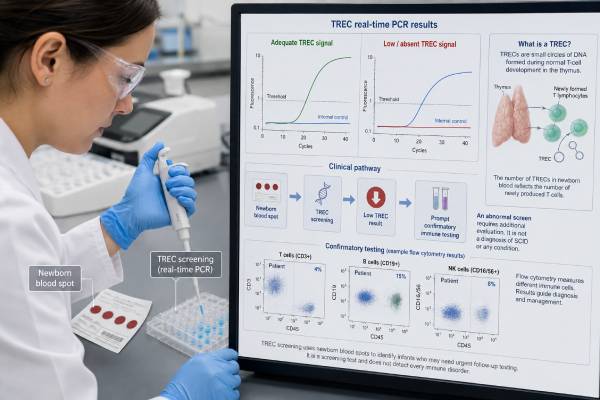
Newborn screening for severe combined immunodeficiency, or SCID, measures tiny DNA circles called TRECs in a dried blood spot. TRECs are produced when new T cells develop in the thymus. Very low or absent TRECs can signal that a baby has too few newly formed T cells and may be unable to fight serious infections. The screen is time-critical because babies with SCID often look healthy at birth, while early protection and treatment can be lifesaving. An out-of-range TREC result does not prove SCID. Prematurity, congenital heart disease, chromosome conditions, thymus problems, illness, medications, and other immune disorders can also reduce TRECs. Follow-up is not another routine screen alone; it usually includes urgent clinical review, a blood count, lymphocyte-subset testing by flow cytometry, and consultation with pediatric immunology. Families should receive clear infection precautions while the diagnosis is being established.
- TRECs reflect production of new T cells, not the presence of one specific SCID gene variant.
- Low or absent TRECs are a screening finding, not a final diagnosis.
- Confirmatory flow cytometry measures T, B, and NK cells and helps define the immune pattern.
- Premature and critically ill newborns have more false-positive results, but they still need the follow-up ordered by the screening program.
- Live vaccines should be delayed after a concerning result until an immune specialist says they are safe.
- A negative screen does not exclude every immune disorder, especially conditions with T cells that are present but function poorly.
Table of Contents
- Why SCID Newborn Screening Matters
- What TRECs Show and How the Test Works
- Reading Normal, Borderline, and Abnormal Results
- Causes of Low TRECs Besides Classic SCID
- Confirmatory Immune and Genetic Testing
- Precautions While Follow-Up Is Underway
- From a Confirmed Diagnosis to Treatment
- Inheritance, Family Testing, and Practical Questions
Why SCID Newborn Screening Matters
SCID is not one disease. It is a group of inborn errors of immunity in which T-cell development or function is severely impaired. B-cell and natural killer, or NK-cell, function may also be affected, depending on the genetic cause. Without working T cells, ordinary viruses, bacteria, and fungi can cause overwhelming or persistent infection.
A baby with SCID may appear completely well during the first days or weeks. Antibodies transferred during pregnancy can provide temporary protection, and exposure to infection may not yet have occurred. By the time a baby develops persistent thrush, pneumonia, severe diarrhea, poor growth, or unusual infections, treatment can be more complicated. Newborn screening aims to identify severe T-cell lymphopenia before this clinical decline.
Early diagnosis changes care immediately. It allows families and clinicians to reduce infection exposure, avoid live vaccines, use appropriate blood products, test for infections, and arrange definitive treatment before an infection causes organ damage. Hematopoietic stem cell transplantation is the main curative treatment for many forms of SCID. Gene therapy or enzyme replacement may be options for selected genetic types at specialized centers.
What the screen is designed to find
The primary target is classic SCID, but TREC screening also identifies “leaky” or atypical SCID and other causes of severe T-cell lymphopenia. This broader detection is both a benefit and a source of false-positive concern. An abnormal report may ultimately identify a different immune condition that still needs care, or it may resolve as a premature baby’s immune system matures.
SCID screening is part of the newborn dried-blood-spot panel in all U.S. states, although collection timing, cutoffs, repeat rules, and referral pathways vary. Families should follow the instructions from the program that issued the result. A TREC number from one jurisdiction cannot be interpreted using another program’s threshold.
Why it is called time-critical
The term does not mean that every baby with an abnormal screen is in immediate danger. It means that a baby with true SCID can become seriously ill from common exposures and should not wait for symptoms before evaluation. A same-day or next-working-day plan for immunology contact and confirmatory testing is appropriate when the program reports a critical result.
A borderline result may follow a different pathway, such as a repeat dried blood spot. Parents should still complete it promptly. A repeat request is not a diagnosis, but losing follow-up can delay recognition of a genuinely low T-cell count.
What TRECs Show and How the Test Works
T cells develop in the thymus. During this process, their DNA is rearranged to create diverse T-cell receptors capable of recognizing many germs. Small circular pieces of DNA are cut out during rearrangement. These byproducts are T-cell receptor excision circles.
TRECs do not replicate when a cell divides. A high concentration in a newborn blood spot generally indicates recent production of new T cells by the thymus. Few or no TRECs suggest that few new T cells are being produced, although the reason is not yet known.
The laboratory method
A punch from the dried blood spot is tested by quantitative polymerase chain reaction, or PCR. The assay measures TREC DNA and usually includes a control gene to show that the specimen contains adequate amplifiable DNA. Some laboratories use a multiplex assay that measures TRECs and also checks another newborn condition, such as the common SMN1 deletion associated with spinal muscular atrophy.
The report may state:
- TREC copies per microliter or per punch
- A cycle-threshold value from the PCR assay
- “Normal,” “borderline,” “low,” “absent,” or “inconclusive”
- Whether the control gene amplified properly
- Whether a repeat specimen or immediate diagnostic evaluation is required
Parents do not need to compare the raw number with values posted online. Specimen volume, blood-spot quality, extraction method, calibration, and cutoff rules differ. The action category on the issuing program’s report is more useful than an isolated number.
TREC is a marker, not a gene test
The test does not sequence IL2RG, ADA, RAG1, RAG2, JAK3, DCLRE1C, or the many other genes that can cause SCID. It measures the output of T-cell development. This is why one screening method can detect several genetic forms and also detect non-genetic T-cell lymphopenia.
A normal TREC screen means enough newly formed T-cell signal was present to pass the program’s cutoff. It does not prove that T cells function normally. Rare forms of combined immunodeficiency, including some defects in T-cell signaling, may have normal or near-normal T-cell numbers and can be missed. Clinical symptoms and family history remain important after a negative screen.
Reading Normal, Borderline, and Abnormal Results
Newborn screening reports are designed to trigger a follow-up pathway. Wording differs, but results usually fall into several practical categories.
| Report category | What it usually means | Typical action |
|---|---|---|
| In range or screen negative | TREC signal met the program’s cutoff | Routine care; evaluate later symptoms if present |
| Borderline or low | TREC level was reduced but not in the most critical range | Repeat dried blood spot or diagnostic blood tests, depending on age and gestation |
| Critical, absent, or very low | Severe T-cell lymphopenia is possible | Immediate immunology consultation and flow cytometry |
| Unsatisfactory or control failure | The laboratory cannot interpret the specimen reliably | Prompt repeat specimen |
| Persistent low TREC in a premature infant | Immune immaturity may contribute, but lymphopenia remains possible | Serial screening and/or diagnostic testing under local protocol |
Screen negative
An in-range result makes classic SCID much less likely. It does not screen for every primary immune deficiency. Antibody disorders, phagocyte disorders, complement deficiencies, and immune conditions with normal T-cell production can have normal TRECs.
A child with persistent thrush, severe or unusual infection, chronic diarrhea, poor growth, absent tonsils or lymph nodes, complications from a live vaccine, or a strong family history needs immune evaluation regardless of the newborn screen. Do not let “screen negative” end a clinically indicated workup.
Borderline or repeat requested
Programs often use repeat testing for premature infants or values just below the cutoff. TREC production tends to increase with gestational maturity. A repeat sample may normalize as the infant grows.
The clinician should confirm the baby’s gestational age, health status, medications, transfusion history, and whether the child remains hospitalized. If the baby has symptoms, congenital anomalies, or a family history of early infant deaths or immune deficiency, direct diagnostic testing may be more appropriate than repeated screening alone.
Critical or out of range
A critical result means the program found little or no TREC signal and considers severe T-cell lymphopenia possible. The baby may still be well. The appropriate response is calm urgency: inform the family that screening is not diagnosis, arrange pediatric immunology involvement, and obtain the recommended blood tests promptly.
A second dried blood spot by itself is usually not enough for a term infant with a clearly critical result. Flow cytometry answers the immediate question of how many T cells are actually present and what types they are.
Unsatisfactory specimen
A failed control gene, insufficient blood, layered drops, contamination, or delayed specimen can prevent interpretation. “Unsatisfactory” is not the same as low TREC and not the same as normal. A replacement specimen should be collected as soon as the newborn screening program directs.
Causes of Low TRECs Besides Classic SCID
Low TREC indicates reduced production of new T cells. SCID is the most urgent cause, but the differential diagnosis is broad.
Other primary immune and thymus disorders
Low TRECs can occur with:
- Complete or partial congenital absence of the thymus
- 22q11.2 deletion syndrome and other causes of thymic hypoplasia
- CHARGE syndrome
- Ataxia-telangiectasia
- Certain combined immune deficiencies that do not meet classic SCID criteria
- Genetic disorders affecting lymphocyte development or DNA repair
A chromosome or syndrome diagnosis does not by itself reveal immune severity. For example, people with 22q11.2 deletion syndrome range from normal T-cell numbers to rare congenital athymia. Flow cytometry and clinical assessment determine the immune plan.
Prematurity and critical illness
Premature infants, especially those born very early, often have lower TRECs. The thymus and immune system are still maturing, and the blood sample may contain fewer lymphocytes. Many values normalize on repeat testing, but prematurity cannot be assumed to explain every critical result.
Severe illness, congenital heart disease, hydrops, major surgery, and loss of lymphocytes into the chest or intestine can also cause secondary T-cell lymphopenia. Infants in a neonatal intensive care unit may therefore have more abnormal screens without having SCID.
Maternal and treatment-related factors
Certain immune-suppressing medicines used during pregnancy may affect fetal or newborn lymphocytes. Corticosteroids alone are not a simple explanation for every low result, but biologic medicines that cross the placenta can be relevant. Clinicians should record maternal autoimmune disease, transplant history, cancer therapy, and pregnancy medications accurately.
A blood transfusion does not reliably “correct” a low TREC screen and can complicate some newborn testing. The program should know the timing and type of transfusion. Cardiac surgery, thymus removal, and treatment that causes lymphocyte loss can also affect later measurements.
A false-positive screen
Sometimes confirmatory testing shows a normal T-cell count and no immune disorder. Biological variation, sampling, technical factors, or a transient condition may explain the result. Families should not be told that a false-positive result means the screening program made a careless mistake; sensitive screening intentionally accepts some false positives to avoid missing a lethal condition.
Confirmatory Immune and Genetic Testing
The first diagnostic goal is to determine whether the baby truly has T-cell lymphopenia and how severe it is. The second is to identify the cause and decide whether protective treatment is needed.
Complete blood count and lymphocyte subsets
A complete blood count with differential reports the absolute lymphocyte count. A normal total lymphocyte count does not fully exclude SCID because it includes B and NK cells, and maternal T cells can occasionally be present in the baby. The count is a starting point, not the final test.
Flow cytometry separately measures:
- Total T cells, commonly marked CD3
- Helper T cells, commonly marked CD4
- Cytotoxic T cells, commonly marked CD8
- B cells, commonly marked CD19 or CD20
- NK cells, commonly marked CD16 and CD56
- Naive and memory T-cell markers, which help show whether T cells are newly produced
The T-B-NK pattern narrows the possible genetic causes. For example, some forms have absent T and NK cells with preserved B cells, while others have absent T cells and present NK cells. These patterns guide urgent management and gene testing but are not perfectly specific.
Functional and infection testing
Lymphocyte proliferation testing asks whether T cells respond to stimulants in the laboratory. Immunoglobulin levels may be measured, although maternal IgG can make early interpretation difficult. The specialist may test for cytomegalovirus, HIV, and other infections that can mimic or complicate immune deficiency.
Additional studies can include enzyme assays for adenosine deaminase deficiency, HLA typing for transplant planning, chest imaging or thymus assessment, and chromosome testing when a syndromic cause is suspected. Testing is selected from the clinical picture rather than ordered as one identical panel for every baby.
Genetic testing
A rapid primary immunodeficiency panel or exome/genome test can identify the molecular cause. The result may confirm inheritance, guide transplant conditioning, identify a gene-therapy option, and clarify risk to siblings or future pregnancies.
A negative panel does not cancel severe immune findings. Some disease-causing variants are not detected by standard sequencing, some genes remain unknown, and acquired or syndromic causes may not be covered. Treatment decisions may need to proceed from the immune phenotype before the genetic report is final.
Interpreting a reassuring workup
If T-cell numbers and naive-cell markers are normal, the out-of-range screen may be considered false positive or resolved. The immunologist decides whether a repeat count is needed. A premature baby with improving but still-low cells may be followed until recovery rather than discharged after one result.
Written documentation should state whether live vaccines are now safe and whether any specialty follow-up remains. The phrase “SCID ruled out” does not necessarily mean every cause of mild T-cell lymphopenia has been excluded.
Precautions While Follow-Up Is Underway
Precautions should match the level of concern and be directed by the newborn screening program or immunology team. Families need practical steps without being told to create a sterile home.
Vaccines
Do not give live vaccines to a baby with a critical TREC result until an immune specialist confirms adequate immunity. In early infancy, the most immediate U.S. concern is oral rotavirus vaccine because it is live and has a limited age window. In countries where BCG or oral polio vaccine is used, those vaccines also require urgent specialist guidance.
Inactivated vaccines cannot replicate, but the timing and expected response may be adjusted after diagnosis. Household vaccination generally protects the infant by reducing infection exposure, though specific live vaccines for close contacts should be discussed with the immunology team.
Infection exposure
Use careful hand hygiene and avoid close contact with people who are ill. Large crowds, daycare, and nonessential healthcare waiting rooms may be restricted while severe lymphopenia is being evaluated. Healthy household members usually do not need to separate from the baby.
Fever, breathing difficulty, poor feeding, unusual sleepiness, persistent diarrhea, rash, thrush, or other signs of infection deserve immediate medical contact. Tell emergency clinicians about the abnormal SCID screen so they can communicate with immunology.
Breastfeeding and CMV
Cytomegalovirus can be transmitted through breast milk and may be dangerous in a baby with SCID. Practices differ because the benefit of breast milk, maternal CMV status, local testing speed, and the degree of suspected immune deficiency all matter. Do not make a permanent feeding change from an internet article. Contact the immunology team immediately for a plan, which may include maternal CMV testing, holding direct breastfeeding while expressing milk, or another temporary feeding strategy.
Blood products
If transfusion is required before immune status is resolved, the treating team should know about the abnormal screen. Cellular blood products for a baby with possible SCID are generally selected to reduce risks of transfusion-associated graft-versus-host disease and CMV, commonly using irradiated, leukoreduced, and CMV-safe components according to local blood-bank policy.
Family-directed blood donation is not automatically safer and may create additional transplant-related concerns. All blood-product decisions should go through the immunologist, transplant team, and transfusion service.
Medications and prophylaxis
Do not start leftover antibiotics or supplements. A specialist may prescribe antimicrobial prophylaxis, immunoglobulin replacement, or isolation measures after reviewing confirmatory results. A symptomatic baby may need hospital assessment rather than outpatient waiting.
From a Confirmed Diagnosis to Treatment
A confirmed SCID diagnosis requires a coordinated pediatric immunology and transplant team. The immediate plan often includes infection testing, protective isolation tailored to risk, antimicrobial prophylaxis, immunoglobulin replacement, nutrition support, and donor or gene-therapy evaluation.
Hematopoietic stem cell transplantation
Transplantation supplies blood-forming stem cells capable of producing a functional immune system. A matched sibling may be an ideal donor, but many babies are treated successfully using matched unrelated, haploidentical family, or cord-blood donors. Donor choice, conditioning chemotherapy, the SCID genotype, infections, age, and center experience influence the approach.
Outcomes are best when treatment occurs early and before serious infection. Newborn screening improves the chance of reaching transplant in that condition. This is why a well-appearing baby with absent T cells is treated as urgent.
Gene-specific treatment
Gene therapy uses the baby’s own blood-forming stem cells, modified to add or correct a working gene. Availability depends on the exact SCID type, regulatory status, country, and specialized program. Adenosine deaminase deficiency may also be managed temporarily with enzyme replacement, and some cases receive gene therapy or transplant.
No single treatment is best for every genetic form. For some DNA-repair disorders, standard chemotherapy conditioning may cause unusual toxicity. Molecular diagnosis can therefore change the treatment plan even when the immune phenotype already confirms SCID.
Other T-cell lymphopenias
A baby with partial thymic deficiency or secondary lymphopenia may not need transplant. Management can range from observation and vaccine planning to prophylaxis or specialized thymus tissue treatment for congenital athymia. The T-cell count, function, cause, infections, and trend over time determine care.
Families should receive an updated diagnosis rather than remaining indefinitely under the broad label “positive SCID screen.” The final category may be SCID, leaky SCID, another combined immunodeficiency, syndromic T-cell lymphopenia, secondary lymphopenia, idiopathic lymphopenia, or a resolved false-positive result.
Inheritance, Family Testing, and Practical Questions
SCID inheritance depends on the gene. X-linked SCID caused by IL2RG variants usually affects boys, while a carrier mother has a chance of passing the variant in each pregnancy. Many other forms are autosomal recessive, meaning an affected child inherits one disease-causing variant from each carrier parent. Rare forms can follow other inheritance patterns.
Once a molecular diagnosis is established, parents and siblings can receive targeted testing. An apparently healthy sibling should not be assumed unaffected when the family has a known SCID variant; age, sex, inheritance, and prior immune testing matter. Testing may also identify relatives who are potential transplant donors, but donor evaluation is separate from ordinary carrier testing.
For future pregnancies, options may include targeted prenatal diagnosis through CVS or amniocentesis, or IVF with preimplantation genetic testing. These options require the familial variant or another reliable diagnostic marker. A genetic counselor can explain residual risk, timing, and test limitations.
Questions to ask after a low TREC result
- Is the result borderline, urgent, or critical under this screening program?
- Does the baby need a repeat dried blood spot, immediate flow cytometry, or both?
- What are the absolute T-cell count and naive T-cell results?
- Are B cells and NK cells present, and what does the pattern suggest?
- Which live vaccines must be delayed, including rotavirus?
- What infection precautions are recommended at home?
- What is the breastfeeding and CMV plan while testing is pending?
- What blood-product specifications should be placed in the medical record?
- Is rapid genetic testing underway, and could the result change treatment?
- Who should the family call after hours for fever or illness?
- When will the result be considered resolved, another T-cell disorder, or confirmed SCID?
Keep the newborn screening notice, immunology laboratory reports, and written vaccine guidance. Make sure the primary care clinician, emergency department, and any hospital caring for the baby can see the precautions. If the family has language, travel, insurance, or transportation barriers, the screening program and social work team should help prevent delay.
The central interpretation is straightforward: TRECs tell clinicians whether new T-cell production may be low; flow cytometry shows the cells that are present; functional and genetic tests identify what those cells can do and why the problem occurred. Fast, orderly follow-up protects babies with SCID while allowing families with transient or false-positive results to receive accurate reassurance.
References
- Severe Combined Immunodeficiencies — 2026, HRSA Newborn Screening Information Center.
- ACT Sheets and Algorithms — 2024, American College of Medical Genetics and Genomics.
- Newborn screening — 2023, Immune Deficiency Foundation.
- Severe combined immunodeficiency (SCID) — 2025, Immune Deficiency Foundation.
- Positive Newborn Screening for Severe Combined Immunodeficiency: What Should the Pediatrician Do? — 2023, peer-reviewed review.
- Newborn screening for SCID and severe T-cell lymphopenia — 2025, peer-reviewed review.
Disclaimer
This article provides general education and cannot interpret an individual newborn TREC result or replace pediatric immunology care. A critical or out-of-range SCID screen requires prompt follow-up even when the baby appears well. Vaccine, breastfeeding, infection-control, transfusion, and treatment decisions should come from the clinicians managing the baby’s specific immune evaluation.





