
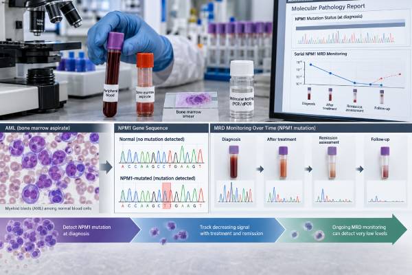
An NPM1 mutation test looks for acquired changes in the nucleophosmin 1 gene that define one of the largest molecular subgroups of acute myeloid leukemia (AML). NPM1 mutations occur in roughly one-third of adult AML and are especially common when the leukemia has a normal karyotype. The result matters for diagnosis, European LeukemiaNet (ELN) risk classification, treatment planning, and measurable residual disease (MRD) monitoring. At diagnosis, an NPM1 mutation can establish a genetically defined AML entity when the rest of the case fits. During and after treatment, the same mutation provides a highly specific molecular marker that can be tracked by sensitive PCR-based methods. NPM1-mutated AML often has a favorable prognosis when FLT3-ITD is absent, but prognosis is not determined by NPM1 alone. Coexisting mutations, cytogenetics, age, treatment response, and especially MRD status can change the risk of relapse and influence decisions such as whether allogeneic stem cell transplantation should be considered in first remission.
- NPM1 mutation is an AML-defining genetic abnormality in current WHO and International Consensus Classification systems, although their blast-count thresholds differ.
- NPM1-mutated AML without FLT3-ITD is generally favorable risk under ELN 2022, while NPM1-mutated AML with FLT3-ITD is generally intermediate risk.
- NPM1 is one of the best molecular markers for AML MRD monitoring because the leukemia-specific mutation can be measured at very low levels.
- A falling or undetectable NPM1 MRD level after treatment is favorable, while persistent or rising molecular disease raises concern for relapse.
- A positive NPM1 result at diagnosis is usually somatic, not inherited, so it generally does not mean relatives carry the same mutation.
Table of Contents
- What the NPM1 Test Detects
- How NPM1 Helps Define AML
- How NPM1 Testing Is Performed
- How to Interpret NPM1 Mutation Status
- NPM1 and AML Prognosis
- NPM1 MRD Monitoring
- Next Steps After an NPM1 Result
What the NPM1 Test Detects
NPM1 encodes nucleophosmin, a protein that normally shuttles between the nucleus and cytoplasm and participates in ribosome production, genomic stability, and other cellular functions. In AML, mutations near the end of NPM1 usually create a new nuclear-export signal and disrupt normal localization. The mutant protein accumulates abnormally in the cytoplasm, a hallmark of NPM1-mutated leukemia.
Most NPM1 mutations occur in exon 12, although clinically important variants can occur outside exon 12. The commonest mutation pattern is called type A, but type B, type D, and many less common insertions have also been described. The exact sequence matters for designing some MRD assays.
An NPM1 mutation is usually somatic, meaning it arose in the leukemic clone. It is not the same as a germline hereditary cancer variant. Rare inherited NPM1 disorders exist, but they are biologically different from the typical AML-associated mutation.
Testing for NPM1 is usually part of an AML molecular workup rather than an isolated test. A broad panel may also assess FLT3, IDH1, IDH2, CEBPA, TP53, and other genes. The companion FLT3 mutation result is especially important because it changes the ELN risk category assigned to NPM1-mutated AML.
How NPM1 Helps Define AML
NPM1 mutation is more than a prognostic marker: it helps define the leukemia itself. Both the fifth edition of the World Health Organization classification and the 2022 International Consensus Classification recognize NPM1-mutated AML as a distinct genetically defined entity.
The classifications differ in how they handle blast percentage. The WHO fifth edition does not require the traditional 20% blast threshold when an AML-defining NPM1 mutation is present and the findings support the diagnosis. The International Consensus Classification generally uses a 10% blast threshold for AML with NPM1 mutation. This difference can affect the diagnostic label in a patient with an NPM1-mutated myeloid neoplasm and a relatively low blast count.
This is why an NPM1 result should not be interpreted separately from the marrow. Pathologists combine:
- Bone marrow and peripheral blood blast percentage
- Cell morphology
- Flow-cytometry immunophenotype
- Chromosome analysis and FISH when appropriate
- NPM1 and other molecular findings
- Clinical history, including prior cytotoxic therapy or a prior myeloid disorder
NPM1-mutated AML often shows monocytic or myelomonocytic differentiation, but its appearance is variable. Some cases have few CD34-positive blasts, which can make flow-based MRD more challenging than in other AML subtypes.
A leukemia flow cytometry panel helps define the abnormal cell population, while NPM1 testing supplies the leukemia-defining molecular information.
How NPM1 Testing Is Performed
At diagnosis, NPM1 mutation status can be determined from bone marrow or peripheral blood when enough leukemic cells are present. Bone marrow is often preferred because it directly samples the main site of AML, but blood can be adequate when circulating blasts are abundant.
Several laboratory methods are used.
DNA sequencing
Targeted next-generation sequencing can detect NPM1 variants along with many other AML genes. This is useful for establishing the complete molecular profile at diagnosis. A sequencing report may give the exact DNA and protein change plus a variant allele fraction.
PCR-based mutation testing
Targeted PCR methods can rapidly detect common NPM1 mutations. Some assays are designed for diagnostic identification, while others are optimized for much more sensitive MRD measurement.
Immunohistochemistry
Because mutant NPM1 protein accumulates in the cytoplasm, immunohistochemistry can show abnormal cytoplasmic NPM1 staining in tissue or marrow biopsy sections. This can be valuable in difficult cases such as myeloid sarcoma. Molecular confirmation is still important when the exact mutation is needed for risk classification or MRD assay design.
There is usually no special preparation for the molecular test itself. Fasting is not required. Turnaround time can range from a few days for targeted testing to one or more weeks for a comprehensive sequencing panel.
Diagnostic testing and MRD testing are not the same
A routine NGS panel may reliably detect a mutation present in 5% or more of sequencing reads, depending on assay validation. MRD testing needs much greater sensitivity because clinically meaningful residual leukemia can exist far below that level. For this reason, a test that is “negative” by routine sequencing after treatment may still be positive by a dedicated NPM1 quantitative PCR assay.
The report should always be read for the assay’s limit of detection and whether the test was intended for diagnosis or MRD.
How to Interpret NPM1 Mutation Status
At diagnosis, the most important question is whether a pathogenic AML-associated NPM1 mutation is detected or not detected. During follow-up, interpretation becomes quantitative: how much NPM1-mutated material remains, how that level changed from baseline, and at what treatment time point it was measured.
| Result | Usual meaning | Important context |
|---|---|---|
| NPM1 mutation detected at diagnosis | Supports/defines NPM1-mutated AML when clinicopathologic criteria fit | FLT3, cytogenetics, and other features are still needed for risk classification |
| NPM1 mutation not detected at diagnosis | The leukemia is not classified as NPM1-mutated by that assay | Other AML-defining genetic abnormalities should be evaluated |
| NPM1 MRD undetectable | No mutant transcript/DNA detected above the assay limit | This is favorable but does not guarantee cure |
| NPM1 MRD persistent | Residual molecular leukemia remains | Risk depends on level, time point, trend, and treatment protocol |
| NPM1 MRD rising after prior decline | Possible molecular relapse or impending hematologic relapse | Usually prompts confirmation and urgent hematology review |
Variant allele fraction at diagnosis
The diagnostic NPM1 VAF reflects the mixture of leukemic and non-leukemic cells in the sample and the clonal structure of the leukemia. It is not a stand-alone severity score. A person with 40% NPM1 VAF does not automatically have twice the disease burden or twice the risk of someone with 20%.
The exact mutation sequence should be retained in the medical record because it can be used to design or select a sensitive MRD assay later. If care is transferred between hospitals, providing the original molecular report can prevent unnecessary repeat diagnostic work and helps the new laboratory confirm that its MRD assay targets the same NPM1 variant. This is particularly useful for uncommon non-type-A mutations.
NPM1 and AML Prognosis
NPM1-mutated AML is often described as “favorable,” but that shorthand is incomplete. NPM1 mutation is favorable only within a broader genetic and response context.
Under ELN 2022 risk stratification:
- NPM1-mutated AML without FLT3-ITD is generally placed in the favorable-risk group.
- NPM1-mutated AML with FLT3-ITD is generally placed in the intermediate-risk group; the older FLT3-ITD allelic-ratio distinction is no longer used in ELN 2022.
Other cytogenetic or molecular abnormalities can modify practical risk, and emerging evidence continues to refine how co-mutations should be interpreted. Recent large studies show that DNMT3A, WT1, certain NPM1 mutation types, adverse cytogenetics, and other features can identify higher-risk subsets, but post-treatment MRD often provides stronger prognostic information than the baseline mutation pattern alone.
That is why two patients with the same NPM1/FLT3 category can have very different treatment plans after induction. One may rapidly clear NPM1 MRD and remain in a low-relapse-risk group. Another may have persistent molecular disease and be considered for intensified post-remission therapy or allogeneic transplantation.
A minimal residual disease test for leukemia therefore becomes a central part of risk refinement rather than an optional add-on.
NPM1 does not determine treatment by itself
Initial AML treatment depends on age, fitness, comorbidities, disease genetics, and whether intensive or lower-intensity therapy is appropriate. NPM1 status can influence the treatment strategy, but it is not a single-drug companion diagnostic in the way some target mutations are.
NPM1-mutated leukemia also has a characteristic dependency on HOX/MEIS signaling. This biology has become therapeutically important because menin inhibitors can disrupt the transcriptional program that supports NPM1-mutated AML. Their role continues to expand, particularly in relapsed or refractory disease and clinical-trial settings.
NPM1 MRD Monitoring
NPM1 is one of the strongest molecular MRD targets in AML because the mutation is leukemia-specific, usually abundant at diagnosis, and often stable through early relapse. Sensitive quantitative reverse-transcription PCR (qRT-PCR) can measure mutant NPM1 RNA over several orders of magnitude.
Another advantage is that NPM1 mutations are generally not typical age-related clonal hematopoiesis markers. Mutations such as DNMT3A, TET2, and ASXL1 can persist in remission because a preleukemic clone remains even when AML has been eradicated, which complicates their use as sole MRD targets. Persistent NPM1, by contrast, is much more closely linked to residual leukemia. That makes the marker especially useful when marrow morphology has returned to normal but a small molecular clone remains.
MRD results before allogeneic transplant are also clinically important. Patients entering transplant with detectable NPM1 MRD generally have a higher relapse risk than those with deep molecular remission, although transplantation can still be curative. A positive pretransplant result may influence conditioning intensity, post-transplant surveillance, donor planning, or consideration of maintenance and clinical trials. The exact strategy varies by age, comorbidities, donor type, and institutional protocol.
The laboratory usually compares the NPM1 signal with a control gene such as ABL1 and reports a ratio, percentage, log reduction from diagnosis, or another validated measure. Results must be compared using the same specimen type and method whenever possible.
Bone marrow versus blood
Bone marrow is generally more sensitive for detecting low-level disease. Peripheral blood is easier to collect and can be useful at defined time points or during serial surveillance, but a negative blood result may not be as sensitive as a negative marrow result.
ELN MRD guidance uses specific time points during and after therapy because the meaning of a positive result changes over time. An early decline after induction is not interpreted the same way as persistent disease at the end of consolidation.
What counts as MRD positive?
There is no single universal number that should be copied from one laboratory report to another. ELN recommendations have defined molecular response categories and clinically relevant thresholds. For example, persistent NPM1 transcripts at 2% or higher in bone marrow at the end of chemotherapy have been used as a high-risk signal that may support considering allogeneic transplantation. Very low-level molecular positivity below such thresholds can have a different meaning, particularly if it is stable and not rising.
The trend is crucial. A one-time low positive result may be repeated for confirmation, while a reproducible increase of one log or more from a prior low level can indicate molecular progression depending on the protocol.
Molecular relapse can precede morphologic relapse
NPM1 MRD can become detectable or rise weeks to months before blasts again meet morphologic relapse criteria. This lead time can create an opportunity for confirmatory testing, donor planning, clinical-trial evaluation, or preemptive treatment strategies in selected patients.
However, MRD should not be interpreted without the full clinical picture. Rare cases can lose the original NPM1 mutation at relapse or evolve in ways that make a single marker less reliable, so marrow morphology, flow cytometry, and broader molecular testing remain important when relapse is suspected.
Next Steps After an NPM1 Result
At diagnosis, an NPM1-positive result should trigger a complete AML risk assessment rather than a simple “good prognosis” label. The oncology team usually reviews FLT3-ITD, chromosome findings, other co-mutations, age, performance status, and whether the patient is eligible for intensive treatment.
During treatment, the emphasis shifts to molecular response. Useful questions include:
- What was the exact NPM1 mutation at diagnosis?
- Is the current test a dedicated MRD assay or a routine sequencing panel?
- Was the sample blood or bone marrow?
- How much has the NPM1 signal fallen from baseline?
- Is the result undetectable, low-level positive, persistently high, or rising?
- Does the result meet the treatment protocol’s threshold for changing post-remission therapy?
Does NPM1 positivity mean transplantation is required?
No. Many patients with favorable-risk NPM1-mutated AML who achieve deep molecular remission do not automatically undergo allogeneic transplantation in first remission. Persistent MRD, FLT3-ITD, adverse genetic features, treatment tolerance, donor availability, and relapse risk all influence that decision.
Can NPM1 be used after transplant?
Yes. When the original leukemia carried a trackable NPM1 mutation, molecular testing can be used before and after allogeneic transplantation to assess residual or recurrent disease. The frequency and specimen type depend on the transplant program and clinical situation.
What does an undetectable result mean?
Undetectable NPM1 MRD is a strong favorable sign, but it means below the assay’s detection limit, not absolute proof that no leukemia cell remains anywhere. Continued surveillance is still needed.
NPM1 testing is unusually valuable because the same mutation can answer different questions over time: it can help define AML at diagnosis, contribute to baseline risk classification, measure treatment response, and provide early warning of molecular relapse. The most useful interpretation always includes the exact assay, treatment time point, specimen source, FLT3 status, and MRD trend. Serial results are more informative than an isolated number because the direction and speed of change can alter the clinical meaning and the urgency of timely follow-up testing.
References
- Diagnosis and management of AML in adults: 2022 recommendations from an international expert panel on behalf of the ELN. 2022 (Guideline)
- 2021 Update on MRD in acute myeloid leukemia: a consensus document from the European LeukemiaNet MRD Working Party 2021 (Position Statement)
- Criteria for Diagnosis and Molecular Monitoring of NPM1-Mutated AML 2024 (Review)
- Biology and Management of Acute Myeloid Leukemia With Mutated NPM1 2025 (Review)
- Molecular, clinical, and therapeutic determinants of outcome in NPM1-mutated AML 2024
Disclaimer
This article provides general information about NPM1 testing in AML and does not replace individualized hematology, pathology, or transplant advice. NPM1 prognosis and MRD results depend on FLT3 status, other genetic findings, the assay and specimen used, treatment phase, and serial trends. Treatment or transplant decisions should be made with an AML specialist using the full clinical record.





