A confirmed parasite in urine is an abnormal finding, but not every object that looks parasitic under a microscope represents a true urinary infection. The two best-known organisms encountered in urine testing are Schistosoma haematobium, whose eggs can be passed in urine after freshwater exposure in endemic regions, and Trichomonas vaginalis, a sexually transmitted protozoan that may be seen as a motile organism in fresh urine sediment. Their diagnosis, risks, and follow-up are very different. Schistosomiasis can cause hematuria, urinary tract scarring, hydronephrosis, and long-term bladder complications, while trichomoniasis primarily affects the genital and lower urinary tract and is now diagnosed more reliably with nucleic-acid amplification tests than routine microscopy. Contamination, artifacts, and rare organisms can complicate interpretation. When a laboratory reports a parasite, clinicians confirm what was seen, review travel, freshwater and sexual exposure, assess symptoms, and use organism-specific testing before treatment whenever possible.
- Any confirmed pathogenic parasite in urine is abnormal, but microscopy artifacts and contamination can mimic parasites and may require confirmation.
- Schistosoma haematobium eggs in urine suggest urogenital schistosomiasis, especially after freshwater exposure in an endemic region and with blood in urine.
- Trichomonas vaginalis may be seen in fresh urine, but nucleic-acid amplification testing is more sensitive than routine wet-mount microscopy.
- The right follow-up depends on the organism: schistosomiasis evaluation focuses on exposure and urinary complications, while trichomoniasis requires STI-focused testing and partner management.
- Fever, severe flank pain, inability to urinate, heavy bleeding, pregnancy with significant symptoms, or severe systemic illness needs prompt medical assessment.
Table of Contents
- What Parasites in Urine Microscopy Mean
- Schistosoma haematobium in Urine
- Trichomonas vaginalis in Urine
- Other Parasites, Contamination, and Microscopy Mimics
- Testing and Confirmation After a Parasite Is Reported
- Treatment, Follow-Up, and When to Seek Care
What Parasites in Urine Microscopy Mean
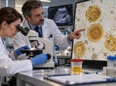
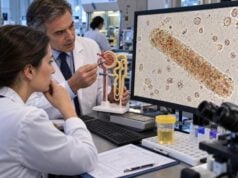
Routine urine microscopy is designed mainly to identify cells, casts, crystals, bacteria, yeast, and other sediment particles. Parasites are uncommon in most routine laboratories, particularly in populations without relevant travel, residence, freshwater, or sexual exposure histories. When a suspected organism appears, the finding deserves attention because a true parasite may require specific treatment, but the image should also be interpreted cautiously.
A parasite can reach a urine sample in several ways. It may truly involve the urinary tract, as with Schistosoma haematobium. It may come from the genital tract and mix with urine during voiding, as often happens with Trichomonas vaginalis. Less commonly, eggs or organisms from the perineal area, stool, skin, or collection container may contaminate the specimen.
This distinction matters because “parasites present” is not a complete diagnosis and should never be interpreted without organism-level clinical context when available. The report should ideally name the suspected organism or describe the morphology. Exposure history is then matched to the finding.
Timing is also part of that history. Schistosomiasis may persist for years after exposure, so a person who has not traveled recently can still have clinically relevant infection acquired long ago. Trichomoniasis, by contrast, is approached as a sexually transmitted infection in which current or recent partner exposure and reinfection are central. Asking only “Have you traveled recently?” or only “Do you have a UTI?” can miss these distinctions. Clinicians may also ask about eosinophilia, genital symptoms, pregnancy, immunosuppression, prior antiparasitic treatment, and whether symptoms began before or after travel. The more closely the epidemiology fits the organism’s biology, the more confidence a microscopy observation carries. Freshwater contact in Africa or another endemic region points toward schistosomiasis; sexual exposure and genital symptoms point toward trichomoniasis. A finding that does not fit either morphology or exposure may need repeat examination or specialist review.
Parasite findings also need to be separated from a bacterial urinalysis for UTI. Pyuria, blood, or dysuria can occur with both parasitic and bacterial disease, but standard antibacterial treatment does not eradicate schistosomes or T. vaginalis.
A complete sediment report may include red blood cells, white blood cells, epithelial cells, bacteria, yeast, crystals, and organisms. Urine sediment microscopy helps describe the pattern, while organism-specific tests provide confirmation.
Schistosoma haematobium in Urine
Schistosoma haematobium is a blood fluke that causes urogenital schistosomiasis. Infection begins when larval parasites released by freshwater snails penetrate human skin during contact with contaminated freshwater. Adult worms eventually live in veins around the bladder and pelvic organs. Their eggs cross into the urinary tract and can be excreted in urine.
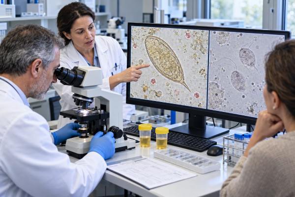
The classic egg is elongated and has a terminal spine. This morphology helps distinguish S. haematobium from Schistosoma mansoni, whose eggs usually have a prominent lateral spine and are more commonly detected in stool. Identification still requires experience because debris, damaged eggs, or atypical orientations can be misleading.
Blood in urine is a characteristic feature of urogenital schistosomiasis. Microscopic hematuria may be found on screening, while some people notice red or brown urine, especially later in the day. White blood cells and protein can also appear. A red blood cell finding in urine is nonspecific, so travel and exposure history are crucial.
Symptoms can include:
- pain or burning with urination;
- urinary frequency;
- lower abdominal or pelvic discomfort;
- visible or microscopic blood in urine;
- genital symptoms in some patients;
- later complications from chronic inflammation and scarring.
Egg deposition triggers inflammation and granuloma formation. Over years, repeated infection can cause fibrosis, bladder-wall calcification, ureteral narrowing, hydronephrosis, and impaired kidney drainage. Chronic urogenital schistosomiasis is also associated with increased risk of squamous cell carcinoma of the bladder in endemic settings.
Microscopy of urine is a traditional diagnostic method, often using filtration to concentrate eggs. Egg shedding can fluctuate, so one negative specimen does not exclude infection, particularly in people with a light parasite burden. Historical collection strategies often emphasize midday urine and collection after activity because egg excretion may be higher then, but current diagnostic approaches can also use serology, antigen tests, or molecular tests depending on availability and whether the question is active infection, prior exposure, or low-intensity disease.
For travelers, the exposure can be remote in time. Symptoms or eggs may appear after return from an endemic area. The incubation period and chronic course mean that a trip taken months or years earlier can still matter. Asking only about recent travel can therefore miss the relevant exposure. People who grew up in endemic regions may have had repeated freshwater contact and may not remember one specific event.
Blood in urine after freshwater exposure is particularly important because it can be mistaken for a routine UTI or stone. Repeated antibacterial courses will not eliminate schistosomiasis. When the exposure history is compatible, clinicians should consider parasite-specific testing even if bacteria are also found, because more than one urinary condition can coexist. A clinician should know not just the destination but whether the person swam, waded, bathed, or worked in freshwater lakes, rivers, canals, or streams. Properly chlorinated pools and seawater are not typical transmission sources.
Trichomonas vaginalis in Urine
Trichomonas vaginalis is a flagellated protozoan transmitted through sexual contact. It primarily infects the urogenital tract. The organism may enter a voided urine sample from the urethra or genital tract and can sometimes be recognized during fresh wet-mount microscopy by its characteristic motility.
In women, trichomoniasis can cause vaginal discharge, itching, irritation, odor, pain with urination, or discomfort during sex. Many infections are asymptomatic. In men, infection is often asymptomatic but may cause urethral irritation, dysuria, or discharge. Symptoms overlap with bacterial UTI and other sexually transmitted infections.
A routine urine microscopy report of motile trichomonads can be a useful clue, but wet-mount microscopy has limited sensitivity. A negative microscope result therefore cannot reliably exclude trichomoniasis when symptoms or exposure make it plausible. This is especially important if the specimen was delayed, chilled, or not examined specifically for motility. The more sensitive molecular test should be chosen when the diagnosis will affect treatment or partner management. The organisms lose motility as the specimen cools or sits, so delays reduce detection. Modern nucleic-acid amplification tests (NAATs) are more sensitive and are preferred for diagnostic confirmation when available. Depending on the validated assay, specimens may include vaginal swabs, urine, or other genital samples.
A dedicated Trichomonas urine test answers a different question from routine sediment microscopy. It detects organism-specific genetic material rather than relying on someone to see a moving protozoan.
Trichomoniasis matters even when symptoms are mild because infection can persist and be transmitted to sexual partners. It is associated with increased risk of acquiring or transmitting HIV and can have reproductive and pregnancy-related implications. Pregnancy does not make a microscopy finding automatically dangerous, but symptomatic infection should be evaluated promptly because treatment choices and obstetric context matter.
The presence of white blood cells without typical bacterial findings may occur with urethritis or genital infection, but this pattern is not specific. A white blood cell finding in urine should therefore be interpreted with STI testing when the sexual history and symptoms support it.
Other Parasites, Contamination, and Microscopy Mimics
True urinary parasitosis beyond schistosomiasis is uncommon, especially in regions where the relevant parasites are not endemic. Rare reports involve other organisms, but an unexpected result should be confirmed before treatment because contamination is a frequent alternative explanation.
For example, Enterobius vermicularis eggs can contaminate urine from the perineal area. This does not necessarily mean the urinary tract itself is infected. Stool material, vaginal secretions, or poorly collected specimens can also introduce helminth eggs or protozoa that originated outside the urinary system.
Artifacts can be surprisingly convincing. Fibers may resemble worms, plant material can look like eggs, and mucus strands can appear elongated or coiled. Air bubbles, starch granules, pollen, skin cells, and crystal aggregates can create geometric shapes that are unfamiliar to someone who does not routinely identify parasites.
A useful distinction is whether the suspected organism has diagnostic structures. Schistosome eggs have a shell and characteristic spine. T. vaginalis has a specific cellular shape and, in fresh preparations, jerky motility. A simple thread that does not show anatomy is more likely to be mucus or a fiber than a helminth.
Sample collection also changes the probability of contamination. Midstream clean-catch urine reduces material from skin and genital surfaces but cannot eliminate it. A catheterized specimen can reduce some external contamination, although catheterization is not justified solely to confirm most parasite findings and has its own risks.
The patient’s geography and exposures should make biological sense. An apparent S. haematobium egg in a person with no freshwater exposure in an endemic region deserves expert review because misidentification may be more likely. Conversely, a compatible egg in a person who spent years swimming in endemic freshwater should not be dismissed simply because the infection was acquired long ago.
When the morphology is unusual, clinical laboratories may send images or specimens to a reference laboratory, parasitologist, microbiologist, or public-health laboratory. The laboratory may also ask for a second specimen collected under more controlled conditions. Reproducibility strengthens confidence: seeing the same characteristic organism again is more persuasive than one isolated, poorly focused object in a contaminated sample. This is particularly valuable when treatment has meaningful side effects or when identifying the species affects broader evaluation.
Testing and Confirmation After a Parasite Is Reported
The first step after an unexpected parasite report is to identify exactly what the laboratory saw. “Parasite present” without a species or description should prompt clarification. The next steps are organism-specific.
For suspected urogenital schistosomiasis, testing may include repeated urine microscopy or filtration for eggs, urinalysis for blood and protein, serology in travelers or people from endemic areas, and molecular or antigen tests where available. The sensitivity of urine microscopy falls when infection intensity is low, so a negative test cannot always end the evaluation.
Imaging may be used when symptoms, long-standing exposure, hematuria, or abnormal kidney function raise concern for urinary tract damage. Ultrasound can identify bladder-wall changes, hydronephrosis, and some structural complications without radiation. More detailed imaging or cystoscopy is reserved for selected cases, particularly persistent hematuria, obstruction, or concern for malignancy.
For suspected trichomoniasis, an NAAT is usually the most useful confirmatory test. Depending on sex and assay, urine or genital swabs may be used. Because co-infection is possible, clinicians often consider testing for other sexually transmitted infections based on exposure, symptoms, pregnancy, and local guidelines.
Testing should answer the clinical question rather than simply repeat microscopy indefinitely. A good evaluation asks:
- Is the organism identification credible? Review morphology, sample quality, and laboratory expertise.
- Does the exposure fit? Consider freshwater contact, sexual exposure, residence or travel, and timing.
- Is there evidence of organ involvement? Check hematuria, urinary obstruction, kidney function, genital symptoms, or other relevant findings.
- What test is most sensitive for this organism? Microscopy, NAAT, serology, antigen testing, or imaging may play different roles.
Treatment should generally follow confirmed or strongly supported diagnosis. Empiric treatment may sometimes be appropriate when the clinical likelihood is high or follow-up is difficult, but that decision depends on the organism and the patient’s circumstances.
Treatment, Follow-Up, and When to Seek Care
Schistosomiasis and trichomoniasis require different medications. Praziquantel is the standard antiparasitic treatment for schistosomiasis. Timing and dosing depend on species and clinical context, and repeat treatment or follow-up testing may be used in some patients. Treatment kills adult worms but does not instantly reverse established scarring or obstruction, so people with chronic urinary complications may also need urologic care.
Trichomoniasis is treated with a nitroimidazole drug such as metronidazole or tinidazole according to current STI guidance. Sexual partners need appropriate evaluation and treatment to prevent reinfection, and patients should avoid sex until the treatment requirements for them and their partners have been met. Follow-up or retesting recommendations differ by sex and situation and should follow current guidance.
Do not use antibiotics intended for routine bacterial UTI as a substitute for parasite-specific treatment. Likewise, do not take veterinary dewormers, leftover antiparasitic medicines, or unverified herbal products after a vague microscopy report. Correct organism identification matters.
After schistosomiasis treatment, persistent hematuria, recurrent urinary symptoms, abnormal imaging, or impaired kidney function may require further assessment for chronic urinary tract damage. People with heavy or long-standing exposure may need specialist follow-up even after parasites are treated.
After trichomoniasis treatment, persistent symptoms can reflect reinfection, incomplete treatment, resistance, or another infection. Reinfection is common enough that partner treatment and recommended retesting are important parts of care, not optional extras. Condom use lowers transmission risk but does not replace treatment of an established infection. Because many infected people have no symptoms, a partner can unknowingly reintroduce the organism after one person has been treated. Repeat testing should follow recommended timing because nucleic-acid tests can sometimes detect residual genetic material too soon after therapy.
Seek prompt care for visible blood in urine that persists, progressive urinary difficulty, recurrent fever, pelvic pain, or significant symptoms during pregnancy. Seek urgent or emergency care for fever with severe flank pain, inability to pass urine, heavy bleeding with clots, fainting, confusion, repeated vomiting, or rapidly worsening illness. These features can signal obstruction, kidney infection, severe bleeding, or another condition beyond uncomplicated parasitic infection.
The main goal is not to treat the word “parasite” on a report. It is to determine whether a true organism is present, identify it accurately, confirm the diagnosis with the best available test, and assess the organ system at risk. That approach prevents both missed parasitic disease and unnecessary treatment of microscopy artifacts.
References
- Diagnostic tests for human Schistosoma mansoni and Schistosoma haematobium infection: a systematic review and meta-analysis 2024 (Systematic Review)
- Schistosomiasis diagnosis: Challenges and opportunities for elimination 2024 (Review)
- WHO guideline on control and elimination of human schistosomiasis 2022 (Guideline)
- Schistosomiasis 2026 (Review)
- Molecular Diagnosis of Trichomonas vaginalis in Women and Men: A Systematic Review 2026 (Systematic Review)
- Recommendations for the treatment of Trichomonas vaginalis, Mycoplasma genitalium, Candida albicans, bacterial vaginosis and human papillomavirus (anogenital warts) 2024 (Guideline)
Disclaimer
This article provides general information about parasites reported in urine microscopy and does not diagnose schistosomiasis, trichomoniasis, or another infection. Suspected parasites should be interpreted with exposure history, symptoms, specimen quality, and organism-specific confirmatory testing. Severe flank pain with fever, urinary blockage, heavy bleeding, or rapidly worsening illness requires urgent medical care.