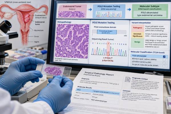
A POLE mutation test for endometrial cancer looks for specific pathogenic changes in the exonuclease, or proofreading, domain of the POLE gene. When a true pathogenic POLE exonuclease-domain mutation is present, the tumor is classified as POLE-mutated, one of the four major molecular subtypes of endometrial cancer. These tumors accumulate extremely large numbers of DNA mutations because the DNA polymerase epsilon proofreading system is impaired. Despite sometimes appearing high grade or aggressive under the microscope, POLE-mutated endometrial cancers generally have an excellent prognosis compared with other molecular groups. The key word is pathogenic: not every POLE variant qualifies. A variant of uncertain significance, a change outside the relevant exonuclease domain, or a variant without established proofreading impact should not automatically be used to assign the favorable POLE-mutated subtype. POLE testing is usually performed on tumor tissue and interpreted with mismatch-repair testing, p53 immunohistochemistry, histology, stage, and clinical risk factors.
- A clinically meaningful POLE result usually requires a pathogenic exonuclease-domain mutation: not every sequence change in the POLE gene defines the POLE-mutated subtype.
- Common pathogenic hotspots include p.P286R and p.V411L: several other validated exonuclease-domain mutations also occur.
- POLE-mutated endometrial cancer generally has an excellent prognosis: this can remain true even when the tumor looks high grade under the microscope.
- A POLE variant of uncertain significance is not equivalent to a positive pathogenic result: treatment or risk classification should not be based on a VUS alone.
- Most tumor POLE mutations are somatic, not inherited: rare germline POLE variants can cause polymerase proofreading-associated polyposis and require separate genetics evaluation.
Table of Contents
- What POLE Testing Measures in Endometrial Cancer
- Which POLE Mutations Count as Pathogenic
- How POLE Defines an Endometrial Cancer Molecular Subtype
- POLE-Mutated Prognosis and Treatment Implications
- POLE in Multiple-Classifier Tumors
- Somatic vs Germline POLE Mutations
- Testing Methods, Limitations, and Next Steps
What POLE Testing Measures in Endometrial Cancer
POLE encodes the catalytic subunit of DNA polymerase epsilon, an enzyme that copies DNA during cell division. The protein has a proofreading function that checks newly synthesized DNA and removes incorrectly inserted bases. This proofreading activity is concentrated in the exonuclease domain.
When a pathogenic mutation disrupts that domain, replication errors accumulate at an unusually high rate. POLE-mutated tumors can therefore have an ultramutated genome, with far more single-base substitutions than most other endometrial cancers. The high mutation burden also generates many abnormal proteins, or neoantigens, that may make the tumor particularly visible to the immune system.
The clinical test is usually performed on DNA extracted from a tumor biopsy, curettage, or hysterectomy specimen. Laboratories may use targeted next-generation sequencing, a focused POLE assay, or a broader tumor panel that includes the relevant exonuclease-domain exons. The report should identify the exact DNA and protein change rather than simply saying “POLE detected.”
POLE sequencing is different from immunohistochemistry. There is no widely used tissue stain that reliably substitutes for sequencing of pathogenic POLE exonuclease-domain variants. By contrast, p53 and mismatch-repair status can often be assessed by IHC. A practical endometrial cancer molecular biomarker panel therefore uses a combination of sequencing and immunostains.
The result is also different from tumor mutational burden (TMB). A pathogenic POLE mutation often causes very high TMB, but high TMB alone does not prove that a tumor belongs to the POLE-mutated molecular group. Other mechanisms, including mismatch-repair deficiency, can also increase mutation burden.
The central interpretive question is not “Does the tumor have any POLE change?” It is “Does the tumor have a validated pathogenic proofreading-domain mutation that defines the POLE-mutated subtype?”
Which POLE Mutations Count as Pathogenic
Not all POLE variants have the same biologic effect. This is one of the most important details in reading a POLE report.
The best-established pathogenic endometrial cancer variants occur in the exonuclease domain and alter proofreading. Two common hotspot mutations are p.P286R and p.V411L. Other recurrent pathogenic changes include variants at residues such as S297, S459, and several additional positions supported by molecular and clinical evidence.
A laboratory may classify a variant as:
- pathogenic;
- likely pathogenic;
- variant of uncertain significance (VUS);
- likely benign; or
- benign.
A pathogenic or accepted likely pathogenic exonuclease-domain variant can support assignment to the POLE-mutated subtype. A VUS should not. The reason is practical: if every rare POLE change were treated as favorable, some biologically ordinary or aggressive tumors could be misclassified and undertreated.
Pathologists and molecular laboratories can use several clues when evaluating a less common variant. These include its location within the exonuclease domain, prior occurrence in established POLE-mutated cancers, the tumor’s mutation spectrum, very high TMB, characteristic base-substitution signatures, and published functional evidence. Specialized classification systems have been developed to distinguish truly proofreading-defective variants from incidental changes.
A POLE mutation outside the exonuclease domain generally does not automatically define the molecular subtype. Such variants can be passengers, have another function, or remain uncertain. The exact laboratory interpretation should be followed rather than relying on the gene name alone.
This distinction is especially important when a broad sequencing report lists dozens of tumor alterations. A patient may see “POLE” in the variant table even though the report’s final molecular interpretation says the tumor is not POLE-mutated. Those statements can both be correct if the detected variant is not a recognized pathogenic exonuclease-domain mutation.
If the report lists a VUS, the usual approach is not to change treatment based on that finding alone. Variant classifications can evolve as more evidence accumulates, so the laboratory may update the interpretation later.
How POLE Defines an Endometrial Cancer Molecular Subtype
Modern endometrial cancer classification divides tumors into four broad molecular groups: POLE-mutated, mismatch-repair deficient (MMRd), p53-abnormal (p53abn), and no specific molecular profile (NSMP). These groups add biologic and prognostic information beyond conventional histologic type and grade.
The POLE-mutated group is distinctive. Tumors can look alarming under the microscope: they may be high grade, have marked nuclear atypia, brisk mitotic activity, or tumor-infiltrating lymphocytes. Historically, morphology alone could place some of these cancers in a high-risk category. Molecular testing showed that many have surprisingly favorable outcomes.
This is why correct POLE classification matters. It prevents the assumption that an aggressive appearance always predicts aggressive behavior.
The POLE result is interpreted alongside mismatch-repair IHC in endometrial cancer and p53 testing for endometrial cancer. A tumor can occasionally show more than one classifier feature, but a validated POLE mutation usually has priority in the final molecular assignment.
Molecular subtype does not erase stage. A small cancer confined to the uterus and a widely metastatic cancer remain clinically different even if both are POLE-mutated. However, subtype can refine recurrence-risk estimates within stage and histologic groups and may influence the intensity of adjuvant therapy.
POLE-mutated tumors are also immunologically interesting because their enormous mutation burden can create many neoantigens. This helps explain the frequent dense lymphocytic response seen microscopically and may contribute to their favorable natural history. It also provides a biologic rationale for sensitivity to immune checkpoint blockade in advanced disease, although treatment choices depend on the full regulatory and clinical setting rather than POLE status alone.
A pathology report may present the final result as “POLEmut” or “POLE-ultramutated.” Those labels should be reserved for qualifying pathogenic variants, not every POLE sequence alteration.
POLE-Mutated Prognosis and Treatment Implications
The most consistent clinical feature of POLE-mutated endometrial cancer is a very favorable prognosis on average. Systematic reviews and clinical cohorts have found low recurrence rates and strong survival outcomes relative to p53-abnormal cancers, even among tumors that would otherwise be considered high risk by grade or histology.
This finding has changed how clinicians think about adjuvant treatment. In the past, high-grade morphology could automatically push a patient toward chemotherapy and/or radiation. Molecularly integrated guidelines now consider whether a qualifying POLE mutation identifies a subgroup that may not benefit from the same treatment intensity as a biologically higher-risk tumor.
The exact recommendation still depends on stage and other pathologic features. De-escalation is most established or actively studied in earlier-stage disease; advanced or metastatic disease requires individualized oncology management. A favorable biomarker should never be interpreted as permission to ignore extrauterine spread.
Treatment planning may consider:
- tumor stage and residual disease;
- depth of myometrial invasion;
- lymphovascular-space invasion;
- cervical or adnexal involvement;
- lymph-node findings;
- histology and grade;
- the final molecular subtype;
- patient health and preferences.
For early-stage POLE-mutated tumors, the molecular result can materially lower the estimated recurrence risk. In selected guideline-defined situations, this may support omission or reduction of adjuvant treatment that would have been recommended based on morphology alone. The decision should be made with the current guideline version because molecularly integrated risk groups continue to evolve.
For recurrent or metastatic disease, the ultramutated phenotype can also be relevant to immunotherapy biology. However, a POLE mutation is not a stand-alone guarantee of response, and the oncology team may also assess mismatch repair, microsatellite instability, TMB, prior therapy, and approved treatment indications.
The important distinction is between prognostic and predictive value. POLE-mutated status is strongly prognostic of favorable outcomes in localized endometrial cancer. Its predictive role for choosing a specific drug is more context-dependent.
POLE in Multiple-Classifier Tumors
Some endometrial cancers meet criteria for more than one molecular group. A POLE-mutated tumor may also show mismatch-repair deficiency or an abnormal p53 pattern. These are called multiple-classifier tumors.
At first glance, a report that says both “POLE pathogenic mutation” and “p53 abnormal” can seem contradictory. It is not. Tumors evolve in steps, and a later subclone can acquire a second molecular abnormality after the original driver process has already shaped the cancer.
In current practical classification, a pathogenic POLE exonuclease-domain mutation generally takes precedence over p53 abnormality and over concurrent MMR deficiency for final subtype assignment. Outcome data suggest that POLE-driven tumors with secondary p53 abnormalities behave more like POLE-mutated cancers than like conventional p53-abnormal cancers.
A related clue is subclonal p53 staining. Instead of the entire tumor showing mutation-type p53, only a sharply defined area may show overexpression or null staining. This can support the idea that TP53 alteration arose later in tumor evolution.
The hierarchy matters because p53-abnormal cancers generally have a much less favorable prognosis. If any focal p53 abnormality automatically overrode POLE status, a biologically favorable tumor could be placed in the wrong risk group.
The same principle explains why complete molecular testing is more informative than ordering one marker at a time. A p53 stain performed alone may suggest high-risk biology, while subsequent POLE sequencing can fundamentally change the integrated interpretation.
Reports may phrase this as “multiple classifier, assigned to POLEmut according to molecular hierarchy.” Patients should focus on the final integrated molecular subtype and ask how it changes the treatment recommendation rather than trying to choose the “worst-looking” individual marker.
Somatic vs Germline POLE Mutations
Most pathogenic POLE exonuclease-domain mutations identified in endometrial tumors are somatic. That means the mutation arose in the cancer and is not present in every cell of the body. A somatic tumor result does not automatically mean children, siblings, or other relatives carry the same variant.
Rare germline POLE pathogenic variants can cause polymerase proofreading-associated polyposis, an inherited syndrome associated particularly with colorectal adenomas and colorectal cancer, and with increased risk of several other cancers in some families. Germline variants can also involve the related POLD1 gene.
The location and nature of the variant matter. The classic recurrent somatic endometrial cancer hotspots are not automatically equivalent to the germline variants seen in hereditary polyposis families. A molecular genetics professional can determine whether a tumor finding has enough evidence to justify confirmatory germline testing.
Germline testing may be considered when:
- the tumor report specifically flags the POLE variant as potentially germline;
- the patient has multiple colorectal adenomas or early-onset colorectal cancer;
- there is a family pattern of polyposis, colorectal cancer, endometrial cancer, or other suspicious tumors;
- paired tumor-normal sequencing suggests the variant is present in constitutional DNA.
If inherited risk is a concern, the appropriate test uses blood, saliva, or another normal-tissue source. Tumor-only sequencing cannot reliably establish inheritance because it analyzes a mixture of cancer-specific and constitutional DNA changes.
A VUS found in germline testing should not be treated as a confirmed hereditary syndrome. Family screening and preventive surgery decisions should be based on pathogenic or likely pathogenic findings plus clinical context, not uncertainty alone.
This germline/somatic distinction is similar to other gynecologic cancer genes: tumor molecular testing may reveal important clues, but hereditary risk requires a separate, carefully interpreted constitutional genetic assessment.
Testing Methods, Limitations, and Next Steps
POLE testing usually needs adequate tumor DNA. A preoperative endometrial biopsy can often provide enough material, which is useful because molecular classification may inform planning before or soon after hysterectomy. If the biopsy is scant, necrotic, or poorly preserved, the hysterectomy specimen may provide better tissue.
Different laboratories test different regions. A focused assay may cover only the most clinically important exonuclease-domain hotspots, while a larger next-generation sequencing panel may sequence broader portions of POLE and many other cancer genes. Broader coverage can discover rare variants but also increases the chance of finding a VUS.
When reviewing a result, check four details:
- Exact variant: the report should name the DNA and/or protein change.
- Location: confirm whether it lies in the exonuclease proofreading domain.
- Classification: pathogenic, likely pathogenic, VUS, likely benign, or benign.
- Integrated subtype: the pathologist or molecular report should state whether the tumor qualifies as POLE-mutated after considering MMR and p53.
If a result is “no mutation detected,” that generally means no qualifying variant was found in the regions the assay examined. It does not automatically place the tumor in NSMP; MMR and p53 results are still needed.
If a VUS is found, treatment should ordinarily follow the tumor’s other validated classifiers rather than assuming favorable POLE biology. In difficult cases, the laboratory may use mutational signature analysis, TMB, literature review, or expert molecular pathology consultation to refine the variant’s significance.
A failed test is also not the same as a negative test. “Insufficient DNA,” “low tumor content,” or “quality-control failure” means the laboratory could not make a reliable call. Another tissue block or specimen may be tested.
After a pathogenic POLE result, useful questions include: Does this variant meet accepted criteria for the POLE-mutated subtype? What are my MMR and p53 results? Does molecular classification change my adjuvant-treatment recommendation? Is the finding clearly somatic, or should I see a genetic counselor about possible germline testing? How does stage modify the otherwise favorable prognosis?
The most useful POLE result is one that is variant-specific, molecularly integrated, and tied to a clinical decision. The gene name alone is not enough.
Clinically, this distinction prevents inappropriate overclassification and overtreatment.
References
- Molecular testing for endometrial cancer: An SGO clinical practice statement 2023 (Position Statement)
- Molecular Classification of Endometrial Cancer and Its Impact on Therapy Selection 2024 (Review)
- Colombian consensus for the molecular diagnosis of endometrial cancer 2024 (Consensus)
- POLE-mutated endometrial cancer: new perspectives on the horizon? 2025 (Review)
- Prognostic Impact of POLE Exonuclease-Domain Mutations in Endometrial Cancer: A Systematic Review and Meta-Analysis 2026 (Systematic Review)
- Characterization of hotspot exonuclease domain mutations in the DNA polymerase ϵ gene in endometrial cancer 2022
Disclaimer
This article is for general education and does not classify an individual endometrial cancer or recommend treatment. POLE results should be interpreted by a pathology and oncology team using the exact variant, its pathogenicity, MMR and p53 findings, tumor stage, and current guidelines. A tumor POLE mutation is not automatically an inherited finding; germline risk requires separate evaluation when clinically indicated.