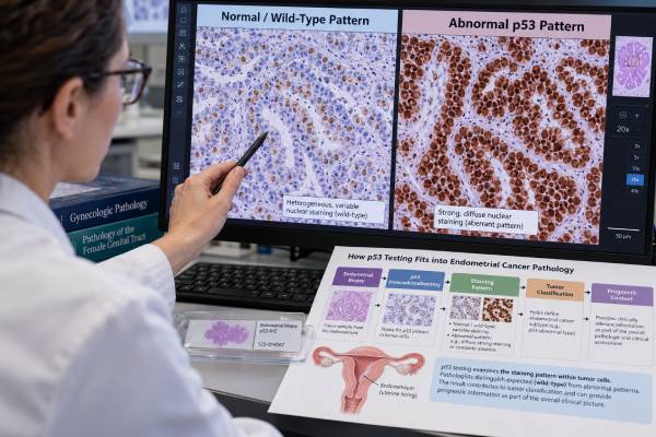
The p53 test for endometrial cancer is usually an immunohistochemistry (IHC) stain performed on tumor tissue to help classify the cancer’s molecular subtype. p53 IHC acts as a practical surrogate for many TP53 gene mutations. A normal, or “wild-type,” pattern shows variable staining from cell to cell. An abnormal, or “mutation-type,” pattern can appear as strong diffuse overexpression, complete absence of staining in tumor cells with intact internal controls, or less commonly abnormal cytoplasmic staining. In modern endometrial cancer classification, p53-abnormal tumors form one of four major molecular groups alongside POLE-mutated, mismatch-repair-deficient, and no-specific-molecular-profile tumors. The p53-abnormal group generally has the least favorable prognosis, but stage, histology, treatment, and overlapping molecular features still matter. An abnormal p53 stain is not an inherited cancer test and does not automatically mean a germline TP53 mutation. Results must be interpreted with morphology and the rest of the molecular workup.
- p53 IHC usually assesses tumor TP53 status indirectly: abnormal staining patterns strongly correlate with TP53 mutations when the stain is technically adequate.
- Wild-type p53 is heterogeneous: a mixture of weak, moderate, strong, and unstained nuclei is expected rather than uniform staining.
- Abnormal p53 has several patterns: diffuse overexpression, complete tumor-cell null staining, and cytoplasmic staining can all represent mutation-type results.
- p53-abnormal is a molecular endometrial cancer subtype: it generally carries higher recurrence risk than POLE-mutated or mismatch-repair-deficient groups.
- Classification follows a hierarchy when biomarkers overlap: a pathogenic POLE mutation or mismatch-repair deficiency can take precedence over p53 abnormality in multiple-classifier tumors.
Table of Contents
- What p53 Testing Measures in Endometrial Cancer
- Normal and Abnormal p53 IHC Patterns
- How p53 Fits Into Molecular Classification
- What p53-Abnormal Status Means for Prognosis and Treatment
- Multiple-Classifier Tumors and Subclonal p53 Staining
- Limitations, Pitfalls, and When TP53 Sequencing Helps
- How to Read the Report and Discuss Next Steps
What p53 Testing Measures in Endometrial Cancer
p53 is a tumor-suppressor protein encoded by the TP53 gene. Under normal conditions, p53 helps damaged cells pause the cell cycle, repair DNA, or undergo programmed cell death. When TP53 is altered in a way that disables this protective system, abnormal cells can survive and accumulate additional genomic damage.
In endometrial cancer, laboratories commonly assess TP53 indirectly with p53 immunohistochemistry. The stain shows how much p53 protein is present and where it is located in tumor cells. Certain staining patterns strongly correlate with underlying TP53 mutations, so IHC can serve as a relatively fast, inexpensive surrogate for sequencing.
This is not the same as a blood genetic test. p53 IHC is performed on the tumor and mainly reflects the biology of that cancer. An abnormal result does not by itself indicate Li-Fraumeni syndrome or an inherited TP53 mutation. Germline TP53 testing is a separate test considered only when personal and family history or specific tumor findings make inherited disease plausible.
p53 IHC also has a different role from serum tumor markers. It does not rise and fall with tumor burden and is not used as a blood marker for recurrence. Instead, it helps classify the tumor at diagnosis and can contribute to risk stratification and treatment planning.
The stain is usually interpreted together with mismatch-repair IHC and POLE mutation testing in endometrial cancer. These biomarkers form the practical backbone of contemporary molecular classification.
Because the classification can alter prognosis and, increasingly, adjuvant treatment decisions, the quality of the pathology specimen and interpretation matters. Tumor tissue from biopsy, curettage, or hysterectomy may be used, provided there is enough viable cancer and appropriate staining controls.
Normal and Abnormal p53 IHC Patterns
A common misconception is that “more p53 staining” always means a worse or more abnormal result. Pathologists instead look at the distribution and pattern of staining.
A wild-type p53 pattern is heterogeneous. Some tumor nuclei are unstained, some stain weakly, and others show moderate or stronger staining. The overall appearance is variable rather than uniform. This pattern generally suggests the TP53 gene is not carrying a typical pathogenic mutation that produces a mutation-type IHC pattern.
Three major abnormal patterns are recognized:
- Overexpression pattern: very strong nuclear staining in nearly all tumor cells, often described as diffuse or involving more than about 80% of nuclei. This commonly reflects a TP53 missense mutation that produces a stable abnormal protein.
- Null pattern: complete absence of tumor-cell nuclear staining while non-tumor internal control cells stain normally. This can occur with truncating, splice-site, or other variants that prevent production of detectable p53 protein.
- Cytoplasmic pattern: abnormal staining is concentrated in the cytoplasm, often with little expected nuclear pattern. This less common pattern can reflect TP53 alterations that affect protein localization.
The internal controls are essential for interpreting a null pattern. If both tumor and normal stromal or inflammatory cells are completely unstained, the slide may have failed technically. A report should not call a tumor “p53 null” unless the stain has demonstrably worked in non-neoplastic cells.
Pathologists may use the term p53-abnormal, p53abn, or mutation-type p53 for any accepted abnormal pattern. “p53 positive” is less precise because wild-type tissue also contains some positive nuclei. A proper report should describe the pattern rather than only the presence or absence of color.
A dedicated p53 IHC interpretation across tumor types follows the same general concept, but endometrial cancer has a particularly important molecular-classification use.
How p53 Fits Into Molecular Classification
Endometrial cancer is now commonly divided into four molecular groups that reflect different tumor biology and prognosis. The framework originated from genomic studies and has been translated into practical pathology testing.
The four groups are:
- POLE-mutated: tumors with a pathogenic mutation in the exonuclease domain of DNA polymerase epsilon. These cancers often have an ultramutated genome and generally excellent outcomes despite sometimes aggressive-looking histology.
- Mismatch-repair deficient (MMRd): tumors showing loss of one or more mismatch-repair proteins or related microsatellite instability. This group can include sporadic tumors and cancers related to Lynch syndrome.
- p53-abnormal (p53abn): tumors with a mutation-type p53 IHC pattern or pathogenic TP53 alteration when sequencing is used. This group resembles the copy-number-high molecular category and generally has higher recurrence risk.
- No specific molecular profile (NSMP): tumors that do not meet criteria for the first three groups. This is a heterogeneous category with intermediate behavior overall.
A practical endometrial cancer biomarker panel therefore often includes MMR IHC, POLE sequencing in appropriate cases, and p53 IHC. The order in which results are interpreted matters because some tumors meet criteria for more than one group.
p53-abnormal status is particularly common in serous endometrial carcinoma, but it is not limited to serous histology. High-grade endometrioid carcinomas and some other morphologic types can also be p53-abnormal. Conversely, morphology that looks serous should not be assumed to be p53-abnormal without molecular confirmation.
The molecular group adds information beyond traditional grade and histologic type. It can explain why two tumors that look similar under the microscope behave differently and can reduce diagnostic ambiguity in high-grade cancers.
What p53-Abnormal Status Means for Prognosis and Treatment
Among the four major molecular endometrial cancer groups, p53-abnormal tumors generally have the least favorable prognosis. They are more likely to be high grade, deeply invasive, or associated with extrauterine disease, although an individual tumor’s outcome still depends strongly on stage and treatment.
Evidence from large clinical-trial cohorts such as PORTEC-3 has shown that molecular subtype can identify groups with different recurrence patterns and treatment effects. p53-abnormal cancers have a substantially higher baseline risk than POLE-mutated cancers, which often have excellent outcomes even when the histology appears high risk.
That does not mean every p53-abnormal tumor requires identical therapy. Treatment planning incorporates:
- FIGO stage and extent of spread;
- depth of myometrial invasion;
- lymphovascular-space invasion;
- lymph-node status;
- histologic type and grade;
- molecular subtype;
- patient age, health, and treatment tolerance;
- other predictive biomarkers, such as HER2 in selected serous or p53-abnormal tumors.
Molecular classification is increasingly integrated into risk groups that guide whether observation, vaginal brachytherapy, pelvic radiation, chemotherapy, combined chemoradiation, or systemic therapy is appropriate. The exact recommendation changes with stage and current guideline version, so p53 should be viewed as one major input rather than a stand-alone treatment order.
A p53-abnormal result can also prompt testing for additional therapeutic markers. For example, HER2 testing in endometrial cancer can be relevant in serous carcinoma and selected other advanced tumors because HER2-targeted treatment may be an option in defined settings.
For a patient reading the pathology report, the key message is that p53-abnormal status identifies a biologically higher-risk group on average, not a predetermined outcome. Early-stage disease that is completely treated can still have a favorable course, while advanced stage remains a major determinant of prognosis.
Multiple-Classifier Tumors and Subclonal p53 Staining
A minority of endometrial cancers show more than one molecular-classifier feature. For example, a tumor may have a pathogenic POLE mutation and also show abnormal p53 staining, or it may be mismatch-repair deficient with a p53-abnormal subclone.
These are called multiple-classifier tumors. They illustrate why p53 cannot be interpreted without the other molecular markers.
Current classification generally uses a hierarchy. A pathogenic POLE exonuclease-domain mutation usually takes precedence because POLE-mutated tumors have a characteristic ultramutated biology and favorable prognosis even when a secondary TP53 alteration is present. Mismatch-repair deficiency also generally takes precedence over a later p53 abnormality in MMRd/p53abn multiple classifiers. Tumors that are neither POLE-mutated nor MMR-deficient and show mutation-type p53 are assigned to the p53-abnormal group.
This hierarchy reflects tumor evolution. Some tumors acquire a TP53 mutation after the initiating molecular process. In such cases, p53 abnormality may involve only part of the tumor rather than every cell.
That creates a pattern called subclonal p53 abnormality. One area may show wild-type heterogeneous staining while another sharply defined area shows diffuse overexpression or a null pattern. Subclonal patterns deserve careful review because they can occur more often in POLE-mutated or MMR-deficient cancers than in conventional p53-abnormal tumors.
The pathology report may mention “subclonal abnormal p53 staining” and still assign the tumor to a different molecular group. That is not contradictory. The final subtype reflects the integrated molecular hierarchy rather than a rule that any abnormal p53 focus automatically defines the cancer.
This is also why ordering p53 alone is insufficient for full molecular classification. A seemingly p53-abnormal tumor could be misclassified if POLE and MMR status are unknown.
Limitations, Pitfalls, and When TP53 Sequencing Helps
p53 IHC is highly useful, but it is not perfect. Most mutation-type patterns correlate well with TP53 sequencing, yet discordant cases occur.
Potential problems include:
- poor fixation or processing, which can weaken staining;
- small biopsies, which may miss a subclonal abnormal area;
- lack of internal control, making a null pattern uninterpretable;
- borderline overexpression, where the proportion and intensity do not fit a classic pattern;
- unusual TP53 variants that retain a wild-type-looking IHC pattern;
- subclonal mutation-type staining, which requires integrated molecular interpretation.
TP53 sequencing can help when the IHC pattern is equivocal, when the morphology and stain are strongly discordant, or when a research or comprehensive genomic panel already includes TP53. Sequencing may identify the specific nucleotide and protein change, but it also creates its own interpretive issues, such as variants of uncertain significance.
A pathogenic TP53 mutation in tumor tissue should not automatically be assumed to be germline. Most TP53 alterations in endometrial cancer are somatic, meaning they developed in the cancer. Germline testing for Li-Fraumeni syndrome is generally based on personal and family cancer patterns or other established criteria rather than routine p53 IHC status.
Another limitation is tumor heterogeneity. A hysterectomy can contain more molecular diversity than a small biopsy. Most studies show high concordance for core molecular classification between preoperative samples and resection specimens, but repeat assessment can be considered when the original tissue is scant or the later specimen looks substantially different.
Finally, p53 status should not be used as a recurrence-monitoring test. Once the tumor is classified, repeating p53 IHC on blood is impossible and repeating it on tissue generally answers classification or evolution questions, not day-to-day disease burden.
How to Read the Report and Discuss Next Steps
When reviewing a pathology report, first find the final molecular subtype rather than focusing only on the p53 line. A complete report may list MMR proteins, POLE status, p53 pattern, histologic diagnosis, grade, stage-related features, and sometimes HER2 or other markers.
Common p53 phrases include:
- “wild-type pattern” — heterogeneous staining, generally not supporting a pathogenic TP53-driven molecular group;
- “abnormal overexpression” — diffuse strong nuclear staining consistent with mutation-type p53;
- “abnormal null pattern” — complete tumor-cell absence with positive internal controls;
- “abnormal cytoplasmic pattern” — unusual cytoplasmic localization consistent with mutation-type p53;
- “subclonal abnormal staining” — only part of the tumor shows a mutation-type pattern.
Then ask how the result fits the other classifiers. If POLE is pathogenic, the tumor may still be classified POLE-mutated despite p53 abnormality. If MMR IHC in endometrial cancer shows deficiency, the case may belong to the MMRd group and may also need evaluation for Lynch syndrome depending on which proteins are lost and whether MLH1 promoter methylation is present.
Useful questions for the oncology team include: What is my final molecular subtype? Is the p53 pattern clearly mutation-type or equivocal? Does the molecular group change my recurrence-risk category? Does it affect the recommendation for chemotherapy or radiation? Should HER2 or another predictive marker be tested? Was the result obtained on biopsy or hysterectomy tissue, and is the sample adequate?
The most clinically meaningful interpretation combines molecular biology with the traditional pathology features. p53 IHC is powerful because it identifies a major endometrial cancer subtype using a simple tissue stain, but its value is greatest when it is one component of a complete classification system.
Molecular classification is especially helpful when traditional pathology features disagree. For example, a high-grade endometrioid carcinoma can resemble serous carcinoma in limited tissue, while a serous-appearing tumor can carry a different molecular driver. p53 helps resolve part of that uncertainty, but the final diagnosis still depends on the whole panel. This reduces the risk of using histologic labels as imperfect substitutes for tumor biology.
The result can also influence how a case is discussed after surgery. A patient with stage I disease may hear both “early stage” and “p53-abnormal,” which can sound contradictory. They describe different dimensions: stage indicates how far the cancer has spread, while molecular subtype describes its biology. An early-stage p53-abnormal cancer can still have a higher recurrence risk than an otherwise similar POLE-mutated tumor, but it is not equivalent to advanced-stage disease. Conversely, a favorable molecular feature cannot erase the importance of metastatic spread. Keeping stage and subtype separate makes prognosis discussions more accurate and prevents a single biomarker from being interpreted as a complete forecast.
For multidisciplinary review, the pathology team can also state whether the p53 result was obtained on the original biopsy or the hysterectomy specimen and whether the pattern was uniform across the sampled tumor. That detail helps explain rare discrepancies and gives the oncology team confidence that the molecular assignment is based on representative tissue.
References
- Molecular testing for endometrial cancer: An SGO clinical practice statement 2023 (Position Statement)
- p53 immunohistochemistry in endometrial cancer: clinical and molecular correlates in the PORTEC-3 trial 2022
- p53 Immunohistochemistry staining patterns and prognosis significance in 212 cases of non-endometrioid endometrial cancer 2024
- Molecular Classification of Endometrial Cancer and Its Impact on Therapy Selection 2024 (Review)
- Colombian consensus for the molecular diagnosis of endometrial cancer 2024 (Consensus)
Disclaimer
This article is for general education and cannot interpret an individual p53 stain or endometrial cancer prognosis. p53 IHC should be reviewed by a pathologist with the tumor morphology, MMR status, POLE result, stage, and treatment context. An abnormal tumor p53 result does not by itself diagnose an inherited TP53 syndrome.