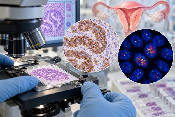
HER2 testing in endometrial cancer looks for excess HER2 protein or extra copies of the ERBB2 gene in tumor tissue. The result can help identify cancers that may respond to HER2-targeted treatment, especially uterine serous carcinoma and some other aggressive, p53-abnormal endometrial cancers. Testing usually begins with immunohistochemistry, or IHC, which shows how strongly and how completely tumor-cell membranes stain for HER2. Equivocal results may be followed by in situ hybridization, commonly FISH, to determine whether the ERBB2 gene is amplified. Endometrial tumors can show uneven or incomplete staining, so interpretation is not identical to breast cancer HER2 testing. A positive result does not describe the entire tumor by itself; HER2 is interpreted with histologic type, stage, molecular subtype, mismatch-repair status, POLE status, and p53 findings. The most useful report states the specimen tested, IHC score, staining pattern, and any amplification result.
- HER2 testing measures HER2 protein overexpression by IHC and, when needed, ERBB2 gene amplification by in situ hybridization.
- HER2 positivity is most established in uterine serous carcinoma, but testing may be relevant in other high-grade endometrial cancers depending on pathology and treatment setting.
- An IHC 3+ result is generally considered positive; an IHC 2+ result often requires reflex amplification testing, but laboratory criteria should be followed.
- Endometrial cancers can show heterogeneous or basolateral HER2 staining, so breast cancer scoring rules should not be applied automatically.
- A positive HER2 result can affect systemic treatment options, but it does not replace stage, molecular classification, or other biomarkers.
Table of Contents
- What HER2 Testing Measures
- Who May Need HER2 Testing
- How HER2 Testing Is Done
- HER2 IHC and Amplification Results
- HER2 and Endometrial Tumor Subtype
- Limitations and Discordant Results
- What HER2 Results Mean for Treatment
What HER2 Testing Measures
HER2 is a receptor protein on the surface of cells. The gene that encodes it is ERBB2. When a tumor has extra ERBB2 copies or produces excessive HER2 protein, signaling through this pathway can promote cell growth and survival.
A HER2 test in endometrial cancer usually asks one of two related questions:
- Is there HER2 protein overexpression on tumor cells?
- Is there ERBB2 gene amplification, meaning an abnormally increased number of gene copies?
These findings often occur together, but not always. IHC assesses protein expression. In situ hybridization, such as fluorescence in situ hybridization, assesses gene copy number. Some next-generation sequencing panels can also identify ERBB2 amplification, although tumor-type-specific IHC and in situ hybridization remain important because sequencing and protein expression are not perfectly interchangeable.
HER2 is a tumor biomarker, not a hereditary cancer test. A positive tumor result usually reflects an acquired change in the cancer. It does not mean that relatives carry an inherited ERBB2 mutation. Hereditary risk in endometrial cancer is more commonly evaluated through mismatch-repair and Lynch syndrome pathways, including MMR IHC testing when appropriate.
The reason to test HER2 is mainly predictive: a positive result can identify a biologic target for therapy. It may also provide information about tumor biology because HER2 overexpression and amplification are enriched in aggressive histologic and molecular subsets.
Who May Need HER2 Testing
HER2 testing is most firmly established for uterine serous carcinoma, a high-grade form of endometrial cancer in which ERBB2 amplification and HER2 overexpression occur often enough to influence treatment planning.
Testing may also be considered in other high-grade endometrial carcinomas, particularly tumors with serous features or p53-abnormal molecular characteristics. Local pathology protocols and oncology guidelines vary, and the indication can depend on whether the cancer is newly diagnosed, advanced, recurrent, or being evaluated for a HER2-directed drug.
A pathologist may test the original hysterectomy specimen, an endometrial biopsy or curettage specimen, or tissue from recurrent or metastatic disease. The best sample is generally one with enough viable tumor and good fixation. If the tumor appears heterogeneous, a larger resection specimen may better represent the full range of HER2 expression.
HER2 is one part of a broader biomarker workup. Modern endometrial cancer classification often incorporates histology with molecular features such as mismatch repair, POLE mutation status, and p53 pattern. A practical overview is provided by the endometrial cancer biomarker panel. These tests answer different questions and should not be viewed as substitutes for one another.
For example, a tumor can be HER2-positive and p53-abnormal, or HER2-positive with other molecular findings that also affect treatment options. The oncology team integrates all relevant results rather than letting a single biomarker define the entire plan.
How HER2 Testing Is Done
HER2 testing is performed on tumor tissue, not a routine blood sample. The tissue may come from a biopsy, curettage, hysterectomy, or metastatic lesion.
Immunohistochemistry is usually the first step
IHC uses an antibody that binds to HER2 protein in preserved tumor tissue. The pathologist evaluates the intensity, completeness, and distribution of membrane staining in tumor cells.
Endometrial serous carcinomas can show a staining pattern that differs from classic breast carcinoma. Strong lateral or basolateral membrane staining may be relevant even when the entire circumference of the cell is not outlined. Tumors may also show striking heterogeneity, with strongly positive areas next to weak or negative areas.
The report commonly assigns a score such as 0, 1+, 2+, or 3+. The exact criteria depend on the validated laboratory algorithm.
In situ hybridization assesses ERBB2 amplification
If IHC is equivocal, usually in the 2+ category, the laboratory may perform FISH or another in situ hybridization method. The test uses labeled DNA probes to count ERBB2 signals and, depending on the assay, compare them with a chromosome 17 reference signal.
Amplification means the tumor has gained extra copies of ERBB2 beyond the laboratory’s validated threshold. This can clarify whether an equivocal protein-staining pattern represents a true HER2-driven tumor.
Sequencing may add information
Comprehensive tumor sequencing can detect ERBB2 amplification and other alterations. However, sequencing measures DNA and may use different thresholds for copy-number calls. A sequencing result may therefore disagree with IHC or FISH, particularly when tumor purity is low or HER2 expression is heterogeneous.
For treatment decisions tied to a specific drug or guideline, clinicians generally use the testing method and positivity definition that matches the supporting evidence.
Pre-analytic handling also matters. Tissue that is poorly fixed, crushed, necrotic, or extremely scant can give unreliable staining or make amplification testing difficult. Pathologists usually select a block with viable invasive tumor and enough cells for interpretation. If the only specimen is a tiny biopsy, the report may note that the result could underrepresent a heterogeneous cancer. A later hysterectomy or metastatic specimen can sometimes provide a better sample for repeat testing.
A useful HER2 pathology report does more than state “positive” or “negative.” It may identify the antibody or platform, specimen and block, IHC score, percentage or pattern of staining, and the result of reflex ISH/FISH when performed. If the tumor shows marked heterogeneity, documenting that feature helps the oncology team understand why another specimen or method might be considered.
HER2 IHC and Amplification Results
A HER2 report may contain several pieces of information. The simplest interpretation is usually negative, equivocal, or positive, but understanding how that conclusion was reached is useful.
| Common result | Typical meaning | Usual next step |
|---|---|---|
| IHC 0 or 1+ | Low or absent HER2 expression by the validated scoring method | Usually considered HER2-negative for classic HER2-positive treatment algorithms |
| IHC 2+ | Equivocal protein expression | Often reflex ERBB2 amplification testing by ISH/FISH |
| IHC 3+ | Strong HER2 overexpression meeting the laboratory’s positive criteria | Usually considered HER2-positive |
| ERBB2 amplified | Extra ERBB2 gene copies detected | Supports HER2-positive classification in the appropriate testing algorithm |
The table is a practical summary, not a replacement for the pathology report. Endometrial cancer scoring is still evolving, and individual laboratories may use specific thresholds validated for uterine serous carcinoma.
A 3+ IHC result generally indicates strong protein overexpression and is considered positive. A 2+ result is usually treated as equivocal rather than automatically positive. If the follow-up in situ hybridization shows amplification, the tumor is commonly classified as HER2-positive. If amplification is absent, it may be considered HER2-negative for regimens requiring amplified or strongly overexpressed disease.
A result of 0 or 1+ means little or no qualifying expression by the assay. Newer HER2-directed drugs can have activity across different expression levels in some cancers, so the treatment meaning of “HER2-low” or low-level staining depends on the drug, cancer type, approval, and evidence. A patient should not assume that terminology developed for breast cancer applies directly to endometrial cancer.
HER2 and Endometrial Tumor Subtype
Endometrial cancer is no longer classified only by how the cells look under a microscope. Molecular classification has become increasingly important because tumors with similar histology can have different biology and outcomes.
Uterine serous carcinoma is usually high grade and often has an abnormal p53 pattern. HER2 overexpression or amplification is enriched in this group. A separate p53 test can help identify the p53-abnormal molecular category, but p53 and HER2 are not the same marker.
Other major molecular groups include mismatch-repair deficient tumors, POLE-mutated tumors, and tumors with no specific molecular profile. POLE mutation testing can identify an ultramutated subtype with distinct prognostic implications in the proper setting. MMR testing can identify tumors with defective DNA mismatch repair and can also trigger evaluation for Lynch syndrome.
HER2 status adds another layer because it can be directly targetable. A tumor can therefore be described simultaneously by histology, molecular subtype, and predictive biomarkers.
This layered approach is especially important in mixed or ambiguous tumors. A carcinoma with serous morphology and strong diffuse p53 abnormality may be a stronger candidate for HER2 assessment than a low-grade endometrioid carcinoma with a clearly different molecular profile. Still, testing policies are evolving, and treatment eligibility should follow current clinical guidance and the patient’s specific disease setting.
Limitations and Discordant Results
HER2 testing in endometrial cancer has several practical limitations that can affect interpretation.
Tumor heterogeneity
One part of the tumor may be strongly positive while another part is negative. A small biopsy may therefore miss a positive clone, or a single block from a hysterectomy may not capture the strongest area. This is one reason pathologists may review multiple blocks or choose tissue carefully.
Specimen differences
HER2 status can differ between the primary tumor and a metastasis or recurrence. Biology can evolve under treatment pressure, and sampling different sites may produce different results. Retesting may be useful if a new treatment decision depends on HER2 and adequate recurrent tumor tissue is available.
Assay and scoring differences
Different antibodies, staining platforms, fixation conditions, and scoring algorithms can affect IHC classification. Studies have shown that commonly used HER2 assays do not perform identically in uterine serous carcinoma. The pathology laboratory should use a validated method and interpret it with an endometrial cancer-specific framework.
IHC, FISH, and sequencing can disagree
Discordance is possible because each method measures something different. IHC measures protein, FISH measures gene amplification in individual cells, and NGS estimates copy number from pooled DNA. Low tumor purity, focal amplification, and heterogeneous expression can all contribute.
When results conflict, clinicians may ask the pathologist to review the slides, verify tissue quality, repeat testing on another block, or use an orthogonal method. The most relevant result is the one generated with a validated method for the treatment decision being considered.
What HER2 Results Mean for Treatment
The main clinical value of a positive HER2 result is that it may make HER2-targeted therapy relevant. In advanced or recurrent uterine serous carcinoma, HER2 status has been used to select patients for trastuzumab-containing treatment, and the field now includes additional antibody-drug conjugates and HER2-directed strategies.
A positive result does not mean a particular drug is automatically appropriate. Treatment depends on stage, prior therapy, performance status, other biomarkers, drug approvals, guideline recommendations, and whether the tumor meets the exact positivity definition used for that therapy.
A negative result similarly does not mean treatment options are limited to one pathway. Endometrial cancer treatment can also be influenced by mismatch-repair status, MSI, hormone receptor status in selected tumors, p53, POLE, and other genomic findings. For example, MMR-deficient disease may have immunotherapy implications that are independent of HER2.
Patients with a HER2-positive result can ask their oncology team several focused questions:
- Was HER2 tested by IHC, FISH/ISH, sequencing, or more than one method?
- What was the exact IHC score and percentage or pattern of staining?
- If the result was 2+, was amplification testing performed?
- Was the tested tissue from the original tumor or a recurrence?
- Does the result meet the criteria for a specific HER2-directed treatment in this disease setting?
- Are other molecular results, such as MMR, POLE, and p53, also available?
These questions help turn a pathology label into a treatment decision. HER2 is most useful when the laboratory result, tumor subtype, and evidence for a specific therapy all line up.
The distinction between HER2-positive and lower levels of HER2 expression is also becoming more important as drug development changes. Historically, many treatment algorithms focused on IHC 3+ tumors or IHC 2+ tumors with confirmed ERBB2 amplification. Some newer antibody-drug conjugates can have activity in tumors that do not fit older binary definitions in certain cancer types. That does not mean every endometrial cancer with faint staining should be treated as HER2-driven. Eligibility depends on the specific drug, regulatory indication, clinical trial evidence, and validated scoring approach used for that setting.
For recurrent disease, retesting can be reasonable when the original result is old, borderline, based on a small sample, or inconsistent with later sequencing. A new biopsy may also provide material for several biomarkers at once. The decision to retest should balance the likelihood that the result will change treatment against the risks and practicality of obtaining additional tissue.
If a report says only “HER2 positive” without the underlying score, patients can ask for the full pathology details. Knowing whether positivity came from strong IHC staining, gene amplification, or both can matter when a treatment protocol uses a specific definition. The same principle applies to a negative result: a clearly adequate IHC 0 result is different from an uninterpretable stain on scant or damaged tissue. When a treatment decision depends on the distinction, pathology review can be more useful than assuming all negative labels are equivalent.
References
- Immunohistochemistry, next generation sequencing (NGS), and whole exome sequencing concordance in HER2 testing in uterine serous carcinoma: a retrospective analysis 2025
- Performance of HER2 DAKO HercepTest and Ventana 4B5 immunohistochemical assays on detecting HER2 gene-amplification in uterine serous carcinomas 2024
- HER2 Status Assessment in Endometrial Serous Carcinoma: Comparative Analysis of Two Proposed Testing and Interpretation Algorithms 2024
- Current Laboratory Testing Practices for Assessment of ERBB2/HER2 in Endometrial Serous Carcinoma and Colorectal Carcinoma 2023
- Endometrial cancer: ESMO Clinical Practice Guideline for diagnosis, treatment and follow-up 2022 (Guideline)
Disclaimer
HER2 testing must be interpreted using the laboratory’s validated endometrial cancer scoring method and the clinical treatment context. A positive or negative result does not determine therapy by itself, and discordant or equivocal findings may require pathology review or additional testing. Treatment decisions should be made with a gynecologic oncologist or medical oncologist familiar with the complete tumor profile.