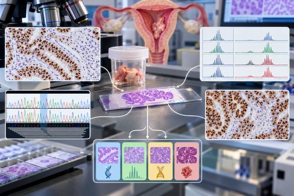
An endometrial cancer biomarker panel helps classify a tumor by its underlying biology, not just by how the cells look under a microscope. The core modern tests are mismatch-repair (MMR) protein immunohistochemistry, microsatellite instability (MSI) testing, POLE mutation testing, and p53 immunohistochemistry. Together with the tumor’s histologic type, grade, stage, and other pathology findings, these markers place most endometrial cancers into a molecular group that can change prognosis and treatment planning. The major groups are POLE-mutated, MMR-deficient, p53-abnormal, and no specific molecular profile (NSMP). MMR testing also serves an important hereditary-cancer role because loss of certain MMR proteins can raise concern for Lynch syndrome. No single abnormal biomarker tells the whole story, and the tests are not interchangeable. Correct interpretation depends on the exact staining pattern, the specific POLE variant, tumor context, and whether more than one classifier is present.
- The four main molecular groups are POLE-mutated, MMR-deficient, p53-abnormal, and NSMP, although newer frameworks further divide NSMP by markers such as estrogen-receptor status.
- MMR immunohistochemistry screens for loss of MLH1, PMS2, MSH2, or MSH6, while MSI testing measures a related genomic consequence of defective mismatch repair.
- Only qualifying pathogenic POLE exonuclease-domain mutations define the POLE-mutated subtype; an incidental POLE variant is not enough.
- Abnormal p53 staining is a surrogate for TP53-pathway alteration and usually identifies a higher-risk molecular group, but staining pattern matters.
- Molecular subtype can affect risk classification, adjuvant therapy, immunotherapy eligibility, and hereditary-cancer evaluation, so results should be read with the full pathology report.
Table of Contents
- What the Endometrial Biomarker Panel Tests
- MMR and MSI: What Deficiency Means
- POLE Mutations and Prognosis
- p53-Abnormal Tumors
- How the Molecular Subtypes Fit Together
- Treatment and Hereditary-Risk Implications
- How to Read a Biomarker Report
What the Endometrial Biomarker Panel Tests
Endometrial carcinoma was historically divided mainly by microscopic type and grade. Those features remain essential, but tumors that look similar can behave differently because they are driven by different molecular processes. Modern classification adds a set of biomarkers that captures those differences.
The most widely used practical panel combines three types of testing. MMR immunohistochemistry examines the proteins MLH1, PMS2, MSH2, and MSH6 in tumor tissue. p53 immunohistochemistry evaluates the pattern of p53 protein expression. POLE sequencing looks for pathogenic mutations in the exonuclease domain of the DNA polymerase epsilon gene. MSI testing may be performed as an additional or alternative assessment of mismatch-repair dysfunction, depending on the laboratory and clinical situation.
The goal is not to produce a long list of unrelated abnormalities. It is to assign the cancer to a biologically meaningful category. A MMR IHC test can identify mismatch-repair deficiency and trigger Lynch syndrome workup. A POLE mutation test can identify a subgroup with unusually favorable outcomes when the variant is a recognized pathogenic exonuclease-domain mutation.
These molecular results are combined with conventional pathology. Histologic subtype, tumor grade, depth of myometrial invasion, lymphovascular space invasion, cervical involvement, lymph-node status, and spread outside the uterus still matter. Current guidelines increasingly integrate molecular classification into staging and risk-stratified treatment rather than treating it as an optional research add-on.
Not every laboratory report uses identical wording. Terms such as “MMRd,” “MSI-H,” “p53 abnormal,” “copy-number high,” “POLEmut,” and “NSMP” may appear. The best interpretation starts by identifying which assay was performed, whether the result is technically adequate, and how the laboratory translated it into a molecular subtype.
MMR and MSI: What Deficiency Means
Mismatch repair is a DNA proofreading system. The proteins MLH1, PMS2, MSH2, and MSH6 work in pairs to correct replication errors. When this system fails, errors accumulate, especially in repetitive DNA sequences called microsatellites.
MMR immunohistochemistry asks whether the four proteins are present in tumor-cell nuclei. Normal surrounding cells provide an internal control. Loss of staining in the tumor suggests mismatch-repair deficiency. The pattern can point toward the affected gene because MLH1 pairs with PMS2 and MSH2 pairs with MSH6.
For example, combined loss of MLH1 and PMS2 commonly leads to testing for MLH1 promoter methylation. Methylation is a frequent sporadic mechanism that switches off MLH1 in an endometrial tumor. If MLH1/PMS2 loss is not explained by the expected methylation pattern, or if MSH2/MSH6, isolated MSH6, or isolated PMS2 loss is present, the findings may raise greater concern for Lynch syndrome and can prompt germline genetic evaluation.
MSI testing approaches the same biological pathway from a different direction. Instead of measuring proteins, it looks for instability in microsatellite sequences. A tumor may be classified as MSI-high when instability exceeds the assay’s threshold. Most MMR-deficient endometrial cancers are MSI-high, but the two methods are not perfectly interchangeable. Discordant results can occur because of technical factors, uncommon biology, low tumor content, or the specific gene involved.
The molecular subtype is usually called MMR-deficient when convincing MMR loss is present, even though the report may also list MSI status. This group generally has an intermediate prognosis as a whole, but individual risk still varies by stage and other pathology factors.
MMR deficiency also has a treatment role. Advanced or recurrent MMR-deficient/MSI-high cancers are particularly responsive to immune-checkpoint approaches, and MMR status is now central to systemic-treatment planning in many settings. That does not mean every early-stage MMR-deficient tumor requires immunotherapy; treatment depends on disease setting and risk.
A tumor result is also not automatically a hereditary diagnosis. Many MMR-deficient endometrial cancers are sporadic. Tumor testing is a screen, while Lynch syndrome is established through appropriate germline evaluation and, when relevant, methylation or other follow-up testing.
POLE Mutations and Prognosis
POLE encodes the catalytic subunit of DNA polymerase epsilon, an enzyme involved in DNA replication and proofreading. Certain mutations in its exonuclease domain disable proofreading and create an “ultramutated” tumor with a very high number of DNA changes.
The clinically important point is specificity: not every POLE variant defines the favorable POLE-mutated molecular subtype. Laboratories must determine whether a detected alteration is a recognized pathogenic exonuclease-domain mutation associated with the characteristic endometrial cancer phenotype. A variant of uncertain significance should not be treated as equivalent to a validated pathogenic POLE mutation.
When a qualifying mutation is present, prognosis is often excellent even when the tumor has features that would traditionally look worrisome, such as high grade. This is one reason molecular classification can change treatment discussions. In selected early-stage cancers, a POLE-mutated result may support treatment de-escalation when current risk algorithms and all clinicopathologic factors agree.
POLE-mutated tumors can also show unusual histology, prominent immune infiltration, or additional molecular abnormalities. Some tumors have more than one classifier—for example, a pathogenic POLE mutation plus abnormal p53 staining. Current molecular algorithms generally give a validated pathogenic POLE mutation priority because outcome data show that these “multiple-classifier” tumors tend to behave according to the POLE-mutated biology rather than the p53-abnormal category.
This hierarchy is important because a report can otherwise appear contradictory. A patient may see “TP53 mutation” or “p53 abnormal” and assume the cancer must belong to the worst-prognosis group. If a pathogenic POLE exonuclease-domain mutation is also present, the final integrated subtype may still be POLE-mutated.
A pathogenic POLE result should therefore be interpreted as part of the classifier algorithm, not as an isolated gene finding. The exact variant, laboratory classification, tumor stage, and current guideline all matter before changing adjuvant treatment.
p53-Abnormal Tumors
p53 immunohistochemistry is used as a practical surrogate for TP53-pathway alteration. The result is based on the pattern of staining, not simply the percentage of positive cells.
A normal, or wild-type, p53 pattern is typically variable and heterogeneous: some tumor-cell nuclei stain, others do not, and intensity differs from cell to cell. An abnormal pattern can appear as strong diffuse nuclear overexpression in most tumor cells, complete absence of nuclear staining with an intact internal control, or an abnormal cytoplasmic pattern in some cases. These patterns support a p53-abnormal classification when interpreted correctly by a pathologist.
The p53 test in endometrial cancer is especially important because the p53-abnormal molecular group generally carries the least favorable prognosis of the four classic groups. It includes many serous carcinomas but is not limited to serous histology. Endometrioid and other tumor types can also be p53 abnormal.
p53 abnormality can affect risk grouping and may support more intensive adjuvant treatment when stage and other factors warrant it. However, an abnormal result does not by itself prove that chemotherapy or radiation is required. Conversely, a wild-type p53 pattern does not automatically mean low risk.
The multiple-classifier issue matters here too. If a tumor has p53-abnormal staining but also a pathogenic POLE exonuclease-domain mutation, it is generally classified as POLE-mutated. If it is p53 abnormal and also clearly MMR deficient without a qualifying POLE mutation, the integrated classification generally follows the MMR-deficient group. The final pathology report should state the resolved molecular category rather than forcing the patient to infer it from individual lines.
How the Molecular Subtypes Fit Together
The classic clinically implemented framework places endometrial cancers into four molecular groups. Newer guidance is beginning to refine the heterogeneous NSMP category further, but the four-group system remains the foundation.
| Group | Typical defining finding | General clinical meaning |
|---|---|---|
| POLE-mutated | Pathogenic POLE exonuclease-domain mutation | Usually excellent prognosis; may support de-escalation in selected early-stage disease. |
| MMR-deficient | Loss of one or more MMR proteins, usually with related MSI phenotype | Intermediate overall prognosis; hereditary evaluation may be needed; strong immunotherapy relevance. |
| p53-abnormal | Abnormal p53 IHC pattern consistent with TP53-pathway alteration | Generally higher-risk biology and poorer prognosis. |
| NSMP | No qualifying POLE mutation, MMR retained, and p53 wild type | Heterogeneous group; prognosis varies and can be refined by histology, grade, ER status, and other features. |
NSMP means “no specific molecular profile.” It is a diagnosis of exclusion rather than a single molecular defect. Many low-grade endometrioid cancers fall here, but NSMP also contains tumors with meaningfully different outcomes. Recent frameworks increasingly consider estrogen-receptor expression and other clinical or molecular factors to separate lower- and higher-risk subsets within NSMP.
The molecular category does not erase stage. A stage I tumor confined to the uterus and a metastatic tumor can share the same molecular subtype but require very different treatment. Likewise, lymphovascular invasion, histology, grade, residual disease, patient age, fertility goals, and medical fitness remain relevant.
The value of the panel is that it improves risk discrimination beyond morphology alone. A high-grade tumor with pathogenic POLE mutation may have a much better outlook than its grade suggests, whereas a p53-abnormal tumor may warrant greater concern despite otherwise limited disease. Molecular classification is therefore best thought of as another major axis of the pathology report, alongside anatomic extent and microscopic features.
There are also practical reasons to complete the classifier early. The best specimen is usually the diagnostic biopsy or hysterectomy tissue with enough viable tumor for reliable immunohistochemistry and sequencing. MMR and p53 staining can often be performed quickly, while POLE sequencing may take longer. If treatment decisions are time-sensitive, clinicians may begin with available results and incorporate the completed molecular profile before finalizing adjuvant therapy. A technically failed or equivocal assay should not be silently treated as normal; repeating the stain, testing another block, or using a complementary molecular method may be appropriate.
Results can also differ between a small biopsy and the final hysterectomy specimen because tumors are heterogeneous and the larger specimen may reveal a different histologic component. Most molecular classifiers are relatively stable, but unusual or discordant cases deserve pathology review. This is especially true when the staining pattern is subclonal, when tumor cellularity is low, or when morphology and molecular findings strongly disagree. The integrated diagnosis should explain meaningful discordance rather than simply list every test result.
Molecular subtype is primarily a tumor characteristic. It should not be confused with the germline genetic result from blood or saliva. A tumor can be MMR deficient because of a purely acquired event, and a patient can carry a hereditary pathogenic variant even if a particular tumor test is not classic. Keeping tumor classification and inherited-risk testing conceptually separate prevents one of the most common interpretation errors.
Treatment and Hereditary-Risk Implications
Molecular results can influence both what treatment is considered and whether inherited cancer risk should be investigated.
For early-stage disease, current guidelines use molecular subtype together with FIGO stage and pathology factors to define recurrence risk and choose among observation, vaginal brachytherapy, external-beam radiation, chemotherapy, or combined approaches. A POLE-mutated tumor may move a patient toward less adjuvant treatment in an appropriate setting, while p53-abnormal biology can move risk in the opposite direction.
For advanced or recurrent disease, MMR deficiency and MSI-high status have major therapeutic relevance because they predict sensitivity to immune-checkpoint therapy. MMR-proficient tumors can also be treated with immunotherapy-containing strategies in certain settings, but the regimen and expected benefit differ. Other tumor markers, including HER2 in appropriate histologies, may add separate treatment options; a HER2 test is not part of the four-group classifier itself.
Hereditary-risk assessment centers on MMR findings. Loss of MSH2/MSH6, isolated MSH6, isolated PMS2, or unexplained MLH1/PMS2 loss can lead to genetics referral and germline testing for Lynch syndrome. A pathogenic germline variant can have implications beyond the treated cancer, including colorectal-cancer surveillance and testing of relatives.
MLH1/PMS2 loss requires special care because many tumors lose these proteins through acquired MLH1 promoter methylation rather than an inherited MLH1 variant. Methylation testing helps distinguish common sporadic cases from those that need additional hereditary evaluation. Family history still matters, especially when tumor and clinical findings do not fit neatly together.
Neither a normal MMR panel nor an NSMP classification rules out every hereditary cancer syndrome. If personal or family history is strongly suggestive, genetics professionals may recommend broader germline testing even when tumor screening is reassuring.
How to Read a Biomarker Report
A useful endometrial biomarker report should let you answer several concrete questions.
First, what was tested? Look for all four MMR proteins, the p53 staining interpretation, and whether POLE sequencing covered the clinically relevant exonuclease domain. If MSI testing was performed, note whether the result is MSI-high, microsatellite stable, or indeterminate.
Second, what is the integrated subtype? The report may list each marker separately, but treatment decisions are easier when the pathologist states the final category: POLE-mutated, MMR-deficient, p53-abnormal, or NSMP. For a multiple-classifier tumor, ask which abnormality has priority and why.
Third, does an MMR abnormality require follow-up? If MLH1/PMS2 are lost, check whether MLH1 promoter methylation was performed or planned. If another loss pattern is present, ask whether genetic counseling or germline testing is recommended.
Fourth, is a POLE variant truly subtype-defining? A report that says “POLE variant” is incomplete for this purpose. The key distinction is whether the variant is recognized as pathogenic and located in the exonuclease domain in a pattern known to define POLE-mutated endometrial carcinoma.
Fifth, how does the molecular result change the treatment recommendation for this stage? Molecular classification is clinically useful only when integrated with surgery, imaging, histology, grade, invasion, nodal findings, and the patient’s goals. The same biomarker can have different implications in stage I disease and recurrent metastatic disease.
Finally, remember that molecular pathology is evolving. Current guidelines increasingly use tumor biology directly in staging and adjuvant decisions, and newer frameworks may split older categories such as NSMP into more precise subgroups. If a pathology report predates molecular testing or contains an indeterminate result, retesting archived tumor tissue may sometimes be useful when the result could change care.
The practical takeaway is simple: MMR/MSI describes DNA mismatch-repair function, POLE identifies an ultramutated favorable subgroup when a qualifying mutation is present, p53 identifies a generally higher-risk molecular pattern, and the combination assigns the tumor’s integrated subtype. That integrated result—not any isolated biomarker—is what should guide prognosis and treatment discussions.
References
- ESGO-ESTRO-ESP guidelines for the management of patients with endometrial carcinoma: update 2025 2025 (Guideline)
- SEOM-GEICO clinical guidelines on endometrial cancer (2025) 2025 (Guideline)
- Integrating molecular classification into endometrial cancer management 2026 (Position Paper)
- Molecular Classification of Endometrial Cancer and Its Impact on Therapy Selection 2024 (Review)
- [Molecular classification of endometrial carcinoma-a short summary for clinical use] 2023 (Review)
Disclaimer
This article is for general educational purposes and does not replace individualized pathology review, genetic counseling, or oncology care. Endometrial cancer biomarkers must be interpreted with tumor stage, histology, grade, surgical findings, prior treatment, and current guidelines. Treatment and hereditary-risk decisions should be made with the appropriate gynecologic oncology, pathology, and genetics professionals.