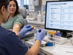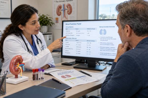
A renal function panel is a group of blood tests used to assess kidney filtration and the body systems the kidneys help regulate. Although the exact panel varies by laboratory, it commonly includes creatinine, estimated glomerular filtration rate (eGFR), blood urea nitrogen (BUN), sodium, potassium, chloride, bicarbonate or total carbon dioxide, calcium, phosphorus, albumin, and sometimes glucose or a calculated anion gap. Looking at these results together gives more information than any single value can provide.
The panel can help detect changes in kidney function, dehydration, electrolyte problems, acid-base disturbances, and mineral abnormalities. It is also commonly used to monitor chronic kidney disease (CKD), acute illness, blood pressure medicines, diuretics, and other treatments that can affect the kidneys. A result outside the laboratory range does not automatically mean kidney disease. The pattern, trend, symptoms, medicines, hydration status, and urine findings all matter. CKD diagnosis usually also requires urine albumin testing and evidence that abnormalities have persisted for at least three months.
- A renal function panel combines filtration markers with electrolytes and minerals, but the exact components differ among laboratories.
- Creatinine and eGFR are the main filtration results, while BUN is a supportive marker affected by hydration, protein intake, and other factors.
- Potassium and bicarbonate are important safety results in kidney disease because severe abnormalities can affect the heart and acid-base balance.
- Calcium, phosphorus, and albumin provide information about mineral and nutritional status, especially as CKD becomes more advanced.
- One abnormal number rarely gives the whole answer; trends and combinations of results are usually more informative than an isolated value.
Table of Contents
- What a Renal Function Panel Includes
- Creatinine, eGFR, and BUN
- Electrolytes and Acid-Base Balance
- Calcium, Phosphorus, Albumin, and Glucose
- Common Renal Panel Result Patterns
- Preparation and Repeat Testing
- How to Interpret the Panel and Next Steps
What a Renal Function Panel Includes
A renal function panel, sometimes called a renal panel or kidney function panel, is not completely standardized. Different laboratories may include slightly different tests. A common version contains BUN, creatinine, sodium, potassium, chloride, bicarbonate or total CO2, calcium, phosphorus, albumin, and glucose. Many laboratories also report an eGFR calculated from creatinine, and some report a BUN-to-creatinine ratio or an anion gap.
This combination reflects several kidney functions at once. The kidneys filter waste products, regulate sodium and water balance, excrete potassium, help maintain acid-base balance, and participate in calcium-phosphorus and vitamin D physiology. A panel therefore gives both a filtration snapshot and a look at consequences that can develop when kidney function is impaired.
Typical adult reference intervals are shown below only as general examples. The range printed by the testing laboratory should be used for an individual result because methods, units, age, sex, and clinical setting can change the expected interval.
| Test | Common example range | What it helps assess |
|---|---|---|
| Sodium | 135–145 mmol/L | Water and sodium balance |
| Potassium | 3.5–5.0 mmol/L | Electrolyte balance and cardiac safety |
| Chloride | 98–107 mmol/L | Fluid and acid-base balance |
| Bicarbonate/total CO2 | About 22–29 mmol/L | Acid-base status |
| BUN | About 7–20 mg/dL | Protein waste handling and hydration context |
| Creatinine | Often about 0.6–1.3 mg/dL | Filtration marker used to calculate eGFR |
| Calcium | About 8.6–10.2 mg/dL | Mineral balance |
| Phosphorus | About 2.5–4.5 mg/dL | Mineral and bone metabolism |
| Albumin | About 3.5–5.0 g/dL | Major blood protein; affects total calcium interpretation |
The name “renal function panel” can be misleading if it is interpreted as a complete kidney evaluation. The panel does not directly show urine albumin leakage, kidney structure, or the cause of reduced kidney function. A complete CKD assessment usually pairs blood-based eGFR with a urine albumin-creatinine ratio and, when indicated, urinalysis, imaging, or other tests.
Creatinine, eGFR, and BUN
Creatinine is produced from normal muscle metabolism and removed largely through kidney filtration. When filtration falls, blood creatinine usually rises. However, creatinine also depends on muscle mass, age, diet, certain medicines, and other non-kidney factors. A small older adult with low muscle mass may have substantial kidney impairment without a dramatically high creatinine, while a muscular person can have a higher creatinine despite good filtration.
The eGFR converts creatinine, age, and sex into an estimate of filtration normalized to a body surface area of 1.73 m². Current adult equations do not use race. In general, persistent eGFR below 60 mL/min/1.73 m² is one criterion for CKD. An eGFR of 60 or higher does not exclude kidney disease if urine albumin, imaging, or other markers show kidney damage.
Creatinine-based eGFR is an estimate, not a direct measurement. It becomes less reliable when creatinine production is unusual, such as with very low or very high muscle mass, amputation, severe malnutrition, some neuromuscular conditions, or rapidly changing kidney function. Cystatin C can be used as an additional filtration marker when greater accuracy is needed. A combined creatinine-cystatin C eGFR is often more accurate than either marker alone.
BUN measures the nitrogen portion of urea, a waste product formed in the liver after protein metabolism. BUN often rises when kidney filtration falls, but it is less specific than creatinine because it can also rise with dehydration, high protein intake, gastrointestinal bleeding, corticosteroid use, fever, or increased tissue breakdown. Low BUN can occur with low protein intake, severe liver dysfunction, or excess body water.
The BUN-to-creatinine ratio may provide context, but it is not a stand-alone diagnostic test. A high ratio can occur with dehydration or upper gastrointestinal bleeding, while a low ratio can reflect low urea production or relatively high creatinine production. Clinical findings remain essential.
The most useful information often comes from trends. A creatinine rising from 0.9 to 1.4 mg/dL over a short period can be clinically important even though 1.4 may not look dramatically elevated on some reports. In acute kidney injury, eGFR equations are also less reliable because creatinine may not yet be in a steady state.
Electrolytes and Acid-Base Balance
Sodium is the major electrolyte in extracellular fluid. Its blood concentration reflects the relationship between body sodium and body water more than total sodium stores alone. Low sodium can occur with excess water retention, diuretics, heart failure, liver disease, adrenal disorders, or severe illness. High sodium usually points toward a relative water deficit. Kidney disease can contribute to either problem when the kidneys cannot appropriately dilute or concentrate urine.
Potassium is particularly important because severe elevations or reductions can disturb heart rhythm. The kidneys normally excrete excess potassium. Hyperkalemia becomes more likely with advanced CKD, acute kidney injury, metabolic acidosis, diabetes, and medicines such as ACE inhibitors, ARBs, mineralocorticoid receptor antagonists, potassium-sparing diuretics, trimethoprim, and some NSAIDs. A falsely high potassium can also result from hemolysis during blood collection.
Chloride generally moves with sodium and participates in acid-base balance. It is often interpreted together with bicarbonate and sodium rather than alone. High chloride can accompany certain types of metabolic acidosis, while low chloride may occur with vomiting, diuretic use, or metabolic alkalosis.
The bicarbonate result may appear on a chemistry panel as “CO2” or “total CO2.” Most of that value represents bicarbonate. The kidneys regenerate bicarbonate and excrete acid, so CKD can lead to metabolic acidosis as functioning nephron mass declines. A persistently low bicarbonate can also have causes unrelated to CKD, including diarrhea, ketoacidosis, lactic acidosis, and some medicines.
Some laboratories calculate an anion gap from sodium, chloride, and bicarbonate. The anion gap can help classify metabolic acidosis, but its reference range depends on the laboratory and whether potassium is included in the calculation. Albumin also influences the gap, so a low albumin can mask an elevation.
Electrolytes can change more quickly than creatinine. A person with stable CKD may develop sudden hyperkalemia or low sodium during dehydration, infection, medication changes, or worsening heart failure. That is why clinicians often repeat a renal panel soon after starting or changing medicines that affect kidney blood flow, potassium, or sodium balance.
Calcium, Phosphorus, Albumin, and Glucose
Calcium and phosphorus are included because the kidneys have a central role in mineral balance. As CKD progresses, phosphate excretion becomes more difficult, active vitamin D production declines, and parathyroid hormone can rise. These changes are part of CKD-mineral and bone disorder, or CKD-MBD.
Phosphorus may remain normal during earlier CKD because hormonal changes increase phosphate excretion through the remaining nephrons. In advanced CKD, phosphorus is more likely to rise. A high result can also be caused by cell breakdown, excess phosphate intake, or certain endocrine problems. Low phosphorus has a different set of causes, including poor nutrition, refeeding, alcohol use, some antacids or binders, and intracellular shifts.
Total calcium includes both protein-bound and free calcium. Because much of circulating calcium is bound to albumin, a low albumin can lower total calcium even when biologically active ionized calcium is normal. Traditional “corrected calcium” formulas can be imprecise, especially in significant illness or advanced kidney disease. When calcium status is clinically uncertain, directly measured ionized calcium can provide better information.
Albumin itself is not a direct measure of kidney filtration. Low serum albumin may reflect inflammation, liver disease, poor nutritional intake, fluid overload, or protein loss in urine. Nephrotic-range urinary protein loss can markedly reduce serum albumin. Importantly, a normal serum albumin does not rule out albuminuria; urine testing is required to detect abnormal albumin leakage.
Glucose is included in many renal panels because diabetes is a major cause of CKD and because severe hyperglycemia affects fluid and electrolyte balance. A random glucose cannot always be interpreted using fasting glucose cutoffs. If diabetes screening or diagnosis is the goal, the clinical context and tests such as fasting glucose or hemoglobin A1c are usually considered separately.
The relationships among these values matter. For example, high phosphorus, low or low-normal calcium, and elevated parathyroid hormone can fit secondary hyperparathyroidism in CKD. Low albumin with edema and high urine protein suggests a different process than low albumin from poor intake or liver disease.
Common Renal Panel Result Patterns
A renal panel is most useful when read as a pattern rather than a collection of independent flags. Several common patterns illustrate this principle.
Rising creatinine with falling eGFR
This pattern suggests reduced filtration, but the cause can be acute or chronic. A rapid change over hours to days raises concern for acute kidney injury, especially after dehydration, infection, obstruction, contrast exposure, or a medication change. A stable abnormal value present for at least three months fits CKD more closely. Previous laboratory results are therefore extremely valuable.
High BUN with relatively smaller creatinine change
This can occur with dehydration, reduced kidney blood flow, high protein intake, gastrointestinal bleeding, corticosteroid use, or increased protein breakdown. It does not prove “prerenal kidney failure.” The physical examination, blood pressure, urine findings, medication history, and trend are needed to determine whether low effective circulating volume is present.
High potassium with low bicarbonate
This combination can appear in advanced CKD or certain forms of renal tubular dysfunction. Diabetes and medicines that reduce aldosterone activity can make the pattern more likely. The potassium level determines urgency because severe hyperkalemia can be life-threatening even before the cause is fully established.
Low sodium with normal or high creatinine
Low sodium may reflect water retention rather than sodium deficiency. Heart failure, advanced kidney disease, diuretics, and several endocrine or medication-related causes can produce this pattern. Treatment depends on the underlying cause and cannot be chosen from sodium alone.
High phosphorus with abnormal calcium
In advanced CKD, this pattern can be part of CKD-MBD and should usually prompt interpretation with PTH, vitamin D status, diet, and treatment history. A single calcium or phosphorus value is less informative than repeated results and the broader mineral pattern.
Normal renal panel despite kidney disease
A person can have kidney disease even when every value on a renal panel falls within the laboratory range. Early glomerular disease may first appear as albumin in the urine. CKD can also be present with eGFR above 60 if there are persistent markers of structural or functional kidney damage. This is why a renal panel should not be used as the only kidney screening tool.
Preparation and Repeat Testing
Most renal function panels require a routine blood draw and often do not require fasting unless glucose or another simultaneously ordered test requires it. Instructions from the ordering clinician or laboratory should take priority. Eating a large meat meal shortly before testing can transiently affect creatinine, and recent heavy exercise may also influence creatinine and potassium in some people.
Hydration can affect results, but deliberately overdrinking water before the test is not useful. Significant dehydration can raise BUN and creatinine, while excess water can lower sodium and dilute some blood measurements. The goal is usually to follow normal hydration unless a clinician gives specific instructions.
Medication and supplement information is important. ACE inhibitors, ARBs, diuretics, NSAIDs, creatine supplements, potassium supplements, salt substitutes, and many other products can change one or more values. Prescription medicines generally should not be stopped just to “improve” the laboratory result unless the clinician specifically advises it.
Repeat testing depends on the reason for the panel. Stable CKD may be monitored every few months or at another interval based on stage and risk. A new medication that affects potassium or kidney blood flow may lead to earlier repeat testing. A sudden creatinine rise, potassium near a dangerous level, severe sodium abnormality, or symptoms of acute illness can require same-day or emergency reassessment.
Laboratory artifacts also matter. Hemolysis can falsely elevate potassium and phosphorus. Delayed sample processing can alter some chemistry values. If a result is surprising and does not fit the clinical picture, clinicians may repeat the test before making a major long-term decision—provided the abnormality is not so severe that treatment must begin immediately.
How to Interpret the Panel and Next Steps
Start with the laboratory’s own reference ranges and compare the panel with previous values. A stable creatinine of 1.2 mg/dL can have a different meaning from a rise from 0.7 to 1.2 mg/dL in two days. Trends in eGFR, potassium, bicarbonate, phosphorus, and albumin can reveal gradual changes that a single panel misses.
Next, connect related tests. Creatinine and eGFR describe filtration. BUN adds hydration and protein-metabolism context. Sodium, potassium, chloride, and bicarbonate show electrolyte and acid-base balance. Calcium and phosphorus reflect mineral regulation, while albumin affects interpretation of total calcium and can signal inflammation, fluid status, nutrition problems, or protein loss.
A renal panel should then be combined with tests that are not on the panel. The urine albumin-creatinine ratio is especially important because albuminuria independently predicts CKD progression and cardiovascular risk. Urinalysis can identify blood, protein, glucose, or other clues. Blood pressure, diabetes status, medications, imaging, and sometimes cystatin C are also part of a complete evaluation.
Urgent assessment may be needed for severe hyperkalemia, rapidly rising creatinine with reduced urine output, severe sodium abnormalities, marked acidosis, confusion, fainting, chest symptoms, or signs of fluid overload such as severe shortness of breath. The level that triggers urgent action depends on symptoms, ECG findings, speed of change, and the person’s underlying disease.
For chronic abnormalities, the next step is usually to identify the cause rather than treating the laboratory number alone. That may mean correcting dehydration, reviewing medicines, managing diabetes and blood pressure, addressing urinary obstruction, treating metabolic acidosis, adjusting dietary phosphorus or potassium when truly needed, or evaluating for a specific kidney disorder.
The renal function panel is best viewed as a multisystem kidney safety panel, not a single pass-or-fail kidney test. Used together with eGFR trends, urine albumin testing, and clinical context, it can show both how well the kidneys are filtering and whether kidney-related disturbances in electrolytes, acid-base balance, or minerals are developing.
References
- A Comprehensive Framework for Kidney Function Assessment: Summary of the National Institute of Diabetes and Digestive and Kidney Diseases Reimagining Kidney Function Assessment Workshop 2025
- Beyond creatinine: New methods to measure renal function? 2025 (Review)
- Recommendations for European laboratories based on the KDIGO 2024 Clinical Practice Guideline for the Evaluation and Management of Chronic Kidney Disease 2024
- Executive summary of the KDIGO 2024 Clinical Practice Guideline for the Evaluation and Management of Chronic Kidney Disease: known knowns and known unknowns 2024 (Guideline Summary)
- RFAMA – Overview: Renal Function Panel, Serum 2026 (Laboratory Test Catalog)
- Understanding your lab values and other CKD health numbers 2023
Disclaimer
Renal function panel results must be interpreted with the laboratory’s reference ranges, prior results, symptoms, medications, and other kidney tests. Do not stop prescription medicines, change electrolyte supplements, or make major dietary restrictions based only on one panel without clinical guidance. Severe potassium or sodium abnormalities, rapidly worsening kidney function, very low urine output, chest symptoms, fainting, confusion, or severe shortness of breath can require urgent medical evaluation.