
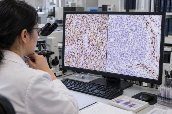
A SMARCB1/INI1 loss test usually refers to immunohistochemistry (IHC) that checks whether tumor-cell nuclei retain the INI1 protein made by the SMARCB1 gene. Complete loss of nuclear INI1 staining can be a powerful diagnostic clue in malignant rhabdoid tumor, atypical teratoid/rhabdoid tumor, epithelioid sarcoma, and several other uncommon cancers. However, INI1 loss is not specific to one diagnosis. The pathologist must confirm that non-tumor cells on the same slide stain normally, then interpret the pattern with tumor location, patient age, microscopic appearance, other IHC markers, and molecular testing. “SMARCB1 loss” can also refer to biallelic gene inactivation detected by sequencing, deletion analysis, or FISH; genetic loss and protein loss are related but not identical tests. In a child with a rhabdoid tumor, identifying SMARCB1 deficiency may also prompt evaluation for a germline SMARCB1 pathogenic variant and rhabdoid tumor predisposition syndrome. In adults, the same IHC finding can point to a different tumor family and different treatment implications.
- Complete tumor-cell nuclear loss of INI1 with retained staining in internal control cells supports SMARCB1 deficiency.
- INI1 loss is not a diagnosis by itself; several rhabdoid, sarcoma, nerve-sheath, renal, and other tumors can show the same pattern.
- A weak or patchy stain is not automatically equivalent to complete SMARCB1 loss and may need repeat or molecular testing.
- Rhabdoid tumors, especially in young children, can be associated with germline SMARCB1 changes and may require genetics evaluation.
- Tazemetostat is an established targeted option for selected advanced epithelioid sarcoma, but INI1 loss does not make every SMARCB1-deficient tumor eligible for the same treatment.
Table of Contents
- What SMARCB1/INI1 Means
- How INI1 IHC Is Performed
- Tumors Associated With INI1 Loss
- How to Interpret the Staining Pattern
- Molecular Testing and Germline Risk
- Treatment and Prognostic Meaning
- Common Pitfalls and Next Steps
What SMARCB1/INI1 Means
SMARCB1 is a tumor suppressor gene on chromosome 22q11.2. It encodes a core component of the SWI/SNF, also called BAF, chromatin-remodeling complex. This complex helps control which parts of DNA are accessible for gene transcription. When SMARCB1 function is lost, cells can develop profound epigenetic dysregulation even without the very high mutation burden seen in some other cancers.
The protein has several names in pathology reports, including INI1, BAF47, and SNF5. In routine surgical pathology, “INI1 IHC” is the most common wording. Normal cells show nuclear staining because INI1 is widely expressed. Therefore, a true deficient tumor shows absent nuclear staining in the malignant cells while nearby lymphocytes, endothelial cells, stromal cells, or other benign elements remain positive.
Genetic inactivation is commonly biallelic. One SMARCB1 copy may be deleted and the other mutated or deleted, or both copies may be lost through a larger deletion. In some tumors, sequencing does not identify an obvious pair of alterations even though protein expression is lost, which is why IHC and genomic testing can be complementary.
Loss of INI1 should also be distinguished from loss of BRG1/SMARCA4, another SWI/SNF component. These abnormalities define overlapping but distinct tumor groups and are not interchangeable markers.
How INI1 IHC Is Performed
INI1 IHC is performed on a tissue section from a biopsy or surgical specimen. An antibody binds to INI1 protein, and a chromogenic reaction makes nuclear staining visible under the microscope. No special patient preparation is required because the test is performed on tissue already obtained for diagnosis.
The pathologist evaluates two things at the same time: tumor cells and internal controls. The controls are crucial. If tumor nuclei are negative but surrounding benign cells are strongly positive, true loss is supported. If both tumor and control cells fail to stain, the test is technically uninterpretable rather than biologically negative.
The classic abnormal pattern is complete loss of nuclear staining in essentially all tumor cells. Some neoplasms show mosaic, heterogeneous, or reduced expression. Those patterns can be biologically meaningful, but they require more caution because fixation problems, tissue damage, or an alternate SWI/SNF abnormality can also alter staining intensity.
A small biopsy can be especially challenging. Necrosis, crush artifact, scant viable tumor, or loss of the relevant component in only part of a heterogeneous tumor may produce an uncertain result. Repeat staining on another block or molecular confirmation may be appropriate when the result would change the diagnosis.
INI1 is usually interpreted as one marker in a panel. Cytokeratins, EMA, S100/SOX10, brachyury, myogenic markers, melanocytic markers, and other stains may be added depending on the differential diagnosis.
Tumors Associated With INI1 Loss
Several distinct tumors are strongly associated with SMARCB1/INI1 deficiency.
Malignant rhabdoid tumor typically occurs in infants and young children, often in the kidney or soft tissue. Histology can show large cells with eccentric nuclei and abundant eosinophilic cytoplasm, but morphology varies. Loss of INI1 is a defining molecular-pathologic feature in most cases.
Atypical teratoid/rhabdoid tumor (AT/RT) is the central nervous system counterpart. Most AT/RTs show SMARCB1 loss; a smaller subset is driven by SMARCA4 deficiency. Modern CNS classification also recognizes other rare SWI/SNF-deficient tumors, so location plus molecular context is essential.
Epithelioid sarcoma is an aggressive soft-tissue sarcoma that often affects distal extremities in younger adults but can occur proximally and in many anatomic sites. More than 90% of conventional and proximal-type epithelioid sarcomas in classic series show complete INI1 loss. Because carcinomas and other epithelioid tumors can mimic it, INI1 is useful but not sufficient alone.
Other tumors that can show loss include poorly differentiated chordoma, subsets of epithelioid malignant peripheral nerve sheath tumor, renal medullary carcinoma, some myoepithelial neoplasms, and rare undifferentiated carcinomas. The spectrum continues to expand as molecular classification improves.
Synovial sarcoma is a useful pitfall. It may show reduced or occasionally lost INI1 staining, but it is defined by an SS18::SSX fusion. When morphology and IHC overlap, an SS18-SSX fusion test can resolve the diagnosis more directly.
Because many entities share INI1 loss, the phrase “SMARCB1-deficient neoplasm” may be used provisionally until the final tumor class is established.
How to Interpret the Staining Pattern
A pathology report may use several phrases:
- Retained INI1 expression: tumor-cell nuclei stain, arguing against complete SMARCB1 protein loss.
- Complete loss of INI1 expression: tumor nuclei are negative while internal controls are positive; this supports SMARCB1 deficiency.
- Reduced or heterogeneous expression: staining is weaker or absent in only part of the tumor; interpretation depends on the entity and technical quality.
- Equivocal or uninterpretable: internal controls fail or tissue quality prevents a confident call.
Complete loss is strongest when it fits the morphology and clinical setting. For example, a pediatric renal mass with rhabdoid morphology and complete INI1 loss is highly suspicious for malignant rhabdoid tumor. The same stain in an adult hand mass with keratin positivity and epithelioid morphology may support epithelioid sarcoma instead.
Retained staining also needs context. A retained result makes classic SMARCB1-deficient rhabdoid tumor less likely, but it does not exclude a rhabdoid-appearing cancer driven by another mechanism. AT/RT with SMARCA4 loss is an example.
The stain should not be translated into a percentage cutoff the way some hormone-receptor or PD-L1 assays are. The clinically important distinction is usually qualitative nuclear retention versus true loss, supported by controls.
A genomic report can add another layer. Homozygous SMARCB1 deletion or clearly biallelic pathogenic alterations strongly support the mechanism. A single heterozygous mutation may be insufficient unless there is evidence that the other allele is also lost or inactivated.
Molecular Testing and Germline Risk
Molecular testing may use targeted next-generation sequencing, copy-number analysis, FISH, or broader genomic profiling. It can confirm SMARCB1 inactivation, identify the exact alteration, and help distinguish tumors with overlapping IHC patterns.
For a newly diagnosed rhabdoid tumor, the germline question is especially important. Rhabdoid tumor predisposition syndrome type 1 is caused by a germline pathogenic SMARCB1 variant. The risk is highest to consider in very young children, patients with multiple primary rhabdoid tumors, bilateral or multifocal disease, or a family history, but genetics evaluation may be appropriate more broadly because family history can be absent.
A tumor mutation is not automatically germline. Confirmatory testing uses a non-tumor sample, typically blood when it is suitable. In certain situations involving constitutional mosaicism or hematologic disease, genetics specialists may choose another tissue.
A germline pathogenic variant can affect parents, siblings, and future children and may lead to specialized surveillance. Some pathogenic SMARCB1 variants are also associated with schwannomatosis phenotypes rather than classic early childhood rhabdoid tumors, so genotype and family phenotype need expert interpretation.
Epithelioid sarcoma in an adult is usually driven by somatic SMARCB1 loss rather than an inherited rhabdoid predisposition syndrome. Germline testing should therefore be guided by age, tumor history, family history, and the exact molecular finding rather than ordered automatically for every INI1-negative sarcoma.
Treatment and Prognostic Meaning
SMARCB1 loss can have direct treatment relevance in selected cancers, but it is not a universal pan-cancer drug biomarker.
The clearest established example is advanced epithelioid sarcoma. Loss of SMARCB1 increases dependence on the epigenetic regulator EZH2. Tazemetostat, an oral EZH2 inhibitor, received approval for selected patients aged 16 years and older with metastatic or locally advanced epithelioid sarcoma that is not eligible for complete resection. Eligibility should be checked against the current drug label and local guidelines.
That does not mean every INI1-negative tumor should receive tazemetostat. Rhabdoid tumors, AT/RT, renal medullary carcinoma, and other SMARCB1-deficient cancers have different standard regimens and levels of evidence. EZH2 inhibition, immunotherapy, and other approaches are being studied across the broader SMARCB1-deficient spectrum, but many uses remain investigational.
Prognosis also depends on tumor type rather than the stain alone. Malignant rhabdoid tumors and AT/RT are aggressive pediatric cancers. Epithelioid sarcoma can recur locally and metastasize, sometimes years after diagnosis. Poorly differentiated chordoma has a different clinical course and treatment strategy. Therefore, “INI1 loss” should never be converted into a generic survival estimate.
The most important practical consequence is often diagnostic accuracy. Correctly identifying the tumor class determines surgery, radiation, systemic treatment, genetic counseling, and referral to an experienced sarcoma, pediatric oncology, neuropathology, or renal cancer team.
Common Pitfalls and Next Steps
The first pitfall is calling a technically failed stain “loss.” Internal control cells must be positive. If they are not, the assay should be repeated or interpreted as noninformative.
The second is assuming INI1 loss is specific for rhabdoid tumor or epithelioid sarcoma. The differential diagnosis depends on anatomic site, age, morphology, and the rest of the immunophenotype. Molecular testing may be decisive.
The third is equating IHC loss with a germline SMARCB1 mutation. IHC is performed on tumor tissue and usually reflects tumor biology. Inherited predisposition requires separate germline evaluation.
The fourth is assuming all SMARCB1-deficient cancers share one targeted therapy. They do not. Treatment evidence is entity specific.
After an abnormal result, useful questions are:
- Were internal control cells positive, confirming true nuclear loss?
- Which tumor diagnoses fit the patient’s age, site, and morphology?
- Is molecular confirmation needed to establish biallelic SMARCB1 inactivation or exclude a fusion-defined mimic?
- Does the tumor type warrant germline SMARCB1 testing?
- Does INI1 loss create an approved treatment option for this exact diagnosis, or only a clinical-trial rationale?
Used this way, SMARCB1/INI1 testing is a highly valuable classifier: it narrows a difficult differential diagnosis, points toward the underlying chromatin-remodeling abnormality, and identifies the patients in whom hereditary evaluation or specific therapy should be considered.
References
- SMARCB1-Deficient Cancers: Novel Molecular Insights and Therapeutic Vulnerabilities 2022 (Review)
- The genetic landscape of SMARCB1 alterations in SMARCB1-deficient spectrum of mesenchymal neoplasms 2022
- SWI/SNF-deficient tumors of the central nervous system: An update 2024 (Review)
- SMARCB1 Deficiency as a Driver of the Hallmarks of Cancer in Rhabdoid Tumours: Novel Insights into Dysregulated Energy Metabolism, Emerging Targets, and Ongoing Clinical Trials 2025 (Review)
- Tazemetostat: EZH2 Inhibitor 2022 (Review)
Disclaimer
INI1 loss is a pathology finding that must be interpreted with morphology, internal staining controls, and the full clinical context. Germline SMARCB1 testing and treatment decisions require specialist evaluation and may differ greatly by tumor type and age. This article is educational and does not replace a pathology report, genetics consultation, or oncology care.





