
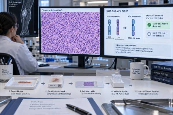
An SS18-SSX fusion test looks for the defining molecular abnormality of synovial sarcoma: a rearrangement involving SS18 on chromosome 18 and an SSX gene on the X chromosome, usually SSX1 or SSX2. More than 90%–95% of synovial sarcomas carry an SS18::SSX fusion, so detecting it strongly supports the diagnosis when the tumor’s location and microscopic features fit. Testing is especially useful because synovial sarcoma can mimic other spindle-cell, epithelioid, or poorly differentiated tumors on a small biopsy. The fusion can be detected by RNA-based next-generation sequencing, reverse-transcription PCR, break-apart FISH, or increasingly by highly specific fusion-directed immunohistochemistry. These methods are not identical: FISH shows rearrangement of the SS18 locus but does not identify the expressed partner, while RNA testing directly demonstrates the fusion transcript. A negative result should be interpreted with assay quality and sample adequacy before synovial sarcoma is excluded. The fusion establishes tumor identity much more reliably than older supportive markers such as TLE1, which is sensitive but not fully specific.
- A confirmed SS18::SSX fusion is a defining molecular feature of synovial sarcoma and strongly supports the diagnosis.
- SS18::SSX1 and SS18::SSX2 are the common fusion types; rare SSX4 partners occur.
- RNA-based NGS directly detects the expressed fusion transcript and can be especially useful when morphology is ambiguous.
- SS18 break-apart FISH can confirm rearrangement but usually does not identify the SSX partner or prove the exact transcript.
- A negative or failed assay does not automatically exclude synovial sarcoma if tissue quality, RNA degradation, or method sensitivity is limited.
Table of Contents
- What the SS18-SSX Fusion Is
- When Testing Is Needed
- Testing Methods
- How to Interpret Results
- Diagnostic Differential and IHC
- Prognosis and Treatment Relevance
- Limitations and Next Steps
What the SS18-SSX Fusion Is
Synovial sarcoma is driven by a characteristic chromosomal translocation, classically written t(X;18)(p11;q11). This rearrangement fuses most of SS18 to the C-terminal region of an SSX gene. The resulting SS18-SSX fusion protein alters the activity of the SWI/SNF, or BAF, chromatin-remodeling complex and rewires gene regulation in ways that promote tumor development.
The common products are SS18::SSX1 and SS18::SSX2. SS18::SSX4 is rare. The fusion is considered the central oncogenic driver rather than an incidental passenger mutation.
The name “synovial sarcoma” can be misleading. The tumor does not need to arise from synovial tissue and can occur in many locations, including the extremities, trunk, head and neck, lung or pleura, abdominal sites, and occasionally visceral organs. It most often affects adolescents and young adults but can occur at a wide range of ages.
Histologically, synovial sarcoma can be monophasic spindle-cell, biphasic with both spindle and epithelial components, or poorly differentiated. The wide morphologic range is exactly why molecular confirmation is so useful.
The SS18-SSX fusion should be distinguished from other fusion-defined sarcomas. An EWSR1 fusion test, DDIT3 testing, or other targeted assay may be more relevant when the morphology suggests another sarcoma family. Broad RNA fusion panels can evaluate several possibilities at once.
When Testing Is Needed
Testing is most useful when synovial sarcoma is in the pathologic differential diagnosis or when a diagnosis needs molecular confirmation before major treatment.
A classic biphasic tumor in a young adult may be recognizable on morphology and immunophenotype, but many cases are less straightforward. Monophasic spindle-cell synovial sarcoma can resemble malignant peripheral nerve sheath tumor, fibrosarcoma, solitary fibrous tumor, leiomyosarcoma, or other spindle-cell neoplasms. Poorly differentiated synovial sarcoma can mimic Ewing sarcoma, lymphoma, poorly differentiated carcinoma, or other round-cell tumors.
Small core biopsies increase the challenge because they may sample only one component. A biphasic tumor can look entirely monophasic if the biopsy misses the epithelial areas.
Molecular confirmation is also valuable when the tumor occurs at an unusual site or age. For example, a spindle-cell tumor in the lung or kidney should not be called primary synovial sarcoma purely because TLE1 is positive. Demonstrating the defining fusion provides much stronger evidence.
Testing can also be performed on recurrent or metastatic tissue if the original diagnosis is uncertain or if archived material is unavailable. The SS18::SSX fusion is generally stable through the disease course because it is an initiating driver.
In modern practice, a solid tumor NGS panel with validated RNA fusion detection may identify SS18::SSX while simultaneously assessing other relevant molecular features.
Testing Methods
Several methods can confirm synovial sarcoma, and the best choice depends on tissue availability, laboratory expertise, and whether broad profiling is also needed.
RNA-based next-generation sequencing
RNA sequencing directly detects the fusion transcript produced by the tumor. Targeted anchored-multiplex or hybrid-capture RNA assays can identify SS18::SSX even when the partner is not specified in advance. This approach is attractive because it confirms that the rearrangement is expressed and often names the exact partner.
RNA quality is the main limitation. Formalin-fixed paraffin-embedded tissue can contain fragmented RNA, especially in old blocks or poorly fixed specimens. A technically failed RNA test should be reported as noninformative, not negative.
Reverse-transcription PCR
RT-PCR can be highly sensitive and specific when primers match the expected fusion transcript. It uses RNA converted to complementary DNA and amplifies the fusion junction. The limitation is that primer design may miss rare breakpoints or unusual SSX partners.
FISH
Break-apart FISH uses probes flanking SS18. A rearranged signal pattern supports disruption of the SS18 locus. FISH works on formalin-fixed tissue and may be useful when RNA quality is poor.
However, FISH generally does not name the SSX partner and can occasionally yield equivocal or technically difficult patterns. It also demonstrates a structural rearrangement rather than the exact expressed fusion transcript.
Fusion-specific immunohistochemistry
A monoclonal antibody directed against the SS18-SSX fusion junction has become a powerful diagnostic tool. Initial validation showed strong diffuse nuclear staining in about 95% of genetically confirmed synovial sarcomas with very high specificity. More recent studies continue to support its diagnostic performance.
Because no single method is perfect, difficult cases may require an orthogonal test. For example, a morphologically convincing tumor with negative fusion-specific IHC can be evaluated by RNA sequencing or FISH.
How to Interpret Results
A positive SS18::SSX fusion result is highly supportive of synovial sarcoma. The report may specify SS18::SSX1, SS18::SSX2, or a rare partner. In the appropriate morphologic setting, this finding is essentially diagnostic.
A positive SS18 break-apart FISH result supports an SS18 rearrangement but may not define the exact partner. If the case is unusual, confirming the transcript can provide additional confidence.
A positive fusion-specific IHC result usually shows strong, diffuse nuclear staining. Focal or weak staining should be interpreted cautiously and may need molecular confirmation, especially if the morphology is discordant.
A negative result has to be qualified by the method. Negative RNA sequencing on a high-quality sample with adequate tumor and validated SS18 coverage is strong evidence against synovial sarcoma. A negative result from degraded RNA, a low-cellularity specimen, or an assay that only detects common transcripts is less definitive.
An equivocal FISH result can occur when signal separation is borderline, tissue is truncated, nuclei overlap, or the rearrangement pattern is complex. Repeating the test on another block or using RNA NGS can resolve many cases.
The fusion type should not be overinterpreted prognostically. Older studies suggested possible differences between SS18::SSX1 and SS18::SSX2, but pooled analyses did not establish a robust overall-survival distinction strong enough to drive routine treatment decisions. Tumor size, site, resectability, grade, necrosis, metastasis, and response to therapy remain more clinically important.
Diagnostic Differential and IHC
Synovial sarcoma often expresses cytokeratins and epithelial membrane antigen, especially in biphasic tumors, but staining can be focal in monophasic or poorly differentiated cases. TLE1 is frequently strong and diffuse, which makes it a useful screening marker. The problem is specificity: TLE1 can be positive in other tumors.
INI1/SMARCB1 staining can show reduced expression in synovial sarcoma, another potential pitfall. Complete INI1 loss is more characteristic of epithelioid sarcoma and other SMARCB1-deficient neoplasms, but variable patterns exist. An INI1 loss result should therefore be integrated with fusion testing rather than used alone to separate overlapping sarcomas.
Other differential markers depend on the site and morphology. S100 and SOX10 may be used when malignant peripheral nerve sheath tumor is considered; STAT6 when solitary fibrous tumor is possible; desmin, myogenin, or MyoD1 for myogenic tumors; and broad keratin panels for carcinoma.
The advantage of SS18::SSX testing is that it targets the defining driver directly. Supportive IHC markers estimate phenotype; the fusion identifies the molecular entity.
This is particularly important in metastatic disease. A lung nodule years after treatment of a thigh synovial sarcoma may morphologically resemble a primary lung tumor. Demonstrating the same SS18::SSX fusion can support metastatic synovial sarcoma and prevent treatment based on the wrong cancer type.
Prognosis and Treatment Relevance
The main immediate value of SS18::SSX testing is diagnostic. Once synovial sarcoma is established, management is usually coordinated through a sarcoma multidisciplinary team.
Localized disease is treated primarily with complete surgical resection when feasible. Radiation is often considered based on size, margins, location, and recurrence risk. Chemotherapy, particularly anthracycline- and ifosfamide-based regimens, may be used in selected high-risk, unresectable, or metastatic settings.
The diagnosis has also become relevant to newer cellular therapy. In the United States, afamitresgene autoleucel (Tecelra) is authorized for eligible patients with unresectable or metastatic synovial sarcoma after prior chemotherapy who meet specific HLA-A*02 criteria and whose tumor expresses MAGE-A4 by an authorized companion diagnostic. The SS18::SSX fusion establishes the synovial sarcoma diagnosis, but it is not the companion biomarker for that therapy; HLA type and MAGE-A4 expression are separate requirements.
This distinction illustrates a broader principle: a diagnostic fusion can be essential for defining the disease without itself being the drug target.
Research continues into therapies that exploit SS18-SSX-driven epigenetic dependencies, transcriptional programs, and immune antigens. These approaches are promising but should be distinguished from established standard-of-care treatment.
Prognosis varies. Localized small tumors that can be completely resected generally do better than large, deep, unresectable, or metastatic disease. Late recurrences and lung metastases can occur, so long-term follow-up is important even after apparently successful treatment.
Limitations and Next Steps
A common mistake is to call a TLE1-positive spindle tumor synovial sarcoma without demonstrating SS18::SSX. TLE1 is supportive, not defining.
Another is to treat a failed RNA test as negative. If RNA quality control fails, the laboratory has not answered the fusion question. FISH, fusion-specific IHC, another block, or a repeat specimen may be appropriate.
A third mistake is assuming all SS18 rearrangements are equivalent. A true in-frame SS18::SSX transcript is the classic driver. Rare or atypical rearrangements need expert review before the diagnosis is finalized.
For a pathology report, the most useful questions are:
- Was the exact SS18::SSX fusion detected, or only an SS18 break-apart pattern?
- Was the assay RNA based, DNA based, FISH, or fusion-specific IHC?
- Did the sample pass tumor-content and nucleic-acid quality controls?
- Does the morphology fit synovial sarcoma?
- If the result is negative but suspicion remains high, is an orthogonal method needed?
- Has the case been reviewed by a sarcoma pathologist if the site or morphology is unusual?
A reliable positive SS18::SSX result can turn a broad differential diagnosis into a specific molecular diagnosis. A reliable negative result can redirect the workup toward other sarcoma or carcinoma families. The key is knowing how much confidence the testing method and specimen quality allow.
References
- Advances of SS18-SSX fusion gene in synovial sarcoma: Emerging novel functions and therapeutic potentials 2024 (Review)
- Synovial sarcoma: characteristics, challenges, and evolving therapeutic strategies 2023 (Review)
- Expanding the Use of an SS18-SSX Antibody for Molecular Assays in Synovial Sarcoma 2022
- Assessment of SS18-SSX Fusion Specific Antibody in Carcinoma and Melanoma 2026
- Afamitresgene autoleucel for advanced synovial sarcoma and myxoid round cell liposarcoma (SPEARHEAD-1): an international, open-label, phase 2 trial 2024
Disclaimer
SS18-SSX testing is a specialized pathology tool, and negative or unusual results may require review by a sarcoma pathologist or confirmation with another method. Treatment depends on stage, resectability, patient factors, and additional biomarkers beyond the fusion itself. This article is educational and does not replace pathology or oncology care.





