
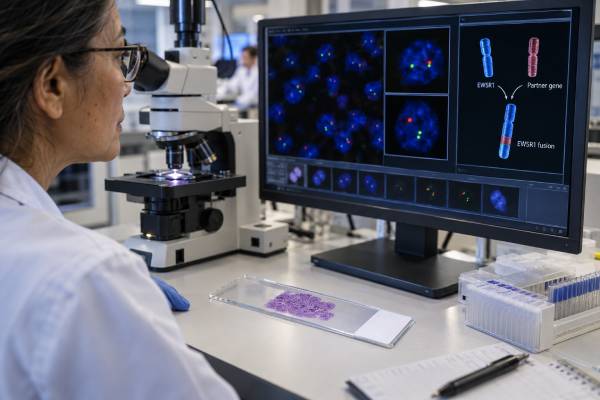
An EWSR1 fusion test looks for a rearrangement involving the EWSR1 gene, a molecular event found in Ewing sarcoma and several other bone and soft-tissue tumors. The key point is that an “EWSR1-positive” result is not a diagnosis by itself. EWSR1 can fuse with many different partner genes, and the partner often determines which tumor entity the result supports. In classic Ewing sarcoma, the most common fusion is EWSR1::FLI1, while other EWSR1 fusions occur in tumors such as desmoplastic small round cell tumor, clear cell sarcoma, extraskeletal myxoid chondrosarcoma, and several newer molecularly defined sarcoma groups. Testing may use fluorescence in situ hybridization (FISH), reverse-transcription PCR, or RNA-based next-generation sequencing. FISH can quickly show that EWSR1 is rearranged, but it usually does not identify the partner gene. Because different EWSR1 rearrangements can point to very different diagnoses, the molecular result must be interpreted with tumor location, microscopic appearance, immunohistochemistry, and the exact fusion partner whenever possible.
- An EWSR1 rearrangement is a molecular clue, not a stand-alone diagnosis, because many distinct tumors can involve EWSR1.
- EWSR1::FLI1 is the most common fusion in Ewing sarcoma, accounting for roughly 85% of cases.
- Break-apart FISH detects EWSR1 rearrangement but usually cannot identify the fusion partner, so RNA sequencing may be needed for precise classification.
- A negative EWSR1 test does not exclude every Ewing-family or round-cell sarcoma because rare tumors use FUS or other molecular drivers.
- The most useful report states the exact fusion, testing method, assay limitations, and whether the finding fits the tumor’s pathology.
Table of Contents
- What EWSR1 Testing Detects
- EWSR1 in Ewing Sarcoma and Related Tumors
- FISH, PCR, and RNA Sequencing
- Interpreting Positive and Negative Results
- Tumor Classification and Differential Diagnosis
- Clinical Meaning and Test Limitations
- Questions to Ask About the Report
What EWSR1 Testing Detects
EWSR1 stands for Ewing sarcoma breakpoint region 1. The gene is located on chromosome 22 and normally encodes an RNA-binding protein involved in gene expression and RNA processing. In certain tumors, a chromosome breaks near EWSR1 and rejoins with another chromosome or genomic region. The rearrangement can create a fusion gene that encodes an abnormal protein capable of driving cancer-cell behavior.
The term gene fusion is more specific than “mutation.” A point mutation changes one or a few DNA letters. A fusion joins parts of two genes. In many sarcomas, that fusion is the central oncogenic event and can be more diagnostically useful than a nonspecific sequence mutation.
EWSR1 is unusually “promiscuous,” meaning it has many possible fusion partners. The biological meaning therefore depends heavily on what is attached to EWSR1. Examples include:
- EWSR1::FLI1 or EWSR1::ERG in Ewing sarcoma;
- EWSR1::WT1 in desmoplastic small round cell tumor;
- EWSR1::ATF1 or EWSR1::CREB1 in clear cell sarcoma and some related neoplasms;
- EWSR1::NR4A3 in a subset of extraskeletal myxoid chondrosarcomas;
- EWSR1::NFATC2 and EWSR1::PATZ1 in molecularly distinct round-cell sarcomas;
- other rare EWSR1 partners that may require expert molecular-pathology review.
This is why a report that says only “EWSR1 rearrangement detected” may be incomplete for final classification. It confirms that the EWSR1 locus is disrupted, but it may not establish which fusion transcript is present.
A broad gene fusion panel can be particularly useful when the microscopic pattern suggests a fusion-driven sarcoma but several entities remain possible.
EWSR1 in Ewing Sarcoma and Related Tumors
Ewing sarcoma is a malignant small round cell tumor that most often affects bone or deep soft tissue in children, adolescents, and young adults, although it can occur at other ages. Modern classification places major weight on the tumor’s fusion biology.
Most Ewing sarcomas harbor a fusion between a member of the FET family, usually EWSR1, and an ETS-family transcription-factor gene. EWSR1::FLI1 is the dominant fusion, present in about 85% of tumors. EWSR1::ERG accounts for a smaller fraction. Rare Ewing sarcomas use other ETS partners or FUS instead of EWSR1.
This molecular definition matters because several tumors can look like Ewing sarcoma under the microscope but are biologically distinct. The World Health Organization framework separates undifferentiated small round cell sarcomas into categories that include:
- Ewing sarcoma;
- sarcoma with EWSR1-non-ETS fusions, such as EWSR1::NFATC2 or EWSR1::PATZ1;
- CIC-rearranged sarcoma; and
- sarcoma with BCOR genetic alterations.
These groups can overlap in age, site, cell shape, and some immunostains, yet they have different molecular drivers and may differ in clinical behavior and treatment sensitivity. Calling every EWSR1-rearranged round-cell tumor “Ewing sarcoma” is therefore incorrect.
Pathologists use morphology and immunohistochemistry to narrow the possibilities before or alongside molecular testing. Classic Ewing sarcoma often shows diffuse membranous CD99 expression and may express NKX2.2 and PAX7, but none of these stains is perfectly specific. The molecular result serves as a high-value confirmation when it matches the histologic picture.
The reverse is also true: a molecular result that does not fit the morphology deserves scrutiny. A technically real EWSR1 fusion may point to a different tumor entity rather than proving the initial suspected diagnosis.
FISH, PCR, and RNA Sequencing
The three most common approaches to EWSR1 testing answer somewhat different questions.
| Method | Main strength | Main limitation |
|---|---|---|
| Break-apart FISH | Fast, widely used, works on formalin-fixed tissue, and detects rearrangement without knowing the partner in advance. | Usually does not identify the partner gene and can yield atypical or equivocal signal patterns. |
| RT-PCR | Highly specific and sensitive for a known fusion transcript. | Requires the laboratory to know which fusion or breakpoint to target; uncommon partners can be missed. |
| RNA-based NGS | Can identify the exact expressed fusion and many possible partners in one assay. | RNA quality can be poor in old or heavily fixed tissue, and assay design differs among laboratories. |
Break-apart FISH
FISH uses fluorescent probes that bind on opposite sides of the EWSR1 locus. In an intact gene, the signals lie close together. When the locus is rearranged, the signals separate or show another abnormal pattern. The laboratory evaluates a defined number of tumor-cell nuclei and applies validated scoring criteria.
A classic positive break-apart result is useful, but EWSR1 break-apart FISH is not tumor-specific. It cannot distinguish EWSR1::FLI1 from EWSR1::WT1, EWSR1::ATF1, or many other fusions. It may also produce unusual signal patterns from deletions, amplifications, complex rearrangements, or technical artifacts.
RNA-based sequencing
RNA sequencing reads expressed transcripts, which makes it especially powerful for fusion detection. Instead of asking only whether EWSR1 is broken, RNA-based NGS can often identify both partners and the fusion junction. This can turn a broad finding such as “EWSR1 rearranged” into a precise result such as “EWSR1::FLI1 detected.”
RNA sequencing is increasingly used when FISH is equivocal, when a rare fusion is suspected, or when the differential diagnosis includes several fusion-defined tumors. A comprehensive solid tumor NGS panel may also capture relevant sequence mutations or copy-number changes alongside the fusion workup.
Interpreting Positive and Negative Results
There is no numeric normal range for an EWSR1 fusion test. Interpretation is categorical and context-dependent.
Positive EWSR1 rearrangement by FISH: This means the EWSR1 locus shows a validated rearranged pattern in the tested tumor cells. It supports a group of EWSR1-rearranged neoplasms but may not identify which one. Partner identification is often the next important step when the diagnosis is not already secure.
Specific EWSR1 fusion detected: A result such as EWSR1::FLI1 provides much stronger classification evidence than a generic break-apart signal. Even then, the finding should fit the specimen’s histology and clinical setting.
Negative result: No target alteration was detected within the assay’s capabilities. A negative EWSR1 FISH result does not exclude every Ewing sarcoma because rare Ewing tumors can involve FUS instead of EWSR1. It also does not exclude CIC-rearranged or BCOR-altered round-cell sarcomas, which are separate entities.
Equivocal or atypical FISH: The signal pattern does not meet clean positive or negative criteria. Follow-up RNA sequencing can be especially valuable in this situation because it may demonstrate whether an expressed oncogenic fusion is actually present.
Insufficient result: There may be too little tumor, degraded nucleic acid, poor hybridization, decalcification damage, or another specimen-quality problem. This is not equivalent to a true negative.
Bone specimens deserve particular attention because strong-acid decalcification can damage DNA and RNA. When a sarcoma is suspected before tissue processing, preserving nondecalcified or gently decalcified material for molecular studies can improve success.
Tumor Classification and Differential Diagnosis
The greatest value of EWSR1 testing is often classification, especially when morphology alone cannot distinguish look-alike tumors.
A small round cell tumor with strong CD99 staining may suggest Ewing sarcoma, but CD99 is also seen in other neoplasms. If FISH shows EWSR1 rearrangement, the diagnosis becomes more likely, yet an EWSR1-non-ETS sarcoma remains possible. Identifying EWSR1::FLI1 or EWSR1::ERG can resolve the question more directly.
In another case, an abdominal tumor with nests of small cells, desmoplastic stroma, and polyphenotypic immunostaining may raise concern for desmoplastic small round cell tumor. Finding EWSR1::WT1 strongly supports that diagnosis and carries different implications from an Ewing fusion.
Likewise, a melanoma-like soft-tissue tumor with melanocytic-marker expression could represent clear cell sarcoma. EWSR1::ATF1 or EWSR1::CREB1 can support that classification. The same broad EWSR1 break-apart result therefore has very different meaning depending on the partner and phenotype.
Pathologists also distinguish EWSR1-associated tumors from fusion-defined sarcomas involving other genes. For example, myxoid liposarcoma is usually driven by a FUS::DDIT3 fusion, while synovial sarcoma is defined by an SS18::SSX fusion. Separate tests such as the FUS fusion test or SS18-SSX fusion test may be more appropriate when those entities are in the differential diagnosis.
This illustrates a practical rule: the best molecular test is the one that answers the actual diagnostic question. A single break-apart probe is efficient when one rearrangement is strongly suspected. A broad RNA panel is more useful when the tumor could belong to several fusion-defined categories.
Clinical Meaning and Test Limitations
An EWSR1 fusion result can affect treatment because accurate tumor classification determines which sarcoma regimen, surgery plan, radiation strategy, or clinical trial is appropriate. In Ewing sarcoma, treatment is generally multimodal and differs substantially from the management of many other EWSR1-rearranged tumors.
However, most EWSR1 fusion proteins are transcription factors rather than easily druggable kinases. The presence of EWSR1::FLI1 currently functions mainly as a diagnostic driver marker, not as a routine marker for an approved fusion-specific targeted drug. Research continues into ways to target fusion-protein biology, transcriptional dependencies, and downstream pathways.
Important limitations include:
- Partner ambiguity with break-apart FISH. A positive EWSR1 signal alone may be too broad for final classification.
- False-negative technical results. Poor tissue quality, low tumor content, decalcification, or unusual breakpoints can reduce sensitivity.
- Rare alternate drivers. Some tumors in the Ewing morphologic spectrum use FUS or non-EWSR1 alterations.
- Incidental or unexpected findings. Broad sequencing can uncover rare fusions whose significance is not fully established.
- Tumor heterogeneity and sampling. A small biopsy may not represent every component of a complex neoplasm.
The report should therefore be read as a synthesis rather than a binary laboratory answer. The strongest diagnostic conclusion usually combines the fusion, morphology, immunophenotype, site, age, imaging findings, and expert pathology review.
Questions to Ask About the Report
When an EWSR1 test is part of a sarcoma workup, useful questions include:
- Was the test break-apart FISH, targeted PCR, RNA sequencing, or a broader NGS panel?
- Did the test identify only an EWSR1 rearrangement, or the exact fusion partner?
- If EWSR1 is rearranged, which tumor entities fit that partner and the pathology?
- If FISH is positive but the partner is unknown, would RNA sequencing improve classification?
- If the result is negative, were FUS, CIC, BCOR, and other relevant fusion drivers evaluated?
- Was the specimen decalcified, and could that have affected test sensitivity?
- Does the molecular result match the microscopic appearance and immunohistochemistry?
- Will the final molecular classification change treatment, prognosis discussions, or clinical-trial eligibility?
For patients and families, the most important distinction is simple: EWSR1 is a gene involved in many fusion-defined tumors, while the exact fusion and pathology determine the diagnosis. A positive result is highly informative when placed in the right context, but it should not be interpreted as synonymous with Ewing sarcoma unless the rest of the evidence supports that conclusion.
A few report details deserve extra attention because they can change the interpretation substantially. The first is whether the laboratory found an in-frame expressed fusion. For many oncogenic sarcoma fusions, the abnormal transcript preserves a functional open reading frame that can encode a chimeric protein. RNA-based assays can provide this information more directly than break-apart FISH. The second is whether the partner is known to define a recognized entity. A familiar partner may strongly support classification, while a novel partner can require review of published cases, domain structure, and other pathologic findings before the laboratory labels it disease-defining.
The wording “rearrangement involving EWSR1” should also be distinguished from “EWSR1 fusion transcript detected.” Rearrangement describes a structural change at the DNA locus. A fusion transcript demonstrates that two genes are actually joined in expressed RNA. In most classic cases the results agree, but complex tumors can produce discordant findings. This distinction helps explain why a pathologist may recommend orthogonal confirmation rather than treating one unusual FISH pattern as definitive.
For Ewing sarcoma specifically, molecular confirmation is especially valuable when the presentation is atypical—for example, an unusual age, organ site, or immunophenotype. Conversely, in a classic tumor with limited tissue, the pathologist may choose a focused test first and preserve material for broader sequencing only if the result is negative or ambiguous. Tissue stewardship matters because small biopsies may also be needed for immunostains, research testing, and future treatment biomarkers.
Finally, molecular classification can affect communication about prognosis without turning the fusion into a simple survival score. Ewing sarcoma, EWSR1::NFATC2 sarcoma, CIC-rearranged sarcoma, and BCOR-altered sarcoma are not interchangeable diseases. Their outcomes and treatment evidence differ. Precise naming therefore reduces the risk of applying outcome statistics or treatment assumptions from the wrong sarcoma group.
A negative EWSR1 result is most useful when the report explains what kind of negative result it is. A high-quality RNA panel that successfully assessed the relevant fusion space gives different reassurance from a failed RNA assay or a narrow test that interrogated only one expected junction. In a small round cell sarcoma, the next step after a negative EWSR1-focused result may be testing for other molecularly defined groups, including CIC- or BCOR-associated tumors, rather than repeating the same assay. The pathology pattern should guide that choice. This layered approach prevents two opposite errors: calling every EWSR1 rearrangement “Ewing sarcoma,” and assuming that an EWSR1-negative round cell tumor lacks a defining molecular alteration.
If tissue is scarce, test selection should be planned with the pathologist before exhausting the block. A targeted assay may answer a strong diagnostic hypothesis quickly, while a broader RNA panel may be more efficient when several fusion-defined sarcomas remain plausible. The best choice depends on the differential diagnosis and specimen quality.
References
- Updates on WHO classification for small round cell tumors: Ewing sarcoma vs. everything else 2024 (Review)
- Ewing and Ewing-like sarcomas: A morphological guide through genetically-defined entities 2022 (Review)
- EWSR1: the promiscuous king of mesenchymal neoplasia. 2024 (Review)
- Identification of Novel/Rare EWSR1 Fusion Partners in Undifferentiated Mesenchymal Neoplasms 2024
- Diagnosis of Fusion-Associated Sarcomas by Exon Expression Imbalance and Gene Expression 2023
Disclaimer
EWSR1 testing is a specialized molecular-pathology tool and should be interpreted together with tumor morphology, immunohistochemistry, imaging, and clinical findings. A positive EWSR1 rearrangement does not by itself establish Ewing sarcoma, and negative or equivocal results may require additional fusion testing. Individual diagnosis and treatment decisions should be made by the treating sarcoma and pathology teams.





