
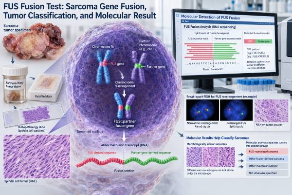
A FUS fusion test looks for an abnormal rearrangement involving the FUS gene, usually in a tumor sample. The result can be highly informative in sarcoma because particular FUS fusion partners are strongly associated with specific tumor types. The best-known example is FUS::DDIT3, the defining molecular alteration in most myxoid liposarcomas. Other FUS fusions, such as FUS::CREB3L2, support different diagnoses.
The important point is that “FUS rearranged” is not a complete diagnosis by itself. The exact partner gene, tumor appearance under the microscope, immunohistochemistry, anatomic site, and clinical setting all matter. A break-apart FISH result may show that FUS is rearranged without identifying the partner, while RNA-based sequencing can often name the full fusion. The pathology report should therefore be read as a molecular finding that helps classify the tumor, not as a stand-alone cancer label.
- A positive FUS fusion result means a tumor-associated FUS rearrangement was detected; the partner gene determines much of its diagnostic meaning.
- FUS::DDIT3 strongly supports myxoid liposarcoma and is present in more than 90% of cases.
- FUS::CREB3L2 is strongly associated with low-grade fibromyxoid sarcoma, usually together with compatible morphology and MUC4 expression.
- A negative result does not rule out sarcoma because not every sarcoma has a FUS fusion and some assays can miss uncommon partners or poor-quality RNA.
- There is no normal numeric range: reports usually state detected/not detected and identify the fusion or rearrangement when possible.
Table of Contents
- What a FUS fusion test detects
- FUS fusions and the sarcoma types they help classify
- How FUS fusion testing is performed
- How to interpret positive, negative, and indeterminate results
- Why the fusion partner and pathology context matter
- Treatment, prognosis, and follow-up implications
- Questions to ask about a FUS fusion result
What a FUS fusion test detects
A FUS fusion test searches for a structural genetic change in which part of the FUS gene becomes joined to part of another gene. This is different from a small DNA mutation that changes one or a few bases. A fusion usually develops because chromosomes break and rejoin in an abnormal arrangement inside a tumor cell.
FUS stands for Fused in Sarcoma. The normal FUS protein helps regulate RNA processing and other cellular functions. In a cancer-associated fusion, the FUS portion may be placed next to a transcription-regulating partner. The resulting fusion protein can switch on an abnormal gene-expression program that drives tumor development.
Many sarcoma-associated fusions are recurrent, meaning the same gene pairing appears repeatedly in a particular tumor type. That makes them valuable diagnostic markers. A pathologist may order a FUS test when the tumor’s appearance suggests a fusion-driven sarcoma or when the microscopic findings overlap among several possible diagnoses.
The report may describe one of several result types:
- A named fusion, such as FUS::DDIT3 or FUS::CREB3L2.
- A FUS rearrangement, meaning the gene is disrupted but the partner was not identified.
- No fusion detected, meaning the assay did not find a reportable FUS event.
- An indeterminate or failed result, usually because the specimen or nucleic acid quality was inadequate.
A FUS test is often part of a broader gene fusion panel rather than a single-gene assay. Broader testing is useful when several fusion-driven tumors are in the differential diagnosis.
There is no “high” or “low” FUS fusion value and no therapeutic range. The clinically meaningful question is whether a rearrangement is present and, ideally, which genes and breakpoints form the fusion.
FUS fusions and the sarcoma types they help classify
The diagnostic meaning of a FUS fusion depends heavily on the partner gene. The same FUS gene can participate in biologically different tumors.
| Fusion or finding | Common association | Key interpretation point |
|---|---|---|
| FUS::DDIT3 | Myxoid liposarcoma | A highly characteristic driver; found in more than 90% of myxoid liposarcomas. |
| FUS::CREB3L2 | Low-grade fibromyxoid sarcoma | Strongly supports the diagnosis when morphology and MUC4 staining fit. |
| FUS::CREB3L1 | Low-grade fibromyxoid sarcoma and related tumors | Less common than FUS::CREB3L2; context remains important. |
| FUS::ERG | Rare Ewing sarcoma | Uncommon alternative to the more typical EWSR1-associated Ewing fusions. |
| FUS rearrangement without partner identification | Several possible fusion-driven neoplasms | Usually requires partner-specific testing or correlation with pathology. |
Myxoid liposarcoma and FUS::DDIT3
Myxoid liposarcoma is the tumor most strongly linked with FUS in routine sarcoma practice. More than 90% harbor FUS::DDIT3, generally produced by a t(12;16) chromosomal translocation. A small minority instead have EWSR1::DDIT3. For this reason, a DDIT3-directed assay may sometimes be more diagnostically complete than testing FUS alone.
The fusion helps separate true myxoid liposarcoma from tumors that may look myxoid under the microscope but have different biology. A related DDIT3 fusion test can focus directly on this defining alteration.
Low-grade fibromyxoid sarcoma and FUS::CREB3L2
Low-grade fibromyxoid sarcoma can look deceptively bland despite its ability to recur or metastasize, sometimes after a long interval. FUS::CREB3L2 is its best-known fusion. MUC4 immunostaining is also highly sensitive in this tumor type, so laboratories commonly integrate morphology, MUC4, and molecular findings rather than relying on one test.
A FUS fusion may therefore answer two different questions: “Is this tumor fusion-driven?” and “Which specific sarcoma does the fusion support?” The second question usually requires identifying the partner.
How FUS fusion testing is performed
FUS fusion testing usually uses tumor tissue from a biopsy or surgical specimen. Formalin-fixed, paraffin-embedded tissue is common, although fresh or frozen material may be available in some centers. The pathology laboratory first selects a block or slide containing enough viable tumor.
Several methods can be used.
Break-apart FISH
Fluorescence in situ hybridization, or FISH, uses labeled probes on either side of the FUS locus. If the probes separate beyond the laboratory’s validated threshold, the result supports a FUS rearrangement.
FISH can be fast and works on small specimens, but it has an important limitation: a break-apart result generally does not identify the partner gene. It can also produce atypical signal patterns caused by complex rearrangements, deletions, copy-number changes, or technical issues. That is why a positive FUS break-apart result may need follow-up testing.
RT-PCR
Reverse-transcription polymerase chain reaction, or RT-PCR, tests RNA for a specific expected fusion transcript. It can be sensitive and efficient when the suspected tumor and likely breakpoint are known. Its weakness is narrow scope: an assay designed for common FUS::DDIT3 variants may miss an uncommon breakpoint or a different partner.
RNA-based next-generation sequencing
Targeted RNA sequencing can examine many fusion genes at once and often identifies both partners. This makes it especially useful when the tumor’s classification is uncertain. RNA sequencing also shows that the rearrangement produces an expressed fusion transcript, which can strengthen interpretation.
RNA is more fragile than DNA, however. Formalin fixation, old tissue, decalcification, low tumor cellularity, and limited material can reduce assay quality. A “no fusion detected” result is most reassuring when the assay passed all quality-control metrics and had adequate tumor input.
A broad solid tumor NGS panel may combine fusion analysis with mutations and other genomic changes when a more comprehensive profile is clinically useful.
How to interpret positive, negative, and indeterminate results
A positive named FUS fusion is generally the most informative result. If the fusion is well established for a tumor type and the pathology fits, it can provide strong molecular support for the diagnosis. For example, finding FUS::DDIT3 in a compatible myxoid sarcoma is powerful evidence for myxoid liposarcoma.
A report that says only FUS rearrangement detected is less specific. It confirms structural alteration of FUS but may not show what gene is attached. In that situation, the next useful question is whether the laboratory can identify the partner with RNA sequencing, targeted PCR, or another method.
A negative result has several possible meanings:
- The tumor truly lacks a FUS rearrangement.
- The tumor is a related entity driven by another fusion gene.
- The specific assay did not cover the relevant breakpoint or partner.
- The tumor fraction was too low.
- RNA or DNA quality was inadequate.
- The sample was affected by fixation, decalcification, necrosis, or other pre-analytic factors.
For example, a suspected myxoid liposarcoma with negative FUS testing could still contain EWSR1::DDIT3. Likewise, a round-cell sarcoma may have an EWSR1 fusion rather than a FUS fusion.
An indeterminate, failed, or quantity-not-sufficient result should not be interpreted as negative. The report may recommend another tissue block, repeat extraction, a different molecular method, or a new biopsy if the result would change diagnosis or management.
Some reports include read counts, percent abnormal nuclei, or other technical measurements. These values are assay-specific and are not comparable across laboratories. The interpretive conclusion matters more than treating a technical number as a universal cutoff.
Why the fusion partner and pathology context matter
Sarcoma diagnosis is an integration task. Molecular data can be decisive, but the same gene can participate in multiple tumors, and a fusion that looks plausible must still fit the specimen.
The pathologist usually considers:
- Tumor architecture and cell shape under the microscope.
- Myxoid, fibrous, lipogenic, epithelioid, or round-cell features.
- Immunohistochemical staining pattern.
- Patient age and tumor location.
- Imaging and clinical behavior.
- Exact fusion partner and predicted transcript.
- Whether the result is a recognized driver or an uncertain event.
This prevents a common error: interpreting the word FUS as though it names one cancer. It does not. FUS is one component of several molecularly distinct diseases.
The exact partner can also resolve look-alike tumors. FUS::DDIT3 points toward myxoid liposarcoma, while FUS::CREB3L2 points toward low-grade fibromyxoid sarcoma. A different sarcoma may instead be defined by SS18::SSX, EWSR1-associated fusions, or another rearrangement.
Laboratories also evaluate whether a detected transcript is biologically credible. Very low read support, an out-of-frame event, a likely read-through transcript, or a fusion not previously associated with the tumor may be reported cautiously. A technically detected fusion is not automatically a pathogenic cancer driver.
This is why the most useful report is not simply “positive.” It explains what was found, how it was detected, whether the event is known to be oncogenic, and how well it fits the proposed diagnosis.
Treatment, prognosis, and follow-up implications
For most FUS-rearranged sarcomas, the immediate value of testing is accurate tumor classification. Correct classification matters because surgery, radiation, chemotherapy sensitivity, surveillance, and expected patterns of spread differ among sarcoma subtypes.
In myxoid liposarcoma, confirming FUS::DDIT3 helps establish a diagnosis with recognized clinical behavior. Myxoid liposarcoma has a distinctive tendency to metastasize to extrapulmonary sites, and it can be relatively sensitive to radiotherapy compared with many other soft-tissue sarcomas. Treatment decisions still depend on tumor size, location, resectability, grade or round-cell component, metastatic status, prior therapy, and the patient’s overall condition.
In low-grade fibromyxoid sarcoma, molecular confirmation can be particularly valuable because the tumor may look low grade but retain long-term metastatic potential. Long follow-up may be needed. The presence of FUS::CREB3L2 itself does not provide a simple numeric prognosis; the clinical course depends on the full disease picture.
A FUS fusion is not usually interpreted as a hereditary finding. These rearrangements are generally somatic, meaning they arise in the tumor rather than being inherited in every cell of the body. If a separate germline concern exists because of personal or family history, that requires dedicated hereditary cancer assessment.
Fusion proteins are active areas of drug development, but a FUS fusion should not be assumed to have an approved fusion-specific therapy. Some sarcoma treatments exploit broader tumor biology rather than directly inhibiting FUS. Clinical-trial eligibility may depend on the specific fusion, tumor type, disease stage, and prior therapy.
When a molecular finding changes the diagnostic label, it may also change which sarcoma specialist, staging approach, or clinical trial is most appropriate. That is one reason difficult cases are often reviewed by a specialist soft-tissue pathologist.
Questions to ask about a FUS fusion result
A short discussion with the pathology or oncology team can clarify what the molecular report means in practice. Useful questions include:
- Was an exact fusion identified, or only a FUS rearrangement? A named partner generally provides more diagnostic information than a break-apart result alone.
- Does the fusion fit the tumor’s appearance and immunostains? Concordance strengthens confidence in the diagnosis.
- What method was used? FISH, RT-PCR, DNA sequencing, and RNA sequencing have different strengths and limitations.
- Did the specimen pass quality-control requirements? This is especially important for a negative or failed result.
- Could another fusion explain the tumor if FUS testing was negative? Broader RNA testing may be useful when suspicion remains high.
- Does the result change the final tumor classification? The molecular finding may confirm, refine, or occasionally overturn the preliminary diagnosis.
- Does the diagnosis change treatment or surveillance? The answer usually depends on the sarcoma subtype and stage rather than the fusion name alone.
- Would expert pathology review help? Rare sarcomas and unusual fusion results often benefit from subspecialty review.
A FUS fusion result is most useful when it is read as one part of a complete pathology interpretation. A recognized fusion in the right morphologic setting can provide strong diagnostic certainty; an isolated or technically limited finding may need confirmation before it drives care.
When repeat or broader testing can be useful
Additional testing is most useful when the molecular result and the pathology do not agree. For example, a pathologist may strongly suspect myxoid liposarcoma but receive a negative FUS break-apart result. Because a minority of these tumors have EWSR1::DDIT3 rather than FUS::DDIT3, a DDIT3-based assay or broader RNA fusion panel may answer the question more directly. Similarly, a FUS rearrangement without a known partner may need RNA sequencing before the final diagnosis is assigned.
Repeat testing can also be reasonable after a technical failure. A different paraffin block may contain more viable tumor or less necrosis. A non-decalcified specimen can be preferable to heavily decalcified bone tissue because some decalcification methods damage nucleic acids. When only a tiny biopsy is available, the laboratory and treating team may need to balance molecular testing against preserving tissue for immunohistochemistry and other essential studies.
The report’s limitation section deserves close attention. It should explain whether the assay detects only selected partners, whether novel partners can be discovered, and what minimum quality thresholds were met. Those details determine how much confidence to place in a negative result. In a rare tumor, a negative narrow assay may simply mean that the next test needs to look more broadly.
A final practical point is that the partner gene often carries more diagnostic weight than the word FUS itself. FUS::DDIT3 strongly supports myxoid liposarcoma in the appropriate setting, while FUS::CREB3L2 is characteristic of low-grade fibromyxoid sarcoma. A laboratory that reports only “FUS rearranged” has answered a narrower question. When classification affects surgery, surveillance, or expected behavior, resolving the partner can be worth the additional testing.
References
- Diagnosis and Treatment of Myxoid Liposarcoma 2024 (Review)
- FUS::DDIT3 Fusion Protein in the Development of Myxoid Liposarcoma and Possible Implications for Therapy 2024 (Review)
- Low-Grade Fibromyxoid Sarcoma and Related Subtypes: A Systematic Review and Pooled Analysis of 773 Cases 2026 (Systematic Review)
- Clinical Utility of an RNA-based Gene Fusion Assay in Sarcoma for Diagnosis and Management: Experience in an Australian Laboratory 2026
Disclaimer
This article is for general educational purposes and does not replace interpretation by a pathologist, sarcoma specialist, or other qualified clinician. FUS fusion results must be interpreted with the tumor’s morphology, immunohistochemistry, specimen quality, testing method, and clinical context. Treatment and follow-up decisions should be based on the final integrated diagnosis, not the fusion result alone.





