
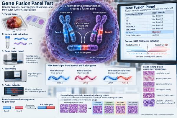
A gene fusion panel tests a tumor for abnormal gene pairings created when chromosomes rearrange. These fusions can act as cancer drivers, help identify a tumor type, or point to a targeted treatment. Modern panels often use RNA-based next-generation sequencing (NGS) because RNA can reveal the actual fusion transcript produced by the tumor and can identify many possible gene partners in one test.
Fusion testing is especially important in lung cancer, sarcoma, thyroid cancer, and several other solid tumors, but its role varies by disease. Some fusions are primarily diagnostic, such as SS18::SSX in synovial sarcoma. Others are treatment biomarkers, such as ALK, ROS1, RET, or NTRK fusions in appropriate cancers. A panel result therefore needs more than a simple positive/negative reading. The exact genes, breakpoint, assay method, specimen quality, tumor type, and whether the fusion is a recognized driver all determine what the result means.
- A gene fusion panel looks for abnormal joined genes that can classify a tumor or reveal a treatment target.
- RNA-based NGS is often preferred for fusion detection because it can identify expressed fusion transcripts and many possible partners.
- A positive result is most useful when the exact fusion is a known cancer driver that fits the tumor type and pathology.
- A negative panel does not exclude every fusion; poor RNA quality, low tumor content, or panel design can cause missed events.
- There is no normal numeric range: results are usually reported as detected, not detected, indeterminate, or failed, with interpretation of any fusion found.
Table of Contents
- What a gene fusion panel tests
- Why gene fusions matter in cancer
- RNA, DNA, FISH, and PCR methods
- How to read gene fusion panel results
- Specimen quality and test limitations
- How results can change diagnosis or treatment
- Questions to ask after fusion panel testing
What a gene fusion panel tests
A gene fusion panel searches for rearrangements that join parts of two genes together. The rearrangement may create a new fusion protein, place a growth-promoting gene under abnormal control, or otherwise activate a cancer pathway. Many such events are acquired only in the tumor and are not inherited.
A panel differs from a single-gene fusion assay because it can examine dozens or hundreds of genes at once. That is useful when several molecular diagnoses are possible or when the cancer could contain one of many actionable fusions.
Depending on the platform, a report may detect:
- Known fusion pairs, such as EML4::ALK.
- A known driver gene joined to an unexpected partner.
- Rearrangements involving genes such as ALK, ROS1, RET, NTRK1, NTRK2, NTRK3, EWSR1, FUS, FGFR2, FGFR3, or others included in the assay.
- Exon-skipping or other RNA events on some panels.
- In-frame versus out-of-frame transcripts, if the laboratory reports that information.
The panel’s exact gene list matters. “Fusion panel” is not one universal test. One laboratory may focus on lung cancer targets, another on sarcomas, and another may use a broad pan-cancer design. The report should state what genes and fusion types the assay can detect.
A broad solid tumor NGS panel may combine fusion analysis with small mutations, copy-number changes, and other biomarkers. A dedicated fusion panel may be preferred when RNA-based fusion detection is the main clinical question or when tissue needs to be conserved.
Because fusion panels are qualitative molecular tests, they usually do not have a reference interval like a blood test. The clinically important output is the identity and interpretation of a detected rearrangement.
Why gene fusions matter in cancer
Gene fusions matter for two major reasons: tumor classification and treatment selection. Some do both.
Diagnostic and classification fusions
Many sarcomas and other rare tumors are defined or strongly supported by characteristic gene fusions. Examples include:
- SS18::SSX in synovial sarcoma.
- FUS::DDIT3 or EWSR1::DDIT3 in myxoid liposarcoma.
- EWSR1::FLI1 in most Ewing sarcomas.
- FUS::CREB3L2 in many low-grade fibromyxoid sarcomas.
These findings can resolve cases in which the microscopic appearance overlaps with other tumors. A specific fusion can confirm a suspected diagnosis, refine an imprecise label, or show that a tumor belongs to a different molecular class.
For example, an EWSR1 fusion result is not meaningful until the partner is considered. EWSR1 can fuse with FLI1 in Ewing sarcoma, WT1 in desmoplastic small round cell tumor, and other partners in unrelated neoplasms.
Predictive fusions
Other fusions create activated kinases that can be targeted by drugs. ALK, ROS1, RET, and NTRK fusions are established examples in specific clinical settings. An NTRK fusion, for instance, can be a tumor-agnostic treatment biomarker when it is a qualifying oncogenic fusion and the clinical criteria for therapy are met.
The same principle applies to RET fusions and ALK fusions in cancers where targeted treatment is established. However, the presence of a rearrangement involving a familiar gene does not automatically mean a drug will work. The fusion must be biologically relevant, and the treatment evidence must apply to that tumor and alteration.
This is why laboratories often classify results as pathogenic/oncogenic, likely pathogenic, uncertain significance, or nonactionable rather than treating all fusion calls equally.
RNA, DNA, FISH, and PCR methods
Several technologies can detect gene fusions. They answer overlapping but not identical questions.
| Method | Main strength | Main limitation |
|---|---|---|
| RNA-based NGS | Detects expressed fusion transcripts and can identify many partners | RNA can degrade in formalin-fixed or old tissue |
| DNA-based NGS | Can combine mutations, copy number, and rearrangements in one assay | Large or repetitive introns can make some fusions difficult to capture |
| Break-apart FISH | Fast, targeted evidence that a selected gene is rearranged | Usually does not identify the partner and may show atypical patterns |
| RT-PCR | Highly targeted and sensitive for a known transcript | May miss unexpected partners or breakpoint variants |
Why RNA is often valuable
RNA sequencing reads the transcript made after a rearrangement. This has several advantages. Many fusion-driving genes contain very large introns, making DNA capture technically difficult. RNA removes most intronic sequence through normal splicing, so the joined exons can be easier to detect. Finding an expressed, in-frame transcript also helps establish that the rearrangement is biologically plausible.
Two common targeted RNA strategies are anchored multiplex PCR and hybrid-capture RNA sequencing. Anchored methods can discover an unknown partner when one side of the fusion is a targeted gene. Hybrid capture can interrogate a broad set of transcripts without requiring every possible gene pairing to be predefined.
RNA is not automatically superior in every specimen. It degrades more easily than DNA. A highly damaged sample may yield a failed RNA assay even when DNA sequencing succeeds. Some laboratories therefore use combined DNA and RNA workflows.
When FISH or PCR still helps
FISH remains useful when a specific rearrangement is strongly suspected and tissue is limited. It can also serve as an orthogonal confirmation method. RT-PCR is useful when a known fusion transcript is expected and rapid, focused testing is appropriate.
The most informative method depends on the diagnostic question. A broad uncertain sarcoma often benefits from RNA sequencing, while a narrowly defined question may be answered efficiently by a validated single-gene assay.
How to read gene fusion panel results
A gene fusion report should be read in layers. Start with the result category, then identify the exact fusion, and finally read the laboratory’s clinical interpretation.
Detected or positive
A positive report may list a fusion in the form gene A::gene B, often with exons or transcript identifiers. The key questions are:
- Is the fusion known to be oncogenic?
- Is it in-frame and expressed?
- Is the gene orientation biologically plausible?
- Does it match the tumor’s pathology and site?
- Is there evidence that it predicts response to a targeted drug?
- Does the result define or strongly support a particular diagnosis?
A named fusion is generally more informative than “rearrangement detected.” For example, the diagnostic meaning of a FUS fusion changes depending on whether the partner is DDIT3, CREB3L2, ERG, or another gene.
Not detected or negative
A negative result means no reportable fusion was found within the assay’s validated scope. It does not mean the tumor has no molecular driver and does not exclude every possible rearrangement.
A negative result is more convincing when:
- Tumor content was adequate.
- RNA or DNA passed quality thresholds.
- The relevant genes and breakpoints are included in the panel design.
- The assay is validated for the specimen type.
- No technical warning limits interpretation.
If the pathology strongly suggests a fusion-driven tumor, a negative DNA panel may lead to RNA testing, or a failed RNA test may lead to FISH or another specimen.
Variant or fusion of uncertain significance
Some panels identify uncommon transcripts whose role is unclear. A fusion can be real but not known to drive cancer. Laboratories may evaluate whether the event preserves the protein reading frame, retains critical functional domains, has been reported in cancer databases or literature, and occurs at sufficient read support.
A fusion of uncertain significance should not be treated as though it has the same evidence as a well-established driver. Clinical decisions should follow the laboratory interpretation and disease-specific evidence.
Specimen quality and test limitations
Fusion testing begins before sequencing. The quality of the tissue strongly affects whether the test can answer the question.
Most panels use formalin-fixed, paraffin-embedded tumor from a biopsy or resection. A pathologist typically selects the area with the highest viable tumor content and may mark tissue for macrodissection. Factors that can reduce success include:
- Very small biopsies.
- Low tumor cellularity.
- Extensive necrosis.
- Heavy cautery artifact.
- Prolonged fixation.
- Old tissue blocks with degraded RNA.
- Acid decalcification of bone specimens.
- Prior treatment that leaves little viable tumor.
In a large prospective clinical experience with targeted RNA sequencing across more than 2,000 cancers, testing was feasible in most formalin-fixed specimens, but failures still occurred. Real-world studies also show that tissue cellularity influences success.
This makes the phrase quantity not sufficient clinically important. It is not a negative molecular result. If fusion status could change diagnosis or treatment, another block, another method, or a new biopsy may be considered.
Panel design also creates limitations. Some assays examine only selected genes or exons. DNA panels may not cover long intronic regions where rearrangements occur. RNA panels can miss fusions if the transcript is poorly expressed or degraded. Bioinformatic filters may intentionally suppress low-confidence calls to reduce false positives.
A result can also be technically complex. Break-apart FISH may show borderline or atypical signal patterns. RNA sequencing may find a low-support transcript. DNA sequencing may detect a structural breakpoint without proving that a functional transcript is expressed. Such cases may need orthogonal confirmation and expert molecular pathology review.
How results can change diagnosis or treatment
The clinical value of a fusion panel is often greater than simply adding another line to the pathology report. In selected patients, the result can change the tumor name, stage-specific treatment options, or trial eligibility.
Recent real-world studies of RNA-based fusion testing have found that fusion results can confirm or revise diagnoses and can also identify alterations that lead to targeted treatment. This is especially useful in poorly differentiated tumors, rare sarcomas, cancers with an unknown primary, and cases in which earlier molecular tests were inconclusive.
A diagnostic fusion can prevent treatment based on the wrong tumor category. For example, confirming SS18::SSX supports synovial sarcoma, while identifying a different fusion may redirect the diagnosis to another sarcoma family. In an ambiguous round-cell tumor, finding the exact fusion partner may be more informative than morphology alone.
A predictive fusion can open a targeted treatment pathway. Whether that happens depends on the gene, tumor type, treatment history, and current regulatory or guideline criteria. A fusion should therefore be matched to current disease-specific evidence, not simply to a drug that targets the same broad pathway.
The absence of an actionable fusion can still be useful. It may narrow the differential diagnosis, prevent inappropriate targeted therapy, or prompt a different molecular strategy. A negative focused panel may lead to broader sequencing when the clinical suspicion remains high.
Fusion testing usually evaluates somatic tumor alterations. A positive cancer fusion generally does not mean the patient inherited that rearrangement or that relatives need testing. Separate germline testing is used when personal or family history suggests an inherited cancer syndrome.
Questions to ask after fusion panel testing
The most useful follow-up questions focus on what the assay actually tested and what the result changes.
- Was RNA, DNA, FISH, or PCR used? The method affects sensitivity and the type of information available.
- Did the panel identify the exact fusion partner? A partner-specific result is often more clinically meaningful than a generic rearrangement.
- Is the fusion considered an established driver, likely driver, or uncertain finding? The evidence level matters.
- Does the fusion fit the pathology diagnosis? Molecular and microscopic findings should be interpreted together.
- Did the sample pass quality control? A negative result with poor RNA quality may need additional testing.
- Does this fusion have an approved treatment in this cancer? Treatment relevance can be tumor-specific even when the same gene is actionable elsewhere.
- Could a broader panel find something this assay does not cover? This is useful when a focused panel is negative but suspicion remains.
- Would confirmation by another method help? Borderline, novel, or unexpected findings sometimes warrant orthogonal testing.
For patients, the main takeaway is that a fusion panel is not simply a “cancer-positive” test. It is a molecular classification tool. Its value comes from connecting an exact rearrangement with the tumor’s histology, clinical setting, and current evidence for diagnosis or therapy.
What to keep from the report
It is useful to keep the complete molecular report rather than only the one-line result. Future treatment decisions may depend on the exact fusion partner, exon structure, specimen source, test date, and assay platform. If the cancer later recurs or spreads, an oncology team can compare the original fusion with newer testing and decide whether repeat profiling is worthwhile.
The report also records whether a finding was considered diagnostic, prognostic, or potentially actionable at the time of testing. Those categories can evolve as new evidence and drug approvals appear. The underlying fusion usually does not change simply because its clinical interpretation changes. Keeping the original report makes it easier for a later specialist to reassess the alteration against current evidence without assuming what the earlier assay did or did not cover.
When a panel is ordered for treatment selection, the report should also make clear whether the assay can detect the fusion architecture required by the relevant evidence. Some therapies are linked to a gene fusion broadly, while others depend on a particular gene, tumor context, or validated companion test. A technically real fusion can therefore be diagnostically important without being drug-actionable. Separating “detected,” “oncogenic,” and “actionable” avoids turning one molecular finding into three assumptions.
References
- Clinical utility of targeted RNA sequencing in cancer molecular diagnostics 2025
- Diagnostic impact of RNA-based next-generation sequencing fusion panel for solid tumors: A single-institution experience 2023
- Practical Considerations for Oncogenic Fusion Detection and Reporting in Solid Tumors. 2024 (Review)
- Consensus Recommendations to Optimize the Detection and Reporting of NTRK Gene Fusions by RNA-Based Next-Generation Sequencing 2023 (Consensus)
- Molecular Testing in Solid Tumors: Best Practices from the Molecular Pathology and Precision Medicine Study Group of the Italian Society of Pathology (PMMP/SIAPeC) 2025
Disclaimer
This article provides general educational information about tumor gene fusion testing. Panel coverage, assay performance, result classification, and treatment relevance differ among laboratories and cancer types. A molecular pathologist and treating oncology team should interpret the result together with the specimen quality, pathology diagnosis, disease stage, and current treatment guidance.





