
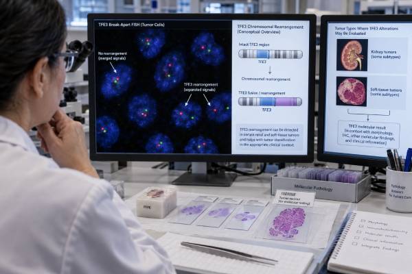
A TFE3 fusion test looks for rearrangements of the TFE3 transcription-factor gene on chromosome Xp11. These fusions define or strongly support several uncommon tumor types, most importantly TFE3-rearranged renal cell carcinoma (RCC), alveolar soft part sarcoma, and a subset of PEComas. The test is primarily a diagnostic and classification marker rather than a universal drug-selection biomarker. TFE3 protein immunohistochemistry can raise suspicion, but molecular confirmation is often important because staining can be technically variable and can occur without a true gene fusion. Break-apart FISH detects many TFE3 rearrangements, while RNA-based next-generation sequencing can identify the exact expressed partner, such as ASPSCR1::TFE3 or PRCC::TFE3. Some intrachromosomal rearrangements can be difficult for standard FISH, so a negative result does not always end the workup when morphology and immunophenotype remain strongly suggestive. In kidney cancer, the 2022 WHO classification places TFE3-rearranged RCC among molecularly defined renal carcinomas, emphasizing that the molecular abnormality—not one specific microscopic appearance—defines the entity.
- A confirmed TFE3 fusion can define TFE3-rearranged RCC and strongly support several fusion-defined soft-tissue tumors.
- TFE3 IHC is a screening clue, not a perfect substitute for molecular testing; strong nuclear staining can be false positive or technically variable.
- Break-apart FISH detects many TFE3 rearrangements, while RNA NGS can identify the exact partner and expressed fusion transcript.
- A negative FISH result can miss certain cryptic or intrachromosomal rearrangements, so discordant cases may need RNA sequencing or another method.
- TFE3 fusion status is mainly diagnostic; treatment still depends on the specific tumor type, stage, and other clinical factors.
Table of Contents
- What a TFE3 Fusion Is
- Tumors Associated With TFE3 Fusions
- How TFE3 Testing Is Performed
- How to Interpret TFE3 Results
- TFE3-Rearranged Kidney Cancer
- Soft-Tissue Tumors and TFE3
- Limitations, Treatment Meaning, and Next Steps
What a TFE3 Fusion Is
TFE3 belongs to the MiT family of transcription factors, which also includes TFEB, TFEC, and MITF. These proteins regulate gene expression involved in lysosomal biology, metabolism, pigmentation, and cellular differentiation.
In a TFE3-rearranged tumor, part of TFE3 becomes joined to another gene. The fusion changes transcriptional control and can act as the main oncogenic driver. Numerous partners have been described. In renal cancer, common examples include ASPSCR1, PRCC, SFPQ, NONO, and others. Alveolar soft part sarcoma is classically associated with ASPSCR1::TFE3. TFE3-rearranged PEComas can involve SFPQ, NONO, DVL2, and additional partners.
The exact partner can influence morphology and possibly biology, but many partner-specific outcome differences remain incompletely defined. For diagnosis, demonstrating a genuine TFE3 rearrangement or expressed fusion is usually more important than trying to predict behavior from the partner alone.
TFE3 fusion should not be confused with TFE3 protein overexpression. IHC detects protein abundance and localization; it does not directly show that two genes are fused. Likewise, TFE3 amplification or other regulatory changes are not equivalent to a canonical fusion.
Because the abnormality is structural, fusion-aware methods are required. A panel that only sequences TFE3 exons for point mutations may not answer the clinical question.
Tumors Associated With TFE3 Fusions
The best-established setting is TFE3-rearranged RCC, historically called Xp11 translocation RCC. The older name came from the TFE3 location on Xp11. The current WHO framework favors a molecularly defined name because tumors can have many different appearances and partners.
TFE3-rearranged RCC occurs in both children and adults. It represents a larger proportion of pediatric RCC than adult RCC, but adult cases are clinically important and can be aggressive. Some patients have a history of prior cytotoxic chemotherapy, although most do not.
Alveolar soft part sarcoma (ASPS) is a rare soft-tissue sarcoma that often affects adolescents and young adults. It commonly arises in deep soft tissue of the extremities or trunk and can metastasize to lung, brain, or bone. ASPSCR1::TFE3 is a defining molecular feature.
TFE3-rearranged PEComa is another recognized group. PEComas show perivascular epithelioid-cell differentiation and often express melanocytic markers. TFE3-rearranged PEComas may occur in younger patients and can differ molecularly from conventional PEComas driven by TSC1/TSC2 pathway alterations.
Rare TFE3 fusions have also been described in other neoplasms, including subsets of epithelioid hemangioendothelioma and unusual renal or soft-tissue tumors. In these settings, the exact partner plus morphology is essential because “TFE3 rearranged” alone may not establish the final entity.
How TFE3 Testing Is Performed
TFE3 immunohistochemistry
TFE3 IHC looks for nuclear protein expression. Strong nuclear staining can support a TFE3-driven tumor, but results are sensitive to fixation and technical conditions. Background or nonspecific staining can create false positives, while poor antigen preservation can create false negatives.
For renal tumors, IHC is usually interpreted with morphology and a broader panel. A PAX8 IHC result can support renal epithelial differentiation, while cathepsin K, melanocytic markers, CAIX, CK7, and others may help separate TFE3-rearranged RCC from mimics. No single IHC panel replaces molecular confirmation in a difficult case.
Break-apart FISH
TFE3 break-apart FISH uses probes on opposite sides of the gene. When the locus is rearranged, the signals separate. FISH works on formalin-fixed tissue and has been widely used as a confirmatory test.
The main limitation is geometry. Some TFE3 fusions result from small intrachromosomal inversions or rearrangements in which the probe signals remain close enough to appear falsely non-rearranged. Borderline signal patterns can also be hard to interpret, especially in truncated nuclei.
RNA-based next-generation sequencing
RNA sequencing detects the actual fusion transcript and can identify the partner. This is particularly valuable when FISH and IHC are discordant, when a cryptic rearrangement is suspected, or when several fusion-defined diagnoses are being considered.
RNA quality can be poor in old or over-fixed tissue. Therefore, a failed RNA assay should be labeled noninformative rather than negative. A broad solid tumor NGS panel that includes validated RNA fusion detection can often answer both diagnostic and treatment-related questions from the same specimen.
How to Interpret TFE3 Results
A positive RNA fusion result naming an in-frame TFE3 partner is strong molecular evidence. In the correct histologic context, it can define the tumor class.
A positive TFE3 break-apart FISH result supports a TFE3 rearrangement but usually does not identify the partner. In classic renal morphology, this can be sufficient for TFE3-rearranged RCC. In an unusual soft-tissue tumor, partner identification may be more informative because different tumor entities can involve TFE3.
A positive TFE3 IHC result raises suspicion but should not automatically be called a fusion. Strong, crisp nuclear staining is more convincing than weak or diffuse background, but molecular confirmation is often preferred when the diagnosis carries major implications.
A negative FISH result does not completely exclude TFE3 rearrangement if the suspected fusion is cryptic or intrachromosomal. RNA sequencing can be a useful reflex test in a highly suspicious case.
A negative high-quality RNA fusion assay is generally stronger evidence against a TFE3 fusion, assuming the assay covers TFE3, the specimen has adequate tumor, and RNA quality passed. Even then, rare breakpoint configurations or assay design limitations should be checked.
An indeterminate result may reflect low tumor content, degraded RNA, failed internal controls, or borderline FISH separation. It should not be converted to “wild type” in the clinical record.
TFE3-Rearranged Kidney Cancer
The 2022 WHO classification moved renal tumor taxonomy further toward molecular definitions. TFE3-rearranged RCC is now grouped among molecularly defined renal carcinomas, along with TFEB-altered, ELOC-mutated, FH-deficient, SDH-deficient, ALK-rearranged, and SMARCB1-deficient renal carcinomas.
This approach reflects the variable appearance of TFE3-rearranged RCC. Tumors can be papillary, nested, alveolar, solid, or mixed. Cells may have clear or eosinophilic cytoplasm, and psammoma bodies can occur. Some cases look similar to clear-cell RCC or papillary RCC.
Age alone should not exclude the diagnosis. Although recognized initially in children and young adults, TFE3-rearranged RCC also occurs in older adults. Adult disease can present at advanced stage and may behave aggressively.
The molecular diagnosis can prevent misclassification. That matters because clinical-trial eligibility, prognosis discussions, and treatment selection increasingly use renal tumor subtype rather than treating all non-clear-cell RCC as one group.
There is no approved drug that directly targets the TFE3 fusion protein. Advanced translocation RCC is generally managed within non-clear-cell RCC strategies, including immune-checkpoint and VEGF-pathway regimens, with growing but still less mature evidence than for clear-cell RCC. Specialist referral and clinical trials are particularly valuable in metastatic disease.
The fusion partner may correlate with certain morphologic patterns—for example, ASPSCR1::TFE3 and PRCC::TFE3 often look different—but partner-specific treatment is not established routine practice.
Soft-Tissue Tumors and TFE3
In soft tissue, the exact fusion can be more entity-defining than simply knowing TFE3 is rearranged.
ASPSCR1::TFE3 supports alveolar soft part sarcoma when morphology fits. ASPS often has organoid nests separated by delicate vascular septa, creating an alveolar appearance. Crystal-like material may be seen, and TFE3 IHC is often strongly nuclear.
TFE3-rearranged PEComas frequently express cathepsin K and melanocytic markers such as HMB45, while epithelial-marker expression can vary. Because TFE3-rearranged RCC can also express melanocytic markers and cathepsin K, renal-site lesions can be particularly difficult. PAX8, epithelial markers, morphology, and the exact fusion partner help separate them.
This distinction may affect treatment. Conventional PEComas with TSC1/TSC2 loss can be sensitive to mTOR-pathway inhibition. TFE3-rearranged PEComas often lack the same TSC-driven biology, so they should not automatically be assumed to behave or respond identically.
ASPS has its own systemic-treatment evidence, including immune-checkpoint therapy in advanced disease. Again, the fusion confirms the tumor identity rather than serving as a direct drug target.
Rare TFE3-rearranged tumors outside these categories should be reviewed carefully because a fusion partner can help distinguish a novel or unusual entity from a more common mimic.
Limitations, Treatment Meaning, and Next Steps
The most common mistake is relying on TFE3 IHC alone. Strong staining can be a useful screening result, but false positivity and technical variability are well documented. A major diagnosis should be molecularly confirmed when possible.
The second mistake is treating FISH as infallible. Standard break-apart probes can miss cryptic rearrangements, especially small inversions. Discordant IHC/FISH cases deserve review of signal pattern, probe design, and possible RNA testing.
The third mistake is assuming every TFE3 fusion means the same cancer. ASPSCR1::TFE3 in a deep soft-tissue mass and a TFE3 fusion in a renal epithelial tumor may define different diseases. Site and morphology remain essential.
The fourth is equating a fusion with an approved targeted therapy. TFE3 is currently more useful for classification than direct treatment selection. Management follows the diagnosed tumor type.
Useful next-step questions include:
- Was the result obtained by IHC, FISH, RNA sequencing, or more than one method?
- If molecularly positive, what is the fusion partner?
- Does the morphology fit TFE3-rearranged RCC, ASPS, PEComa, or another entity?
- Did a negative test pass quality controls, and can the method detect cryptic rearrangements?
- Would expert renal or soft-tissue pathology review change classification?
- Does the final tumor subtype qualify for a disease-specific clinical trial or systemic therapy?
TFE3 fusion testing is therefore best used as a molecular classification tool. A well-validated positive result can define an otherwise confusing renal or soft-tissue tumor; a carefully interpreted negative result can redirect the diagnostic workup.
References
- The WHO 2022 Classification of Renal Neoplasms (5th Edition): Salient Updates 2024 (Review)
- The new WHO 2022 category of molecularly defined renal carcinomas: Accessible to practicing pathologists 2023 (Review)
- TFE3 Rearrangement and Expression in Renal Cell Carcinoma 2023 (Review)
- Management of translocation carcinomas of the kidney 2024 (Review)
- A review of neoplasms with MITF/MiT family translocations 2022 (Review)
Disclaimer
TFE3 results require expert pathology interpretation because IHC, FISH, and RNA assays have different limitations and several distinct tumors can carry TFE3 fusions. Treatment depends on the final tumor diagnosis and stage rather than the fusion alone. This article is educational and does not replace pathology or oncology care.





